


주메뉴
- About IBS 연구원소개
-
Research Centers
연구단소개
- Research Outcomes
- Mathematics
- Physics
- Center for Underground Physics
- Center for Theoretical Physics of the Universe (Particle Theory and Cosmology Group)
- Center for Theoretical Physics of the Universe (Cosmology, Gravity and Astroparticle Physics Group)
- Dark Matter Axion Group
- Center for Artificial Low Dimensional Electronic Systems
- Center for Quantum Nanoscience
- Center for Exotic Nuclear Studies
- Center for Van der Waals Quantum Solids
- Center for Relativistic Laser Science
- Center for Trapped Ion Quantum Science
- Chemistry
- Life Sciences
- Center for Memory and Glioscience (Cognitive Glioscience Group)
- Center for Memory and Glioscience (Learning and Memory Group)
- Center for Synaptic Brain Dysfunctions
- Center for RNA Research
- Center for Genomic Integrity
- Center for Vascular Research
- Center for Genome Engineering
- Center for Microbiome–Body–Brain Physiology
- Earth Science
- Interdisciplinary
- Center for Neuroscience Imaging Research (Neuro Technology Group)
- Center for Neuroscience Imaging Research (Cognitive and Computational Neuroscience Group)
- Center for Algorithmic and Robotized Synthesis
- Center for Nanomedicine
- Center for Biomolecular and Cellular Structure
- Center for 2D Quantum Heterostructures
- Institutes
- Korea Virus Research Institute
- News Center 뉴스 센터
- Career 인재초빙
- Living in Korea IBS School-UST
- IBS School 윤리경영

주메뉴
- About IBS
-
Research Centers
- Research Outcomes
- Mathematics
- Physics
- Center for Underground Physics
- Center for Theoretical Physics of the Universe (Particle Theory and Cosmology Group)
- Center for Theoretical Physics of the Universe (Cosmology, Gravity and Astroparticle Physics Group)
- Dark Matter Axion Group
- Center for Artificial Low Dimensional Electronic Systems
- Center for Quantum Nanoscience
- Center for Exotic Nuclear Studies
- Center for Van der Waals Quantum Solids
- Center for Relativistic Laser Science
- Center for Trapped Ion Quantum Science
- Chemistry
- Life Sciences
- Center for Memory and Glioscience (Cognitive Glioscience Group)
- Center for Memory and Glioscience (Learning and Memory Group)
- Center for Synaptic Brain Dysfunctions
- Center for RNA Research
- Center for Genomic Integrity
- Center for Vascular Research
- Center for Genome Engineering
- Center for Microbiome–Body–Brain Physiology
- Earth Science
- Interdisciplinary
- Center for Neuroscience Imaging Research (Neuro Technology Group)
- Center for Neuroscience Imaging Research (Cognitive and Computational Neuroscience Group)
- Center for Algorithmic and Robotized Synthesis
- Center for Nanomedicine
- Center for Biomolecular and Cellular Structure
- Center for 2D Quantum Heterostructures
- Institutes
- Korea Virus Research Institute
- News Center
- Career
- Living in Korea
- IBS School


News Center

| Title | Explorers of our own minds, brain scientists | ||||
|---|---|---|---|---|---|
| Name | Department of Communications | Registration Date | 2017-07-24 | Hits | 5645 |
| att. |
 thumb.jpg
|
||||
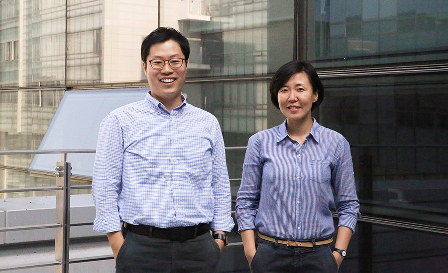
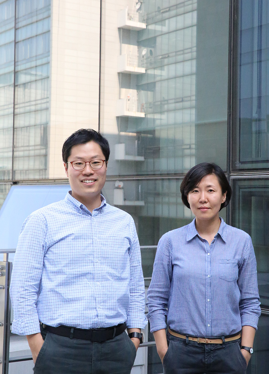
Explorers of our own minds, brain scientists- Research fellows at the Center for Neuroscience Imaging Research, SHIM Won Mok and WOO Choong-Wan - "In ancient Egypt, hearts were kept separate from the bodies when making mummies. This is because hearts were believed to be a place where minds were housed. However, if those mummies were brought back to life now, they won't be capable of anything because they no longer have brains; where human's thoughts and ideas really come from." By telling the story of mummies, research fellow SHIM Won Mok (Global Biomedical Engineering Professor at Sungkyunkwan University) at the Center for Neuroscience Imaging Research explained that the brain makes up who we are and therefore studying the brain is the same as studying ourselves. Before mankind figured out that cognition through senses is made possible by brain functions, people believed that our minds were in our hearts. This belief has led us to call emotional pain heartache. With the introduction of imaging techniques, the field of neuroscience has shown exponential growth. Now modern neuroscience researchers can identify brain structure and explain its functions using MRI and fMRI*. The researchers at the Center for Neuroscience Imaging Research are also committed to studying neuroscience by taking advantage of the most advanced neuroscience imaging technologies. SHIM Won Mok, a seasoned explorer who unlocks the secrets of the brain using fMRI and research fellow WOO Choong-Wan (Global Biomedical Engineering Professor at Sungkyunkwan University) have gathered at N Center, Sungkyunkwan University for an interview. *fMRI(functional magnetic resonance imaging)
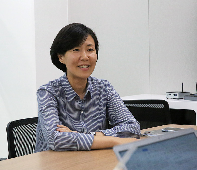
Eyes are the window to the soul, delving into the brain through vision researchShim is in charge of PECON (Perceptual and Cognitive Neuroscience) Lab at the Center for Neuroscience Imaging Research. She studies how the human brain recognizes and perceives sensory information, including visually perceived information. As a psychology major, she was fascinated by classes on cognition and perception. "I was always curious how human minds work. You have to disassemble a clock to know how it works and as such, I thought you have to look at the brain to find out how a mind works. Looking back to my undergraduate years, psychology books didn't have a lot of information on the brain. It was when I went to the University of Illinois as an exchange student that I saw the importance of understanding the brain," she said thinking back.
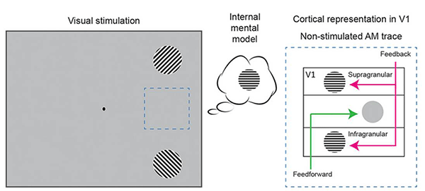
Out of all five senses, Shim focuses on sight. Humans are sight-oriented. Some research even suggests that 90% of all information is perceived through our eyes. Brains process visually perceived information in many ways and Shim's main research is centering on top-down information processing. "The image on the retina is degraded and incomplete. Such limited images go through multi-step processing and create the world as we see it. For the process, our brains interpret information perceived through senses and make guesses not just with what's coming from the eyes, but also experiences and knowledge accumulated over the years and the circumstances they are in. In other words, our brains offer the best guess using the incomplete information identified by our senses," she explained. At her lab, researchers study the interaction between top-down and bottom-up information processing (information processing using the input through sensory organs) used to process visual information, changes in cognition and perception relevant to emotions, working memory mechanism (which temporarily holds information to plan a variety of cognitive processes and helps executing plans in order), and more. Shim received her PhD in experimental cognitive psychology at Harvard for her research on visual perception. Then she established a foundation for fMRI research with Professor Nancy Kanwisher at MIT. After researching at a lab which studies the brain's mechanism for recognizing and processing faces and objects, Shim moved to Dartmouth College as Assistant Professor to explore visual perception processing in more depth. Later she experimented and demonstrated the feedback mechanism which showed that the brain reconstructs information that is absent in the physical input but internally generated. The work was published in the Proceedings of the National Academy of Sciences of the United States (PNAS) in 2016. For the experiment, subjects were flashed a grating tilted in a 2'o clock position and another grating tilted in an 11'o clock position each placed at a different location and shown at a different time. "Unlike when seeing two individual stationary gratings, when perceiving a grating that is apparently rotating from one location to another, the brain fills in details not present in the retinal input corresponding to the apparent motion path. What was fascinating is that the information never presented in the retinal input was reconstructed in the primary visual cortex, which is known to process low-level, rudimentary visual information, via top-down processes," Shim explained.
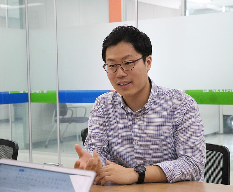
There were times where discussions with students inspired her to land on a research topic. One of her research topics is built on a student's idea. "Before a student's suggestion, the relationship between mood and perceptual process wasn't something I really thought about, but the experimental result was truly intriguing. Subjects in a positive mood showed less selectivity when asked to identify basic visual features when compared to subjects in a negative or neutral mood." said Shim. She shared an example of berry picking. When a person has to pick strawberries while being chased by a lion, she will try to pick only perfectly ripe strawberries. But if the same person is under non-threatening circumstances, she could take her time and look around to find berries that are not perfect red but look good enough. Happy mood may encourage explorative behaviors, which makes us more likely to try unfamiliar food (or stimulus) when we are not in imminent danger. The paper on Shim's research was published in Emotion in 2015. Helping patients with pain research from basic to clinicalWoo, who joined the Center this March, studies physical pain and emotions, which are both subjective experiences. "My lab is called Cocoan lab (Computational Cognitive Affective Neuroscience Laboratory). It is in an infancy stage currently. At the lab, we try to understand human emotions, pain, thoughts and more using fMRI. Particularly, we are working to decode and predict thoughts and emotions using cutting edge technologies, such as machine learning and artificial intelligence," explained Woo. Woo, biology major, became interested in psychopathology while taking psychology classes. The more he explored psychopathology, the more he realized the human mind is intricately connected to the brain. After completing an MA in clinical psychology, Woo started his residency in the department of psychiatry at Seoul National University Hospital. "During the residency, I met roughly 400 patients and listened to their stories. I was able to indirectly experience their walks of life. The experience allowed to me to direct my research interests to studying the brain to help people suffering from pain and emotional distress" said Woo.
After the residency, he moved to the University of Colorado Boulder to study cognitive and affective neuroscience under the supervision of Professor Tor D. Wager. During his PhD and postdoc at the Department of Psychology and Neuroscience and Institute of Cognitive Science, he enjoyed the open research environment in addition to beautiful nature of Rocky Mountains. Starting from 2011, he participated in research projects decoding pain from neural activity and examined the relationship between emotions similar to pain using fMRI. Those projects led him to determine his research goals. Woo is still working on developing new models with which we can read out an individual's pain and emotions using fMRI and other behavioral and physiological signs. Woo's career as a researcher began when he co-authored a research paper that was published in the New England Journal of Medicine in 2013. A year later, he published another paper in Nature Communications which garnered attention. The research findings contradicted previous studies that suggested that the neural representations for social rejection and physical pain are similar. "The emotional pain caused by social isolation, exclusion, bereavement, etc. and physical pain activate the same brain regions. However, brain activation patterns varied depending on the kind of pain. In the research, we emphasized the importance of studying emotional and physical pains and understanding the differences at the level of their activation patterns," he explained.
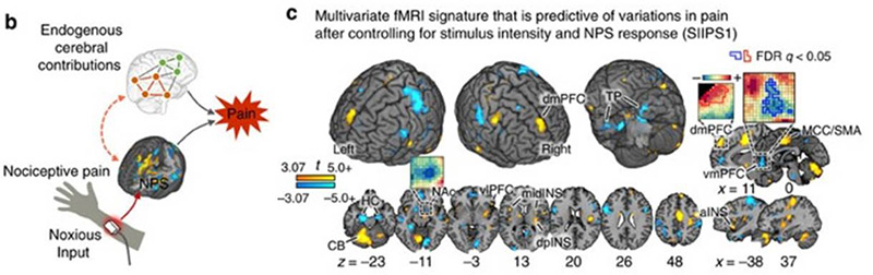
Following the research, Woo introduced a study which demonstrated brain systems which mediate the self-regulation of pain when participants cognitively regulate pain. According to his study, pain can be modulated by how a person interprets and gives meaning to it and the pain modulation effects are mediated by a brain system different from primary pain system that were previously known to serve as the mediator of pain. The study was published in PLOS Biology in 2015 and the outcome of research obtained using machine learning techniques was published in Nature Communications this year. The research discovered that the same level of pain stimulation can be perceived differently depending on the current emotional and physical status and the pain ratings can be captured by the fMRI activation patterns. Woo recently published a review paper on accumulated neuroimaging research outcome and its clinical applications in Nature Neuroscience. From his medical experience, Woo learned how much people suffer from unexpected accidents and trauma, such as post-traumatic stress disorder (PTSD) and complex regional pain syndrome (CRPS). Even since working as a researcher, he is reminded of individual patients. This is why his research goal does not stop at improving basic science. "Building on basic scientific research, I plan to help patients in pain and emotional problems by creating pain-emotion models and tools applicable to clinical practice," explained Woo. Opening the era of post connectome with convergence research centering on fMRIThe technology allows researchers to read the approximately 100 billion neurons of the brain; which has greatly contributed to the advancement of neuroscience. While the procedure is helpful, some believe that fMRI-produced brain activity images should not be thought of as a flawless map. Both research fellows Shim and Woo explained that as they are fully aware of the limitations of fMRI, they want to further improve interpreting and analyzing techniques and create synergy through collaborative research. Accurate data analysis and interpretation are essential in taking full advantage of fMRI. Data provided by fMRI creates three-dimensional images called voxels (volume pixel). A single fMRI voxel is 2mm to 3mm in size and a group of such voxels constructs 2D maps and 3D structures which help researchers find patterns. The size and number of voxels vary based on research topic. For example, Shim's studies typically involve around ten to twenty subjects with 9 million voxels. When a person is scanned every two seconds by fMRI, roughly 200,000 voxels are created. Just five minutes of scanning will produce 30 million voxels. As Woo handles diverse data sets, his studies are composed of countless voxels. "When conducting fMRI studies, researchers always have to keep in mind that the brain is an extensive intelligence system which activates all regions at the same time for various tasks," he explained. "To understand the sophisticated brain, we have to take advantage of algorithms such as AI, machine learning, and deep learning. A large-scale study which involves many data sets is a must."
Woo stressed that we need to go beyond the generation of connectome and should prepare ourselves for the generation of brain predictome. A connectome is a map of neural connections, just like a genome map. There are many ongoing research projects on connectome as it allows researchers to locate brain regions that are responsible for memory, character, talent formation, etc. and understand brain functions and observe brain activities. Woo mentioned the importance to study a predictome that predicts brain activities using the connectome. "Ongoing projects on connectome discovered the network of neural connections on the map. Building on the outcome, researchers should learn to read what the patterns from functional neuroimaging data actually mean, that's predictome," he said. "One of my research goals is to understand the neural processing of complex information, which is distributed throughout the brain in a hierarchical manner, by using machine learning and computational modeling." Shim said the research on fMRI is at a mature stage. The research should move on from simple associations between basic brain functions and cortical regions to sophisticated analyses on how information is processed in the brain in depth. She saw the need for researchers to analyze the brain data at the network level and focus on functional connections, including inter-regional interaction. "An unimaginable amount of data has accumulated. Now finding meaningful information out of the vast amount of data will be the next important brain research goal. I want to contribute by analyzing and predicting information processed in the brain through modeling techniques, investigating how this information is used for our cognition and behavior, and engaging in collaborative work for creating synergy," said Shim. Brain scientists traveling the 1.4 kg microscopic universeWhat would the brain mean to two brain explorers harnessing the power of fMRI? "I believe the brain represents who I am as a person. Psychology offers an opportunity to understand how our minds work and the way we understand the world through scientific methods. That's why I'm fascinated by it," explained Shim. "Starting with my analysis on how our minds work, I want to keep developing hypotheses and testing them to expand our understanding of ourselves and the world. Science does not always guarantee successful results, but I want to stay motivated and have fun," shared Woo. Woo calls the brain the 'meaning maker'. "Our thoughts and emotions are meanings constructed by our brains. Understanding how the brain perceives the outside input, creates meaning out of it, and finding the patterns for that by using machine learning and visualizing techniques would be a cool research project that would satisfy my intellectual curiosity, " explained Woo. |
|||||
| Next | |
|---|---|
| before |
- Content Manager
- Public Relations Team : Suh, William Insang 042-878-8137
- Last Update 2023-11-28 14:20

55, Expo-ro, Yuseong-gu, Daejeon, Korea, 34126
Tel. +82-42-878-8114 | Fax. +82-42-878-8079 | E-mail. webmaster@ibs.re.kr
Copyright(c) IBS. All Rights Reserved.
