


주메뉴
- About IBS 연구원소개
-
Research Centers
연구단소개
- Research Outcomes
- Mathematics
- Physics
- Center for Underground Physics
- Center for Theoretical Physics of the Universe (Particle Theory and Cosmology Group)
- Center for Theoretical Physics of the Universe (Cosmology, Gravity and Astroparticle Physics Group)
- Dark Matter Axion Group
- Center for Artificial Low Dimensional Electronic Systems
- Center for Theoretical Physics of Complex Systems
- Center for Quantum Nanoscience
- Center for Exotic Nuclear Studies
- Center for Van der Waals Quantum Solids
- Center for Relativistic Laser Science
- Chemistry
- Life Sciences
- Earth Science
- Interdisciplinary
- Center for Neuroscience Imaging Research (Neuro Technology Group)
- Center for Neuroscience Imaging Research (Cognitive and Computational Neuroscience Group)
- Center for Algorithmic and Robotized Synthesis
- Center for Genome Engineering
- Center for Nanomedicine
- Center for Biomolecular and Cellular Structure
- Center for 2D Quantum Heterostructures
- Center for Quantum Conversion Research
- Institutes
- Korea Virus Research Institute
- News Center 뉴스 센터
- Career 인재초빙
- Living in Korea IBS School-UST
- IBS School 윤리경영

주메뉴
- About IBS
-
Research Centers
- Research Outcomes
- Mathematics
- Physics
- Center for Underground Physics
- Center for Theoretical Physics of the Universe (Particle Theory and Cosmology Group)
- Center for Theoretical Physics of the Universe (Cosmology, Gravity and Astroparticle Physics Group)
- Dark Matter Axion Group
- Center for Artificial Low Dimensional Electronic Systems
- Center for Theoretical Physics of Complex Systems
- Center for Quantum Nanoscience
- Center for Exotic Nuclear Studies
- Center for Van der Waals Quantum Solids
- Center for Relativistic Laser Science
- Chemistry
- Life Sciences
- Earth Science
- Interdisciplinary
- Center for Neuroscience Imaging Research (Neuro Technology Group)
- Center for Neuroscience Imaging Research (Cognitive and Computational Neuroscience Group)
- Center for Algorithmic and Robotized Synthesis
- Center for Genome Engineering
- Center for Nanomedicine
- Center for Biomolecular and Cellular Structure
- Center for 2D Quantum Heterostructures
- Center for Quantum Conversion Research
- Institutes
- Korea Virus Research Institute
- News Center
- Career
- Living in Korea
- IBS School


News Center

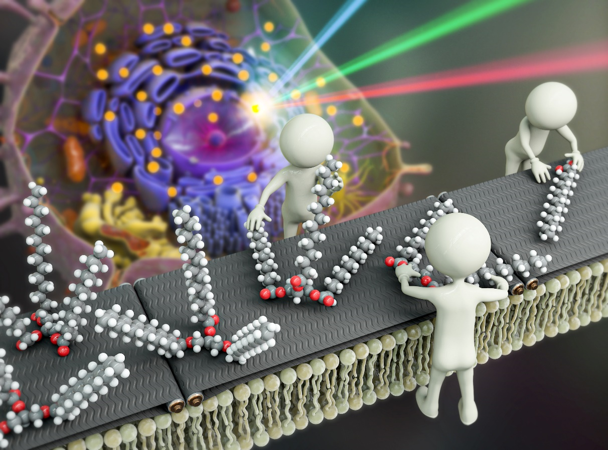
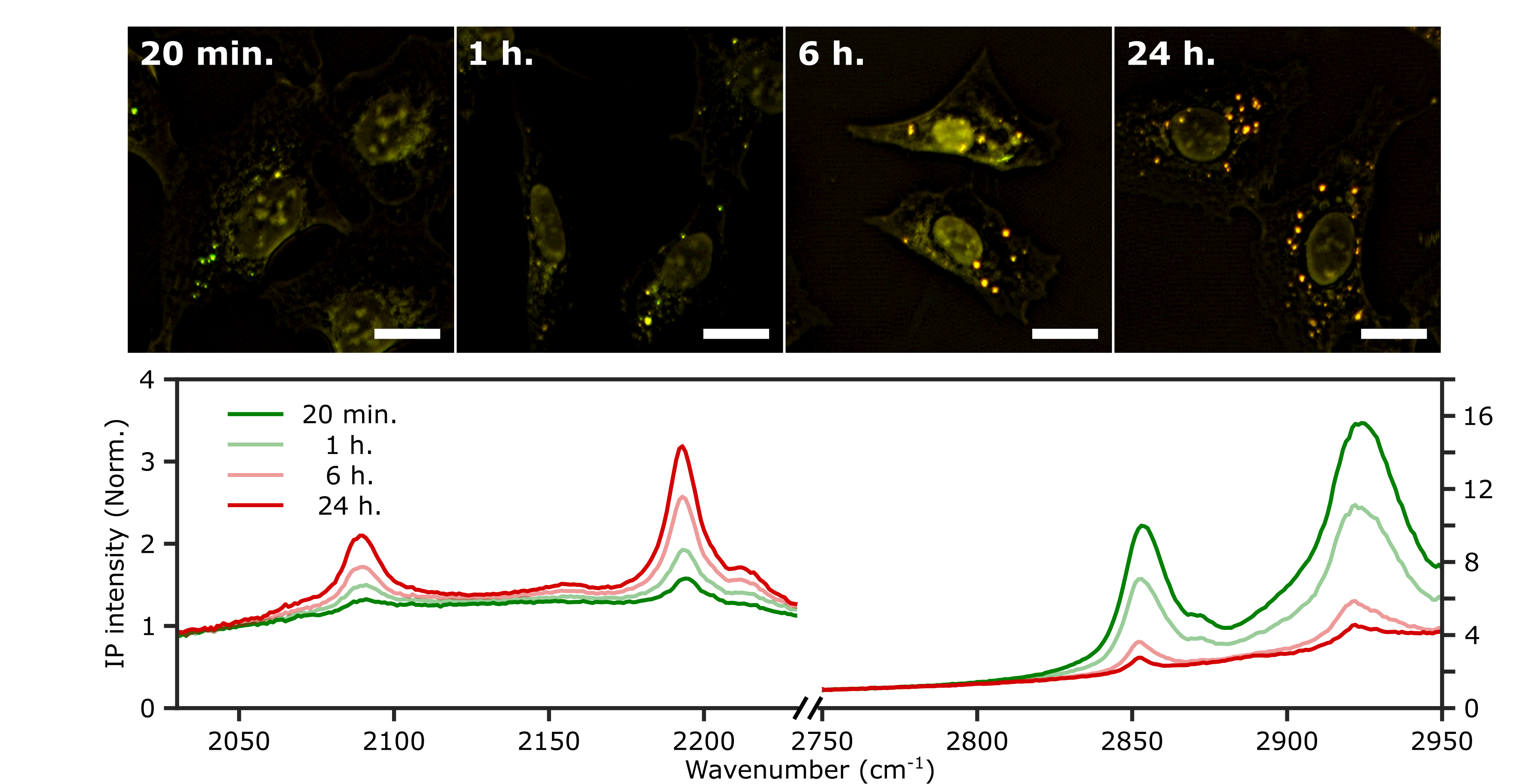
Innovative Microscopy Technique Reveals Secrets of Lipid Synthesis Inside Cells- Two-Color Infrared Photothermal Microscopy (2C-IPM) Opens New Avenues for Long-Term Study of Lipid Metabolism in Living Cells - South Korean researchers led by Director CHO Minhaeng at the IBS Center for Molecular Spectroscopy and Dynamics (IBS CMSD) made a pivotal discovery in the field of cellular microscopy. The team has successfully developed Two-Color Infrared Photothermal Microscopy (2C-IPM), a novel technology designed to investigate neutral lipids within lipid droplets of living cells. This new microscopy can be used with isotope labeling, which allows for the detailed monitoring of neutral lipid synthesis within individual lipid droplets. Lipid droplets (LDs) are structure that consists of pockets of neutral lipids (triglycerides) encapsulated in a monolayer of phospholipids. They have long been thought of as relatively uninteresting organelles, whose only purpose is to store excess energy in the form of neutral lipids. However, recent studies indicate these droplets actually are dynamic players in various cellular metabolic activities. It was found they are actively involved in regulating lipid toxicity and cell communication, as well as their correlation with prevalent diseases like obesity and non-alcoholic fatty liver disease. Understanding the functions of LDs is thus imperative for the diagnosis and treatment of these conditions. Researchers have traditionally stained the cells with lipophilic dyes and employed fluorescence microscopy to study LDs within cells. However, this method has several significant limitations. First is photobleaching of the dyes, which restricts the observation duration of the LDs to very short time windows. Another limitation is the fluorescent dyes themselves. The currently used fluorescent dyes simply target the hydrophobic environment within LDs, utilizing unspecified binding mechanisms. As such, they are incapable of accurately analyzing the composition and quantity of neutral lipids. Unlike previous methods that relied on fluorescent microscopy, the new 2C-IPM technology uses an infrared (IR) spectroscopic method and does not require the use of fluorescent dyes. The new method monitors neutral lipids within LDs directly by detecting changes in IR absorbance. Importantly, this advantage allows researchers to observe the synthesis of neutral lipids within individual LDs in living cells over a long period. Employing the newly developed method, the research team studied the synthesis of neutral lipids in cells when they were exposed to excess fatty acids. The researchers were able to distinguish freshly synthesized neutral lipids from pre-existing neutral lipids within cells by subjecting deuterium-labeled fatty acids, which have distinct spectroscopic properties from non-deuterated forms. The analysis results verified excess fatty acids caused lipid toxicity, and cells respond by increasing the synthesis of neutral lipids. The lead author, researcher PARK Chanjong, remarked, "This study serves as a fundamental example demonstrating the possibility of long-term research on lipid droplets and internal neutral lipids in living cells. The analytical method established in this research can be applied to the diagnosis and treatment of diseases closely associated with lipid metabolism, such as non-alcoholic fatty liver disease." Director CHO Minhaeng commented, "By successfully observing the process of neutral lipid synthesis in living cells, we have laid a new foundation for studying the functions of lipid droplets within cells at the molecular level. This method is expected to be widely used in the investigation of various cellular metabolic phenomena."
Notes for editors
- References
- Media Contact
- About the Institute for Basic Science (IBS)
|

| Next | |
|---|---|
| before |
- Content Manager
- Public Relations Team : Yim Ji Yeob 042-878-8173
- Last Update 2023-11-28 14:20

55, Expo-ro, Yuseong-gu, Daejeon, Korea, 34126
Tel. +82-42-878-8114 | Fax. +82-42-878-8079 | E-mail. webmaster@ibs.re.kr
Copyright(c) IBS. All Rights Reserved.
