


주메뉴
- About IBS 연구원소개
-
Research Centers
연구단소개
- Research Outcomes
- Mathematics
- Physics
- Center for Underground Physics
- Center for Theoretical Physics of the Universe (Particle Theory and Cosmology Group)
- Center for Theoretical Physics of the Universe (Cosmology, Gravity and Astroparticle Physics Group)
- Dark Matter Axion Group
- Center for Artificial Low Dimensional Electronic Systems
- Center for Theoretical Physics of Complex Systems
- Center for Quantum Nanoscience
- Center for Exotic Nuclear Studies
- Center for Van der Waals Quantum Solids
- Center for Relativistic Laser Science
- Chemistry
- Life Sciences
- Earth Science
- Interdisciplinary
- Center for Neuroscience Imaging Research (Neuro Technology Group)
- Center for Neuroscience Imaging Research (Cognitive and Computational Neuroscience Group)
- Center for Algorithmic and Robotized Synthesis
- Center for Genome Engineering
- Center for Nanomedicine
- Center for Biomolecular and Cellular Structure
- Center for 2D Quantum Heterostructures
- Center for Quantum Conversion Research
- Institutes
- Korea Virus Research Institute
- News Center 뉴스 센터
- Career 인재초빙
- Living in Korea IBS School-UST
- IBS School 윤리경영

주메뉴
- About IBS
-
Research Centers
- Research Outcomes
- Mathematics
- Physics
- Center for Underground Physics
- Center for Theoretical Physics of the Universe (Particle Theory and Cosmology Group)
- Center for Theoretical Physics of the Universe (Cosmology, Gravity and Astroparticle Physics Group)
- Dark Matter Axion Group
- Center for Artificial Low Dimensional Electronic Systems
- Center for Theoretical Physics of Complex Systems
- Center for Quantum Nanoscience
- Center for Exotic Nuclear Studies
- Center for Van der Waals Quantum Solids
- Center for Relativistic Laser Science
- Chemistry
- Life Sciences
- Earth Science
- Interdisciplinary
- Center for Neuroscience Imaging Research (Neuro Technology Group)
- Center for Neuroscience Imaging Research (Cognitive and Computational Neuroscience Group)
- Center for Algorithmic and Robotized Synthesis
- Center for Genome Engineering
- Center for Nanomedicine
- Center for Biomolecular and Cellular Structure
- Center for 2D Quantum Heterostructures
- Center for Quantum Conversion Research
- Institutes
- Korea Virus Research Institute
- News Center
- Career
- Living in Korea
- IBS School


News Center

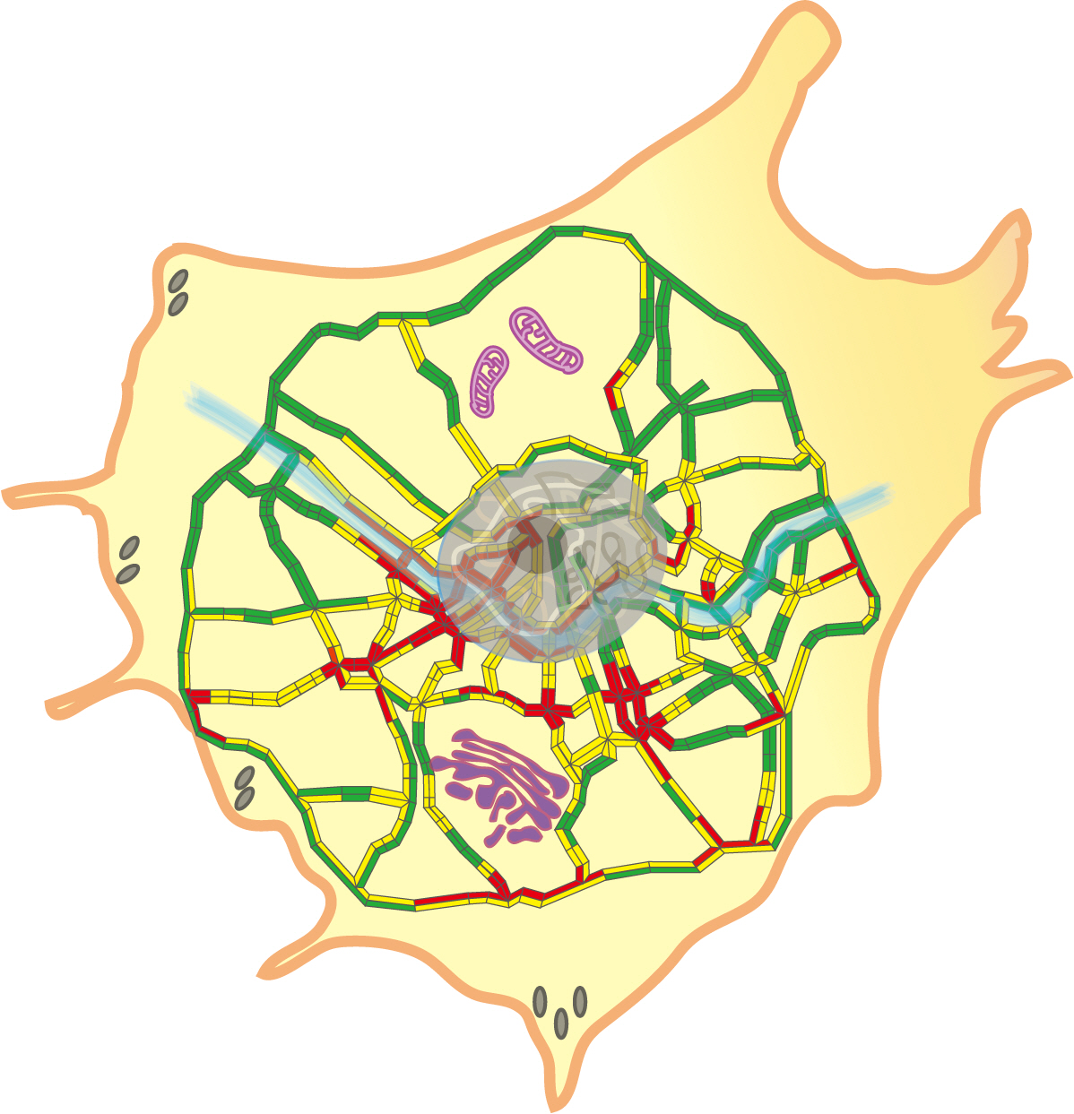
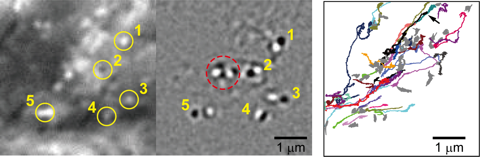
Visualizing “traffic jams” inside living cells- Revolutionary microscopy technique unlocks secrets of intracellular cargo transport - Researchers at the IBS Center for Molecular Spectroscopy and Dynamics (IBS CMSD), led by Director CHO Minhaeng and Professor HONG Seok-Cheol, have unveiled a revolutionary label-free microscopy technique – the “Cargo-Localization Interferometric Scattering (CL-iSCAT) Microscope.” This novel optical imaging method opens new routes in real-time tracking of intracellular cargo movement within living cells without the need for traditional fluorescent labeling. Understanding how intracellular cargo moves is crucial for unraveling the mysteries of a living cell, from its function and metabolism to its ultimate fate. Until now, scientists have relied on fluorescent microscopy to image intracellular cargoes and how they are localized within the cell’s cytoskeleton. However, traditional technology was able to observe only a limited number of specific cargos and is limited by the photobleaching of fluorescent labels. Consequently, visualizing the overall transport phenomena of countless cargos traveling along the intricate cellular scaffold using fluorescence-based methods has proven extremely challenging. The lack of a label-free microscopic technique capable of tracking millions of cargo indefinitely has long hindered our ability to understand the cellular cargo transport phenomena. The newly developed CL-iSCAT Microscope addresses these challenges, allowing for label-free, real-time observation of cargo trafficking in the submicron cellular environment. One feature that sets CL-iSCAT apart is its dual-modality system that integrates fluorescence imaging with iSCAT microscopy. This combination enabled separate observation of specifically labeled cargos or sub-cellular structures against countless unmarked cargos moving along the microtubular networks. The integration of the two complementary techniques is expected to facilitate innovative research in cell biology, thus deepening our understanding of biological phenomena that occur within the cells. Director CHO Minhaeng lauded the significance of CL-iSCAT microscopy, stating, “Through the achievement of observing live cells at an ultra-high resolution independent of fluorescence, we have established a novel paradigm for elucidating the intricate details of biological processes.” Prof. HONG Seok-Cheol, co-corresponding author of the study and professor at Korea University, added, “The development of an imaging technology enabling high-resolution and rapid observation of biological processes allows for an in-depth understanding of life from a molecular dynamics perspective. Our new approach of long-term visualization holds great potential for groundbreaking medical discovery.” With this new tool at their disposal, the IBS researchers were able to selectively monitor the dynamic movement of active cargos within living cells. They used time-differential image analysis to precisely monitor the movements of hundreds of cargos simultaneously over an extended period of time. By utilizing the huge localization data acquired from all cargo positions, they demonstrated the ability of CL-iSCAT microscopy to reconstruct the spatial distribution of microtubular networks in a high spatial resolution beyond the diffraction limit, allowing it to perform with a resolution of down to 15 nm. The potential of this microscope is far-reaching. One of the grand challenges of our time is to investigate viral infection and monitor the effects of anti-viral vaccines and drugs in real time. Since the size of typical viruses is a few tens of nanometers, it will be possible for the CL-iSCAT to visualize the whole process from the onset of viral infection to cell death. Surprisingly, the research team observed that cellular traffic phenomena remarkably mirror the real-life roadway traffic observed in human society. They uncovered many intriguing transport phenomena taking place during cargo movement, including traffic jams within a cell, collective migration, and hitchhiking for efficient cargo transport in uncharted cellular territories. Director Cho said, “It is particularly fascinating to discover several typical traffic events experienced by city commuters in the highly complex cellular world but at micrometer scales. In the future, we aim to delve deeper into the efficient transport strategies adopted by cells to overcome these challenges in transportation and their relevance to cellular phenomena.”
Notes for editors
- References
- Media Contact
- About the Institute for Basic Science (IBS)
|

| Next | |
|---|---|
| before |
- Content Manager
- Public Relations Team : Yim Ji Yeob 042-878-8173
- Last Update 2023-11-28 14:20

55, Expo-ro, Yuseong-gu, Daejeon, Korea, 34126
Tel. +82-42-878-8114 | Fax. +82-42-878-8079 | E-mail. webmaster@ibs.re.kr
Copyright(c) IBS. All Rights Reserved.
