Researchers led by Associate Director CHOI Wonshik of the Center for Molecular Spectroscopy and Dynamics within the Institute for Basic Science, Professor KIM Moonseok of The Catholic University of Korea, and Professor CHOI Myunghwan of Seoul National University developed a new type of holographic microscope. It is said that the new microscope can achieve “see through” the intact skull, and is capable of high-resolution 3D imaging of the neural network within a living mouse brain without removing the skull.
*As a reference, the mouse skull has a similar thickness and opacity as a human fingernail.
In order to scrutinize the internal features of a living organism using light, it is necessary to A) deliver sufficient light energy to the sample and B) accurately measure the signal reflected from the target tissue. However, in living tissues multiple scattering effects and severe aberration1) tend to occur when light hits the cells, which makes it difficult to obtain sharp images.
In complex structures such as living tissue, light undergoes multiple scattering, which causes the photons to randomly change their direction several times as they travel through the tissue. Because of this process, much of the image information carried by the light becomes ruined. However, even if it is a very small amount of reflected light, it is possible to observe the features located relatively deep within the tissues by correcting the wavefront2) distortion of the light that was reflected from the target to be observed. However, the above-mentioned multiple scattering effects interfere with this correction process. Therefore, in order to obtain a high-resolution deep-tissue image, it is important to remove the multiple-scattered waves and increase the ratio of the single-scattered waves.
All the way back in 2019, for the first time the IBS researchers developed the high-speed time-resolved holographic microscope3) that can eliminate multiple scattering and simultaneously measure the amplitude and phase of light. They used this microscope to observe the neural network of live fish without incisional surgery. However, in the case of a mouse which has a thicker skull than that of a fish, it was not possible to obtain a neural network image of the brain without removing or thinning the skull, due to severe light distortion and multiple scattering occurring when the light travels through the bone structure.
The research team managed to quantitatively analyze the interaction between light and matter, which allowed them to further improve their previous microscope. In this recent study, they reported the successful development of a super-depth, three-dimensional time-resolved holographic microscope that allows for the observation of tissues to a greater depth than ever before.
Specifically, the researchers devised a method to preferentially select single-scattered waves by taking advantage of the fact that they have similar reflection waveforms even when light is input from various angles. This is done by a complex algorithm and a numerical operation that analyzes the eigenmode of a medium (a unique wave that delivers light energy into a medium), which allows the finding of a resonance mode that maximizes constructive interference (interference that occurs when waves of the same phase overlap) between wavefronts of light. This enabled the new microscope to focus more than 80 times of light energy on the neural fibers than before, while selectively removing unnecessary signals. This allowed the ratio of single-scattered waves versus multiple-scattered waves to be increased by several orders of magnitude.
The research team went on the demonstration of this new technology by observing the mouse brain. The microscope was able to correct the wavefront distortion even at a depth that was previously impossible using existing technology. The new microscope succeeded in obtaining a high-resolution image of the mouse brain's neural network under the skull. This was all achieved in the visible wavelength without removing the mouse skull and without requiring a fluorescent label.
Professor KIM Moonseok and Dr. JO Yonghyeon, who have developed the foundation of the holographic microscope, said, “When we first observed the optical resonance of complex media, our work received great attention from academia. From basic principles to practical application of observing the neural network beneath the mouse skull, we have opened a new way for brain neuroimaging convergent technology by combining the efforts of talented people in physics, life, and brain science.”
Associate Director CHOI Wonshik said, “For a long time, our Center has developed super-depth bioimaging technology that applies physical principles. It is expected that our present finding will greatly contribute to the development of biomedical interdisciplinary research including neuroscience and the industry of precision metrology.”
This research was published in the online edition of the journal Science Advances (IF 14.136) on July 28th.
Glossary:
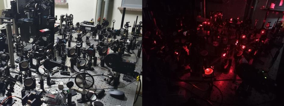
Figure 1. Super-depth 3D holographic microscope
A super-depth 3D holographic microscope developed by researchers at the IBS Center for Molecular Spectroscopy and Dynamics. It is possible to observe the neural network of living organisms by increasing the target optical signal ratio and increasing the image acquisition speed and depth.
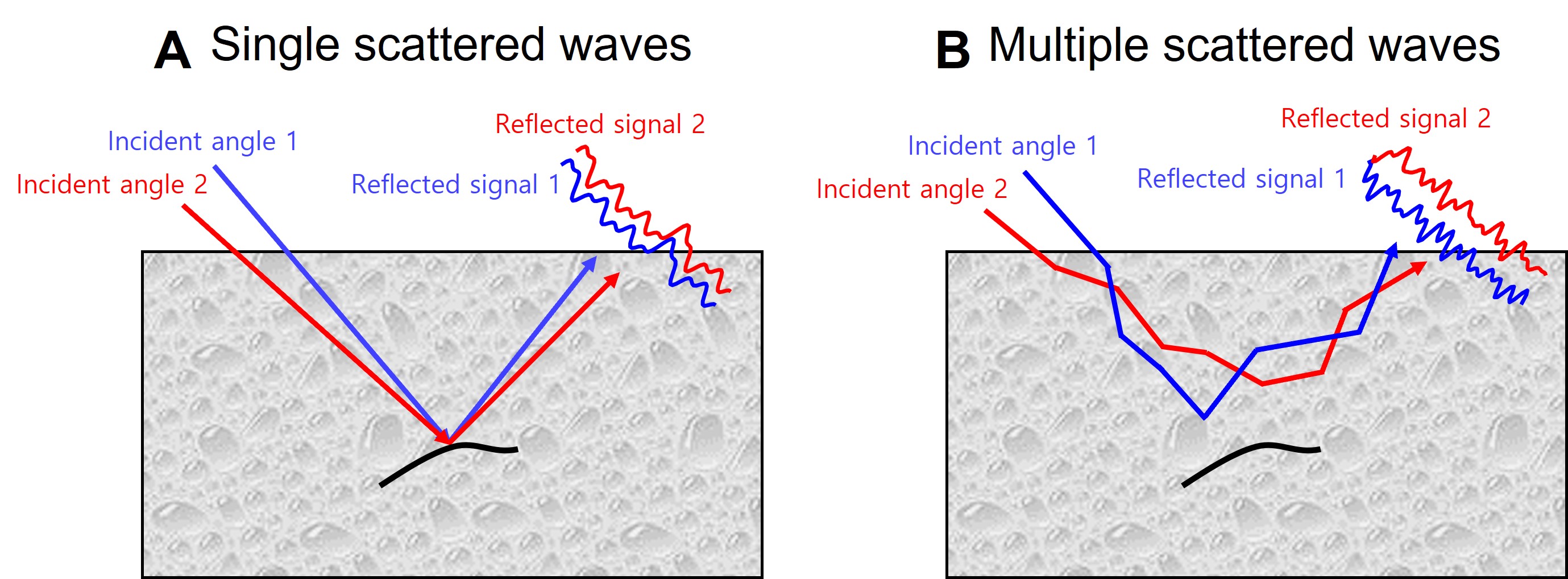
Figure 2. Characteristics of the reflected signal according to the incident angle
(A) If the object is small or has a linear structure, the waveform of the reflected signal of the single scattered waves remains similar even when the incident angle is changed. (B) However, the waveform of the reflected signal of the multiple-scattered waves changes without similarity even with a slight change in incident angle. Using these inter-wavefront properties, single scattering components and multiple scattering components can be separated from each other.
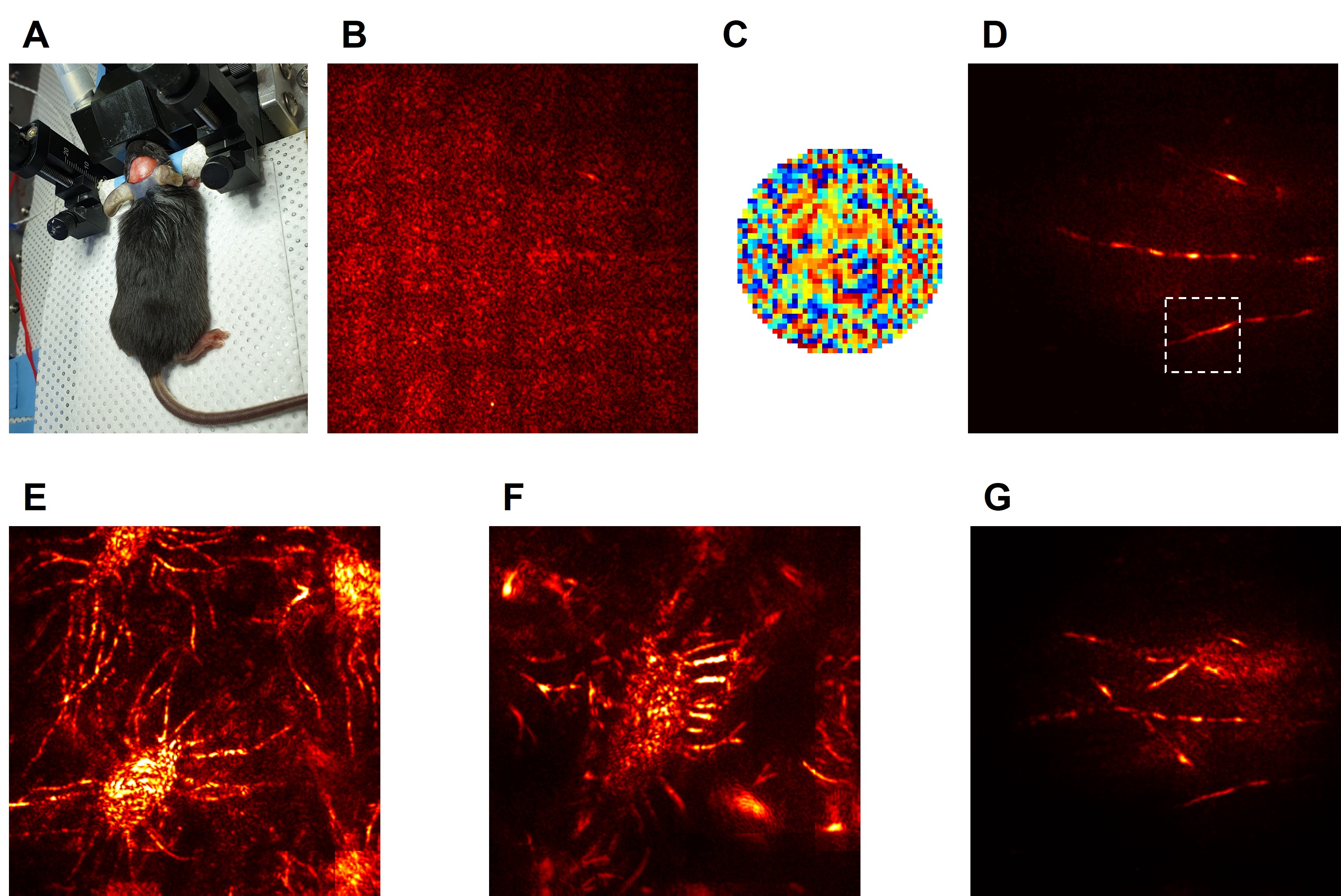
Figure 3. A neural network in the brain of a living mouse was observed without removing the skull
(A). The brain’s neural network was successfully imaged using a light source in the visible wavelength region. Only the skin of a living mouse was removed and the skull was left intact. (B) Using the previous technology, it was not possible to correct the complex aberration due to the severe multiple scattered waves generated in the skull, which makes it impossible to obtain any coherent image. (C) However, the algorithm developed by the research team allowed selective removal of multiple scattering components among the reflected signal, which allows the wavefront aberration to be corrected. (D) This allowed them to resolve the fine structure of neural fibers within the brain. E, F) High-resolution projection images visualize osteocytes inside the skull of the mouse, which flourish between bone layers and dura matters and G) neural network obtained by the microscope.
Notes for editors
- Reference
Yonghyeon Jo, Ye-Ryoung Lee. Through-skull brain imaging in vivo at visible wavelengths via dimensionality reduction adaptive-optical microscopy. Science Advances. DOI: 10.1126/sciadv.abo4366
- Media Contact
For further information or to request media assistance, please contact Wonshik Choi (wonshik@korea.ac.kr) and Yonghyeon Jo (gogogogo2003@korea.ac.kr) at the Center for Molecular Spectroscopy and Dynamics, Institute for Basic Science (IBS) or William I. Suh at the IBS Public Relations Team (willisuh@ibs.re.kr).
- About the Institute for Basic Science (IBS)
IBS was founded in 2011 by the government of the Republic of Korea with the sole purpose of driving forward the development of basic science in South Korea. IBS has 4 research institutes and 35 research centers as of August 2022. There are eleven physics, three mathematics, seven chemistry, nine life science, two earth science, and three interdisciplinary research centers.


