


주메뉴
- About IBS 연구원소개
-
Research Centers
연구단소개
- Research Outcomes
- Mathematics
- Physics
- Center for Underground Physics
- Center for Theoretical Physics of the Universe (Particle Theory and Cosmology Group)
- Center for Theoretical Physics of the Universe (Cosmology, Gravity and Astroparticle Physics Group)
- Dark Matter Axion Group
- Center for Artificial Low Dimensional Electronic Systems
- Center for Quantum Nanoscience
- Center for Exotic Nuclear Studies
- Center for Van der Waals Quantum Solids
- Center for Relativistic Laser Science
- Center for Trapped Ion Quantum Science
- Chemistry
- Life Sciences
- Center for Memory and Glioscience (Cognitive Glioscience Group)
- Center for Memory and Glioscience (Learning and Memory Group)
- Center for Synaptic Brain Dysfunctions
- Center for RNA Research
- Center for Genomic Integrity
- Center for Vascular Research
- Center for Genome Engineering
- Center for Microbiome–Body–Brain Physiology
- Earth Science
- Interdisciplinary
- Center for Neuroscience Imaging Research (Neuro Technology Group)
- Center for Neuroscience Imaging Research (Cognitive and Computational Neuroscience Group)
- Center for Algorithmic and Robotized Synthesis
- Center for Nanomedicine
- Center for Biomolecular and Cellular Structure
- Center for 2D Quantum Heterostructures
- Institutes
- Korea Virus Research Institute
- News Center 뉴스 센터
- Career 인재초빙
- Living in Korea IBS School-UST
- IBS School 윤리경영

주메뉴
- About IBS
-
Research Centers
- Research Outcomes
- Mathematics
- Physics
- Center for Underground Physics
- Center for Theoretical Physics of the Universe (Particle Theory and Cosmology Group)
- Center for Theoretical Physics of the Universe (Cosmology, Gravity and Astroparticle Physics Group)
- Dark Matter Axion Group
- Center for Artificial Low Dimensional Electronic Systems
- Center for Quantum Nanoscience
- Center for Exotic Nuclear Studies
- Center for Van der Waals Quantum Solids
- Center for Relativistic Laser Science
- Center for Trapped Ion Quantum Science
- Chemistry
- Life Sciences
- Center for Memory and Glioscience (Cognitive Glioscience Group)
- Center for Memory and Glioscience (Learning and Memory Group)
- Center for Synaptic Brain Dysfunctions
- Center for RNA Research
- Center for Genomic Integrity
- Center for Vascular Research
- Center for Genome Engineering
- Center for Microbiome–Body–Brain Physiology
- Earth Science
- Interdisciplinary
- Center for Neuroscience Imaging Research (Neuro Technology Group)
- Center for Neuroscience Imaging Research (Cognitive and Computational Neuroscience Group)
- Center for Algorithmic and Robotized Synthesis
- Center for Nanomedicine
- Center for Biomolecular and Cellular Structure
- Center for 2D Quantum Heterostructures
- Institutes
- Korea Virus Research Institute
- News Center
- Career
- Living in Korea
- IBS School


News Center

How to enable light to switch on and off therapeutic antibodiesThe split-rejoin technique allows optobody, a novel optogenetic tool to fine-tune the activation of antibodies When antigens such as a viruse or bacteria invade our body, the immune system springs into action: it creates antibodies that stick to the antigens so that it can identify and destroy the intruders. Did you know that these Y-shaped proteins, AKA antibodies, have been revolutionizing the treatment of cancer, inflammatory disease and autoimmune disease, and many other? Therapeutic antibodies generate immediate immune responses against their target antigens, saving time and energy for patients. Antibodies, or their fragments thereof, can find various medical applications, but few options are available to switch on and off their activity. Though chemical induction can regulate their expression or degradation, it has remained quite elusive to fine-tune their activity when or where needed.
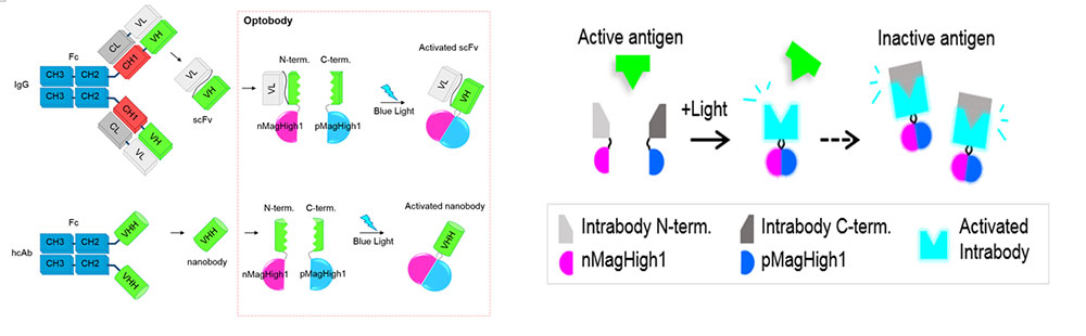
Led by professor Won Do Heo, and researchers at the Center for Cognition and Sociality within the Institute for Basic Science (IBS) and Korea Advanced Institute of Science and Technology (KAIST) in Daejeon, South Korea have developed a new biological tool that activates antibody fragments via a blue light. This optogenetic platform called “optogenetically activated intracellular antibody, or optobody for short” enables the precise control of target protein’s functions in living cells. By this split-rejoin technique, the researchers controlled the activation of optobodies. Inserted as two splits for each antibody fragment into the body, the optobody system at first remains inactive in the body. With light-illumination, optobody on each split links together and makes a whole optobody to “get into action”. Existing approaches do not allow for any temporal control as they induce an instant expression of antibodies immediately after the insertion of DNAs.
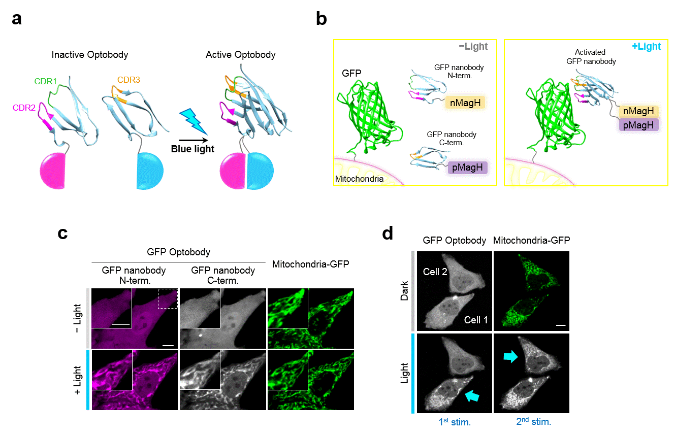
Researchers used two types of antibody fragments –single-chain variable fragment (scFv) and single-domain antibody (VHH, a so-called nanobody) – for their high target-specificity and stability. The research team found the most suitable split site on the GFP nanobody for a temporary inactivation and regaining of the functions. As GFP photoreceptors are triggered by blue light, split GFP nanobody fragments, which were roaming freely in the cell, reassemble. These now whole activated GFP nanobodies move toward their target proteins. The research team confirmed the proper reassembly of fragments by comparing the expression patterns in activated GFP nanobodies and mitochondria GFPs as shown in Figure 2.
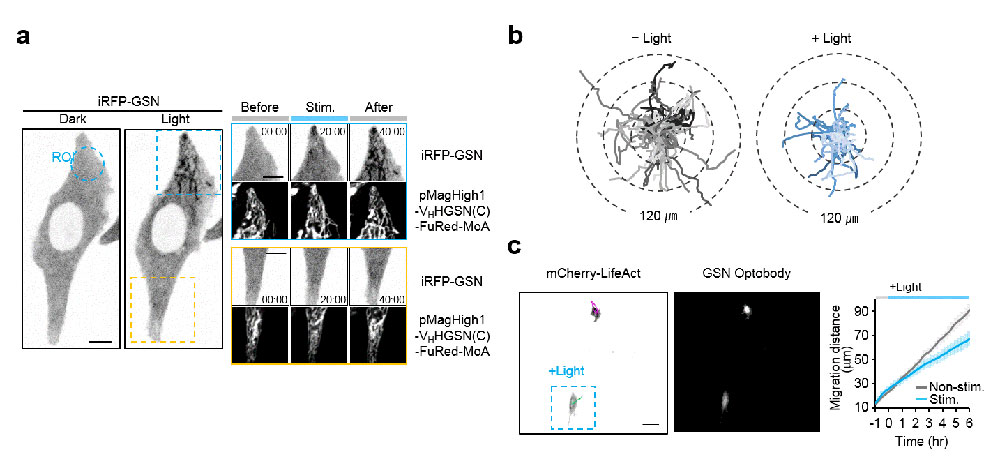
This optobody platform, the split-protein system and light-responsive proteins, is proved to be functional in other well-known antibody fragments targeting endogenous proteins. They succeeded in generating several optobodies derived from three nanobodies and one scFv. All of the optobodies clearly bind to their target proteins (see Figure 3), highlighting the versatility of the optobody system. Specifically, several antibody fragments function as inhibitors of endogenous protein. The researcher investigated the cell movement and the receptor signaling when the cells were stimulated by the blue light. Interestingly, the photo-activated optobodies induced the decrease in cell movement or the disruption of signaling transduction. In addition, through excellent optical control, they could spatiotemporally activate the optobodies at cellular and subcellular level (see Figure 3). The first author, Ph.D. student Daseuli Yu says, “The optobody system is an innovative biological technique in both the fields of antibody engineering and optogenetics.” Purified antibody fragments have been already approved for clinical uses aganist such issues as macular degeneration or Crohn’s disease. Notably, as this new tool allows more precise control of target protein activity in spatial and temporal manner than conventional approach, there seems to be no limit in finding more applications for therapeutic antibodies. Not only being an inhibitor of endogenous protein functions, this optobody system may be able to play other roles for versatility of antibodies. Moreover, a broad pool of antibodies will make this optogenetic manipulation all the more valuable for various clinical applications. Designing potentially inducible drugs is one of the possibilities. “Our optobody system is a great tool for the study of the roles of endogenous proteins in live cells and animals, and also show great clinical promise for therapeutic strategies in the future,” explains professor Heo. Dahee Carol Kim Notes for editors - References - Media Contact - About the Institute for Basic Science (IBS) |
|||
Center for Cognition and SocialityPublication Repository |
|||
|
|

| Next | |
|---|---|
| before |
- Content Manager
- Public Relations Team : Suh, William Insang 042-878-8137
- Last Update 2023-11-28 14:20

55, Expo-ro, Yuseong-gu, Daejeon, Korea, 34126
Tel. +82-42-878-8114 | Fax. +82-42-878-8079 | E-mail. webmaster@ibs.re.kr
Copyright(c) IBS. All Rights Reserved.
