
Lipid Droplets and Diseases
What happens when cells are exposed to excessive energy sources? Similar to how animals store excess nutrients as fat, cells convert surplus energy sources (e.g., glucose, fatty acids) into neutral lipids, storing them in cellular organelles called lipid droplets. Previously, lipid droplets were understood as mere energy reservoirs storing excess neutral lipids.
However, recent discoveries have shown that lipid droplets play key roles beyond energy storage, such as regulating lipid metabolism, cell protection, signal transduction, and protein storage and regulation. Notably, the quantity, size, and distribution of lipid droplets are closely linked to metabolic diseases prevalent in modern society, such as obesity and alcoholic fatty liver disease. This connection positions lipid droplet research as a crucial tool for early diagnosis and monitoring the progression of diseases.
Studying Lipid Droplets with Microscopy
The most intuitive way to analyze lipid droplets in living cells is through direct observation using microscopy. Fluorescence microscopy is the most widely used method for observing lipid droplets. Since lipid droplets are hydrophobic, they can be selectively stained with fluorescent dyes, which generate strong fluorescent signals, making it easy to quickly observe their quantity and size.
Unfortunately, fluorescence microscopy has limitations in providing information on the lipid composition and quantity within droplets. Due to the properties of fluorescent dyes, quantifying specific lipid components is challenging. Moreover, fluorescent dyes suffer from photobleaching, where the fluorescent signal weakens or disappears over time, making it difficult to study lipid droplet metabolism for extended periods. To overcome these limitations, alternative microscopic techniques are being explored.
Infrared Photothermal Microscopy: A New Approach to Analyzing Lipid Droplets
Infrared (IR) microscopy is an effective technique that analyzes the absorption of infrared light by samples to provide information on their composition and quantity. The IBS Center for Molecular Spectroscopy and Dynamics has been working on developing a novel form of infrared microscopy capable of precisely analyzing lipid droplets smaller than a micrometer in living cells. This research focused on the photothermal effect, which is a process where absorbed light energy is converted into heat.
How does the photothermal effect improve the spatial resolution of IR microscopy? To understand this, a basic understanding of optics is required. In optical microscopy, the ability to resolve two particles depends on the size of the focal spot created when light is focused on a sample. The smaller the focal spot, the better the resolution, and the focal size is proportional to the wavelength of light used. Conventional IR microscopy, which uses long-wavelength infrared light, is limited to a spatial resolution of several micrometers, making it difficult to resolve small lipid droplets in cells.
Infrared photothermal microscopy overcomes this limitation by using two laser light sources with different wavelengths. One is an infrared source that induces the photothermal effect, while the other is a visible light source that measures the extent of the photothermal effect. Here, the spatial resolution is determined by the shorter visible light wavelength, allowing the detection of lipid droplets as small as a few hundred nanometers.
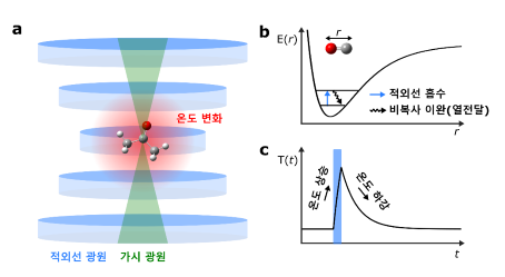
[Figure 1] Principle of Infrared Photothermal Microscopy
(a) Comparison of focal sizes when focusing infrared and visible light sources on the same optical system. Visible light (400–700 nm) creates much smaller focal spots compared to mid-infrared light.
(b) Diagram showing energy changes during infrared absorption and non-radiative relaxation in molecular chemical bonds. Specific chemical bonds have specific energy levels, and they can only selectively absorb infrared light matching their energy levels.
(c) Changes in sample temperature over time upon exposure to a pulsed infrared light source. Infrared photothermal microscopy improves signal sensitivity by inducing photothermal effects in short pulses and selectively isolating the generated signal in the frequency domain.
Real-Time Observation of Neutral Lipid Synthesis in Lipid Droplets
The Center for Molecular Spectroscopy and Dynamics successfully observed the neutral lipid synthesis in individual lipid droplets in real time when living cells were exposed to excessive fatty acids, using infrared photothermal microscopy. To effectively track neutral lipid synthesis, the research team used deuterium-labeled fatty acids (containing carbon-deuterium bonds). By introducing a dual-color photothermal microscopy technique, the team simultaneously analyzed newly synthesized deuterium-labeled neutral lipids and pre-existing neutral lipids without interference by generating photothermal signals at different frequency ranges. Based on these signals and IR absorption coefficients, the team quantitatively tracked changes in the relative ratios of the two lipid components within individual lipid droplets over time.
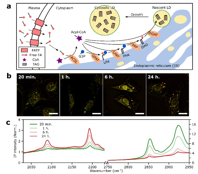
[Figure 2] Observation of Lipid Synthesis in Droplets Using Dual-Color Infrared Photothermal Microscopy
(a) Mechanism of neutral lipid synthesis upon exposure to excessive fatty acids.
(b) Time-lapse dual-color infrared photothermal images after treatment with deuterium-labeled fatty acids. Pre-existing neutral lipids are shown in green, and newly synthesized neutral lipids in red. Over time, the number of lipid droplets increases, and their color shifts to red, indicating the promotion of neutral lipid synthesis.
(c) Representative infrared spectra of lipid droplets over time following deuterium-labeled fatty acid treatment. The infrared absorption signal in the 2000–2200 cm⁻¹ range (carbon-deuterium bonds in newly synthesized neutral lipids) increases, while the signal in the 2800–3000 cm⁻¹ range (carbon-hydrogen bonds in pre-existing neutral lipids) decreases.
The Future of Lipid Droplet Research: New Pathways for Disease Diagnosis and Treatment
Lipid droplets are now recognized as vital organelles that perform critical physiological functions beyond energy storage. Research has shown that lipid droplets are closely associated with modern metabolic diseases such as obesity and alcoholic fatty liver disease. Consequently, lipid droplet research holds great promise for early disease diagnosis and monitoring disease progression.
The IBS Center for Molecular Spectroscopy and Dynamics has demonstrated that infrared photothermal microscopy enables real-time observation of neutral lipid synthesis within lipid droplets, providing precise analysis capabilities. This technological advancement is expected to make substantial contributions to understanding diseases and developing personalized treatment approaches in the future.


