


주메뉴
- About IBS 연구원소개
-
Research Centers
연구단소개
- Research Outcomes
- Mathematics
- Physics
- Center for Theoretical Physics of the Universe(Particle Theory and Cosmology Group)
- Center for Theoretical Physics of the Universe(Cosmology, Gravity and Astroparticle Physics Group)
- Center for Exotic Nuclear Studies
- Center for Artificial Low Dimensional Electronic Systems
- Center for Underground Physics
- Center for Axion and Precision Physics Research
- Center for Theoretical Physics of Complex Systems
- Center for Quantum Nanoscience
- Center for Van der Waals Quantum Solids
- Chemistry
- Life Sciences
- Earth Science
- Interdisciplinary
- Institutes
- Korea Virus Research Institute
- News Center 뉴스 센터
- Career 인재초빙
- Living in Korea IBS School-UST
- IBS School 윤리경영

주메뉴
- About IBS
-
Research Centers
- Research Outcomes
- Mathematics
- Physics
- Center for Theoretical Physics of the Universe(Particle Theory and Cosmology Group)
- Center for Theoretical Physics of the Universe(Cosmology, Gravity and Astroparticle Physics Group)
- Center for Exotic Nuclear Studies
- Center for Artificial Low Dimensional Electronic Systems
- Center for Underground Physics
- Center for Axion and Precision Physics Research
- Center for Theoretical Physics of Complex Systems
- Center for Quantum Nanoscience
- Center for Van der Waals Quantum Solids
- Chemistry
- Life Sciences
- Earth Science
- Interdisciplinary
- Institutes
- Korea Virus Research Institute
- News Center
- Career
- Living in Korea
- IBS School


News Center

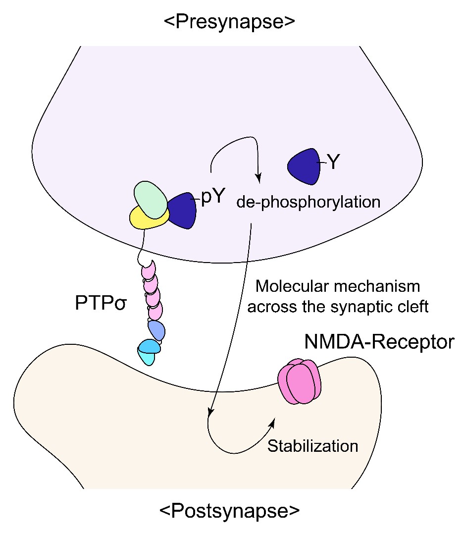
How the brain recognizes changeSynaptic adhesion molecule PTPσ regulates postsynaptic responses and novelty recognition in mice Have you ever gotten a dramatic haircut? Certainly for some who had their heart broken. That is like a silent shouting to call people to spot change in ourselves and find “the new us”. Then how does our brain spot change? Recognizing new objects, new people, new environments, and new rules is critical for survival. Though animal studies found that the hippocampus and NMDA receptors, which mediates and regulates excitatory synaptic transmission, are considered important for novelty recognition, the underlying neural circuit and synaptic mechanisms remain largely unclear. Led by Professor KIM Eunjoon, a research team of the Center for Synaptic Brain Dysfunctions within the Institute for Basic Science (IBS) in Daejeon, South Korea revealed in an animal study a previously unknown role of a presynaptic adhesion molecule to tell the new change by regulating postsynaptic NMDA-type receptor responses at excitatory synapses. “In order to form a synapse and mediate synaptic transmission, postsynaptic receptors should cluster at sites of new synaptic formation and maturation. Little has been known about what “matures” new synapses and whether synapse maturation affects cognitive brain functions such as novelty recognition”. Our data suggest that presynaptic PTPσ promotes postsynaptic NMDA receptor responses, thus allowing to recognize new change” explains Kim. The brain is composed of a large number of neurons, and these neurons are connected through submicron-size structures known as “synapses”. Each individual synapses are composed of two parts; the presynaptic structure that releases neurotransmitter, and the postsynaptic structure that responds to the released neurotransmitter through neurotransmitter receptors. Cell adhesion molecules bridge pre- and postsynaptic specializations. Since there are many different types of synaptic adhesion molecules, it is important for correct pairs of pre- and postsynaptic adhesion molecules to form a complex (bridge) and connect correct partners of neurons. After the initial connections, pre- and postsynaptic adhesion molecules organize the maturation of pre- and postsynaptic structures to mediate synaptic transmission. One of the key synapse maturation processes is the recruitment of postsynaptic neurotransmitter receptors. However, whether and how presynaptic adhesion molecules trans-synaptically regulate the localization and stabilization of postsynaptic neurotransmitter receptors remained largely unclear. Hypothesizing that there is a key presynaptic adhesion molecule that trans-synaptically regulates postsynaptic receptor responses, the research team knocked out PTPσ, a presynaptic adhesion molecule at excitatory synapses in mice to see whether and how this deletion affects synapse formation and function and mouse behaviors.
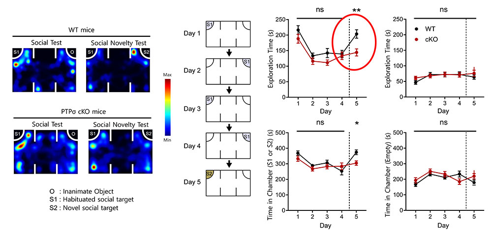
As described in Figure 1, (Left) researchers tested social ability and social novelty recognition ability in the three-chamber apparatus. APTP-sigma KO mice, or a WT mouse, was exposed to a social target (S1; stranger mouse) and a non-social target (O; object) for 10 min, where both WT and PTP-sigma KO mice showed normal preference for S1 over O, indicative of normal social interaction. In the next session for the test of social novelty recognition, where O was replaced with S2 (a novel social stranger), whereas the WT mouse showed normal preference for S2 over S1, PTP-sigma KO mice showed impaired social novelty recognition, suggesting that PTPsigma promotes normal social novelty recognition. The heat maps represent mouse movements during social approach and social novelty recognition behaviors indicated: locations with red colors indicate the sites of longer stay of the mouse. The graphs indicate quantitative analyses of social approach (left) and social novelty recognition (right). (Right) In the second set of experiments (in Figure 1), a PTP-sigma KO, or WT, mice were exposed to a stranger mouse (social target, S1) for four consecutive days, during which the subject mouse spent less and less time exploring the social stranger because of the increasing habituation to the stranger and indicative of normal social recognition and memory. On day 5, when S1 is replaced with a novel stranger mouse (S2), the WT subject showed strongly increased social exploration, as shown by time spent in target exploration or chamber, indicative of normal social novelty recognition and exploration, whereas the PTP-sigma KO mouse showed unchanged exploration of S2 (relative to that for S1 on day 4) (shown by a red circle), indicative of impaired social novelty recognition and exploration. In control experiments, time spent for exploring the empty container was unaffected by experimental conditions. In sum, they found that PTPσ deletion did not affect excitatory synapse formation but strongly suppressed NMDA receptor responses in the hippocampus, a brain region known to regulate learning and memory. In addition, mice lacking PTPσ showed strongly suppressed novelty recognition in various behavioral tests. For instance, PTPσ-mutant mice failed to recognize new objects, new stranger mice, and new rules. These results suggest that presynaptic PTPσ trans-synaptically regulates postsynaptic NMDA receptor responses and novelty recognition in mice.
“The findings suggest that dephosphorylation of some other presynaptic adhesion molecules and certain trans-synaptic mechanisms may underlie the presynaptic PTPσ-dependent regulation of postsynaptic NMDA receptors. However, the underlying molecular mechanisms still need to identified,” notes the first author, KIM Kyungdeok. These results were corroborated by the essentially similar results reported by the group of Dr. Thomas Sudhof at Stanford University in the same journal eLife almost at the same time. Dahee Carol Kim Notes for editors - References - Media Contact - About the Institute for Basic Science (IBS) |
|||
|
|
| Next | |
|---|---|
| before |
- Content Manager
- Public Relations Team : Suh, William Insang 042-878-8137
- Last Update 2023-11-28 14:20

55, Expo-ro, Yuseong-gu, Daejeon, Korea, 34126
Tel. +82-42-878-8114 | Fax. +82-42-878-8079 | E-mail. webmaster@ibs.re.kr
Copyright(c) IBS. All Rights Reserved.
