


주메뉴
- About IBS 연구원소개
-
Research Centers
연구단소개
- Research Outcomes
- Mathematics
- Physics
- Center for Underground Physics
- Center for Theoretical Physics of the Universe(Particle Theory and Cosmology Group)
- Center for Theoretical Physics of the Universe(Cosmology, Gravity and Astroparticle Physics Group)
- Center for Exotic Nuclear Studies
- Dark Matter Axion Group
- Center for Artificial Low Dimensional Electronic Systems
- Center for Theoretical Physics of Complex Systems
- Center for Quantum Nanoscience
- Center for Van der Waals Quantum Solids
- Center for Relativistic Laser Science
- Chemistry
- Life Sciences
- Earth Science
- Interdisciplinary
- Center for Neuroscience Imaging Research(Neuro Technology Group)
- Center for Neuroscience Imaging Research(Cognitive and Computational Neuroscience Group)
- Center for Algorithmic and Robotized Synthesis
- Center for Genome Engineering
- Center for Nanomedicine
- Center for Biomolecular and Cellular Structure
- Center for 2D Quantum Heterostructures
- Center for Quantum Conversion Research
- Institutes
- Korea Virus Research Institute
- News Center 뉴스 센터
- Career 인재초빙
- Living in Korea IBS School-UST
- IBS School 윤리경영

주메뉴
- About IBS
-
Research Centers
- Research Outcomes
- Mathematics
- Physics
- Center for Underground Physics
- Center for Theoretical Physics of the Universe(Particle Theory and Cosmology Group)
- Center for Theoretical Physics of the Universe(Cosmology, Gravity and Astroparticle Physics Group)
- Center for Exotic Nuclear Studies
- Dark Matter Axion Group
- Center for Artificial Low Dimensional Electronic Systems
- Center for Theoretical Physics of Complex Systems
- Center for Quantum Nanoscience
- Center for Van der Waals Quantum Solids
- Center for Relativistic Laser Science
- Chemistry
- Life Sciences
- Earth Science
- Interdisciplinary
- Center for Neuroscience Imaging Research(Neuro Technology Group)
- Center for Neuroscience Imaging Research(Cognitive and Computational Neuroscience Group)
- Center for Algorithmic and Robotized Synthesis
- Center for Genome Engineering
- Center for Nanomedicine
- Center for Biomolecular and Cellular Structure
- Center for 2D Quantum Heterostructures
- Center for Quantum Conversion Research
- Institutes
- Korea Virus Research Institute
- News Center
- Career
- Living in Korea
- IBS School


News Center

| Title | Reliable diagnostics at the tip of your finger | ||
|---|---|---|---|
| Embargo date | 2022-05-18 00:00 | Hits | 612 |
| Press release |
![docx 파일명 : [v8] v2 PR_2022-0503_y_영문_0516 JSR.docx](/images/mimetype/docx.gif) [v8] v2 PR_2022-0503_y_영문_0516 JSR.docx
[v8] v2 PR_2022-0503_y_영문_0516 JSR.docx
|
||
| att. |
 Figure 1.png
|
||
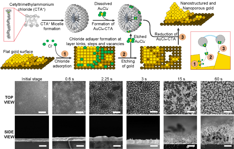
Reliable diagnostics at the tip of your fingerDirect and sensitive analysis of clinical samples for point-of-care diagnostics Biomarkers are components that may be present in biological samples and are related to specific diseases. Therefore, doctors can analyze biological samples from a patient to check their health condition or to monitor the progress of a specific therapy. Typically, these samples need to be purified and diluted before the analysis, and current medical diagnostic techniques rely on healthcare facilities and laboratories for these routine analyses. This is a lengthy process that requires trained personnel and expensive instrumentation to extract, transport, store, process, and analyze the samples in centralized locations. Moreover, during a period of global crisis like the ongoing pandemic, the pressure of thousands of analysis requests can saturate and collapse the healthcare system. On the other hand, point-of-care devices, which are small automated instruments, are capable of performing diagnostics in decentralized locations and can provide quick answers. One example of such a device is the glucose meter that people with diabetes use to monitor their sugar levels in the blood. These devices can overcome the inherent limitations of having to process a sample through a centralized system, empowering anyone to be able to monitor their health from home, simply using a tiny blood sample extracted with a fingerprick. However, the development of these devices has been burdened by the technical challenges related to measuring biological samples. Biomarkers for some diseases and infections are only present in the samples in very small amounts, which in turn imposes the challenge to develop extremely sensitive detection techniques. While increasing the surface area of the biosensor can increase the sensitivity of the instrument, these surfaces tend to be quickly clogged and contaminated, rendering them unusable. To this end, the team led by Professor CHO, Yoon-Kyoung at the Center for Soft and Living Matter within the Institute for Basic Science (IBS) in Ulsan, South Korea recently developed a biosensor using a method to generate nanostructured and nanoporous surfaces. This combined strategy not only provides the sensor with an unprecedented sensitivity but also makes it resistant to fouling by proteins. While previously there has been no known method to reliably create electrodes using such nanostructured and nanoporous substrates, the team reported a simple method to generate such materials. The mechanism is based on the application of electric pulses to a flat gold surface in the presence of sodium chloride and a surfactant that can form micelles in solution. These electric pulses drive a preferent reaction to etch and redeposit gold from the surface and, in turn, grow nanostructures and form the nanopores (Figure 1). The use of surfactant in the form of micelles is essential to the success of this strategy since it prevents the material that is being etched from diffusing away during the process, so it can be redeposited. The formation of these nanostructures yielded a large surface area which was beneficial for increasing the sensitivity of the assays, whereas the formation of nanopore substrates was ideal to prevent contamination from the biological samples. Both the nanostructures and the nanopores' combined benefits were key to the success of this strategy, which could be applied for the direct analysis of clinical plasma samples. The researchers further demonstrated this new technology by building a biosensor for the detection of prostate cancer. The electrode was sensitive enough to discriminate between a group of prostate cancer and healthy donors using only a tiny amount of blood plasma or urine samples. No dilution or preprocessing steps were used, which means that the technology could easily be used for the point-of-care diagnosis of cancer. Professor Cho stated, “We believe that this technology is essential for the future development of point-of-care devices and diagnostic tests that work with biological samples. The capability to detect low concentrations of relevant biomarkers with robust performance opens a door to possibilities in the field of diagnostics for cancer, pathogens, and other diseases.” The findings of this research have been published in Advanced Materials (IF: 30.849) on May 17th, 2022 and the associated illustration was selected for the frontispiece in the current issue.
Notes for editors
- References
- Media Contact
- About the Institute for Basic Science (IBS)
|
|||
|
|
|||
| Next | |
|---|---|
| before |
- Content Manager
- Communications Team : Kwon Ye Seul 042-878-8237
- Last Update 2023-11-28 14:20

55, Expo-ro, Yuseong-gu, Daejeon, Korea, 34126
Tel. +82-42-878-8114 | Fax. +82-42-878-8079 | E-mail. webmaster@ibs.re.kr
Copyright(c) IBS. All Rights Reserved.
