


주메뉴
- About IBS 연구원소개
-
Research Centers
연구단소개
- Research Outcomes
- Mathematics
- Physics
- Center for Theoretical Physics of the Universe(Particle Theory and Cosmology Group)
- Center for Theoretical Physics of the Universe(Cosmology, Gravity and Astroparticle Physics Group)
- Center for Exotic Nuclear Studies
- Center for Artificial Low Dimensional Electronic Systems
- Center for Underground Physics
- Center for Axion and Precision Physics Research
- Center for Theoretical Physics of Complex Systems
- Center for Quantum Nanoscience
- Center for Van der Waals Quantum Solids
- Chemistry
- Life Sciences
- Earth Science
- Interdisciplinary
- Institutes
- Korea Virus Research Institute
- News Center 뉴스 센터
- Career 인재초빙
- Living in Korea IBS School-UST
- IBS School 윤리경영

주메뉴
- About IBS
-
Research Centers
- Research Outcomes
- Mathematics
- Physics
- Center for Theoretical Physics of the Universe(Particle Theory and Cosmology Group)
- Center for Theoretical Physics of the Universe(Cosmology, Gravity and Astroparticle Physics Group)
- Center for Exotic Nuclear Studies
- Center for Artificial Low Dimensional Electronic Systems
- Center for Underground Physics
- Center for Axion and Precision Physics Research
- Center for Theoretical Physics of Complex Systems
- Center for Quantum Nanoscience
- Center for Van der Waals Quantum Solids
- Chemistry
- Life Sciences
- Earth Science
- Interdisciplinary
- Institutes
- Korea Virus Research Institute
- News Center
- Career
- Living in Korea
- IBS School


News Center

|
Opening the door to early diagnosis with high resolution, deep depth imaging technology
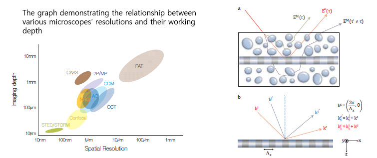
Center for Molecular Spectroscopy and Dynamics Over the last few decades, optical microscope has been revolutionized over and over in an attempt to find a way to look into smaller objects with increased precision. There have been prominent advances in super-resolution optical microscopy that led to the Nobel Prize in Chemistry for the year 2014. On the other hand, the progress in the imaging deep inside biological tissues has so far been incremental. Existing optical approaches have been near-sighted, and important biological and biomedical reactions occurring deep within tissues have been out of reach. The major reason why the depth of optical imaging is too shallow is that biological tissues are not optically transparent. Light waves experience multiple scattering events while they pass through biological tissues. Therefore, the intensity of single-scattered waves carrying visual information about the target objects decrease as they are located deep. Singlescattered waves are the waves scattered only once by the target object, but not scattered at all while they pass though the scattering medium. Therefore, the precise image of the target object can be obtained when the single-scattered wave intensity is strong enough. In general, the intensity of single-scattered waves is exponentially attenuated each time they travel by the mean free path in scattering medium. For example, when imaging a target object located 500μm deep in a biological tissue whose mean free path is 100μm, the intensity of the single-scattered wave is attenuated to 0.005 percent of the intensity of the incident light. In short, most of the reflected light in the scattering medium can be considered noise, while a singlescattered wave carrying the visual information about the target object is covered by multiplescattering waves. As a consequence, highresolution imaging that can detect intercellular structure can work only a few hundred micrometers deep The CMSD in IBS has successfully overcome this limitation through a recent study. The team developed an ultra-deep imaging technology using collective accumulation of ingle-scattered waves, and the microscope applying this technology is called the collective accumulation of single-scattering (CASS) microscope. Here are the two properties used. First, in the case of single-scattered waves, the time of flight from the target object to the camera are determined by the depth of the object. On the other hand, multiple-scattered waves can be scattered to any direction, and the time of flight from the target object to the camera varies as it passes various paths (see Figure 2). Therefore, multiple-scattered elements can be effectively removed by selectively collecting waves reaching the camera after a certain period of time with regard to the depth of the target object. This is called a time-resolved measurement. However, as the Figure 2a shows that there still exist multiple-scattered waves with the same flight time as the single-scattered waves, timeresolved measurement alone therefore cannot sufficiently increase the possible imaging depth. An additional method is required, and that is to use the law of conservation momentum in a horizontal direction that only single-scattered waves hold.
▲ Left) 2P/MP: two-photon/multiphoton microscopy, SHG:Second Harmonic Generation Microscope, AO: adaptive optics microscope, OCT/OCM: optical coherence microscopy, Confocal: confocal microscopy, STED/STROM: stimulated emission depletion/Stochastic Reconstruction microscopy, PAT: Photoacoustic Tomography, CASS: the microscope that was developed by the researchers
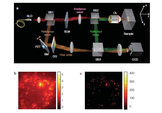
To make it simple, think about an object with a periodic structure, represented in Figure 2b. When plane waves are induced on the object, the reflected waves' momentum in a horizontal direction is the incidence waves’ momentum in a horizontal direction plus the momentum created by the lattice structure. Therefore, the changes in momentum between the reflected waves and the incidence waves are determined by the periodicity of the structure without any regard to the angle of incidence. In the case of multiple-scattered waves, a change in the incidence angle has no such relation to the change in the direction of the reflected waves. In this study, to utilize the law of momentum conservation for single-scattered waves, the momentum of reflected waves were mapped at different incidence angles. In the meantime, elements whose momentums are conserved are added in phase. As a result, the intensity of the single-scattered waves increased proportionally to the square of the number of measurements while that of the multiple-scattered waves grew linearly since they are added randomly in phase. Using this method, noise created by multiple-scattered waves could effectively be reduced. We designed the CASS microscope in order to meet the two requirements explained above (Figure 3a). After constructing interferometric microscope using low-coherence light source, incidence angle of light to the target object were modulated by an optical spatial light modulator. At the different angles of incidence, interfere images between the reference light and the reflected light from the sample were measured to obtain the timeresolved complex image, and then it was mapped in the two-dimensional spatial frequency space using two-dimensional Fourier transform. Figure 3b shows a time-resolved image obtained under the normal illumination. Since multiple-scattering waves are far stronger than single-scattering waves, clear image information cannot be obtained, even after the image underwent timeresolved measurements. Therefore, the image was photographed 2500 times at different angles of incidence and, from the photographs, singlescattering elements were extracted, and eventually clear image as shown in Figure 3c could be acquired. The CASS microscope, with 1μm resolution, has the deepest depth amongst all the existing microscopes. Recognized for this accomplishment, the research paper has been published in Nature Photonics, the most influential journal in the field of optical science. The findings of this research are expected to contribute to developing medical imaging devices and diagnosis instruments with far greater precision. In particular, the findings would help identify cancer at early stages. Cancer cells are initially formed around 1~2mm deep in biological tissue, and changes in the size of the cell nucleus, which is only a few micrometers, begin. However, these changes cannot be detected using the existing medical imaging methods, but found later when the cancer cells become polypus. Additionally, there have been difficulties in determining the area where cancer cells have spread in the surgical operation. The further development of CASS microscope is expected to revolutionize the way cancel cells at an early stage are diagnosed and the way they are treated. Published paper Sungsam Kang et al, “Imaging deep within a scattering medium using collective accumulation of single-scattered waves”, Nature Photonics, 9, pp. 253–258 (2015) |
| before | |
|---|---|
| before |
- Content Manager
- Public Relations Team : Suh, William Insang 042-878-8137
- Last Update 2023-11-28 14:20

55, Expo-ro, Yuseong-gu, Daejeon, Korea, 34126
Tel. +82-42-878-8114 | Fax. +82-42-878-8079 | E-mail. webmaster@ibs.re.kr
Copyright(c) IBS. All Rights Reserved.
