2025 IBS Art in Science(10th Anniversary Special Exhibition with MPG)
*Discovery, Exploring the Unknown
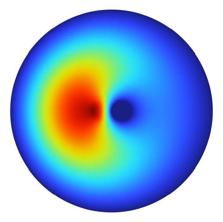
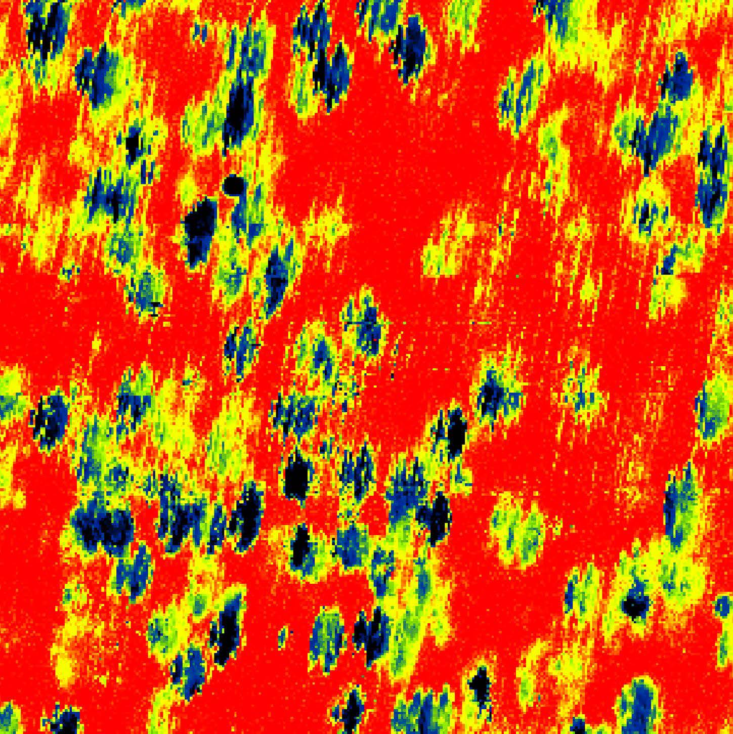
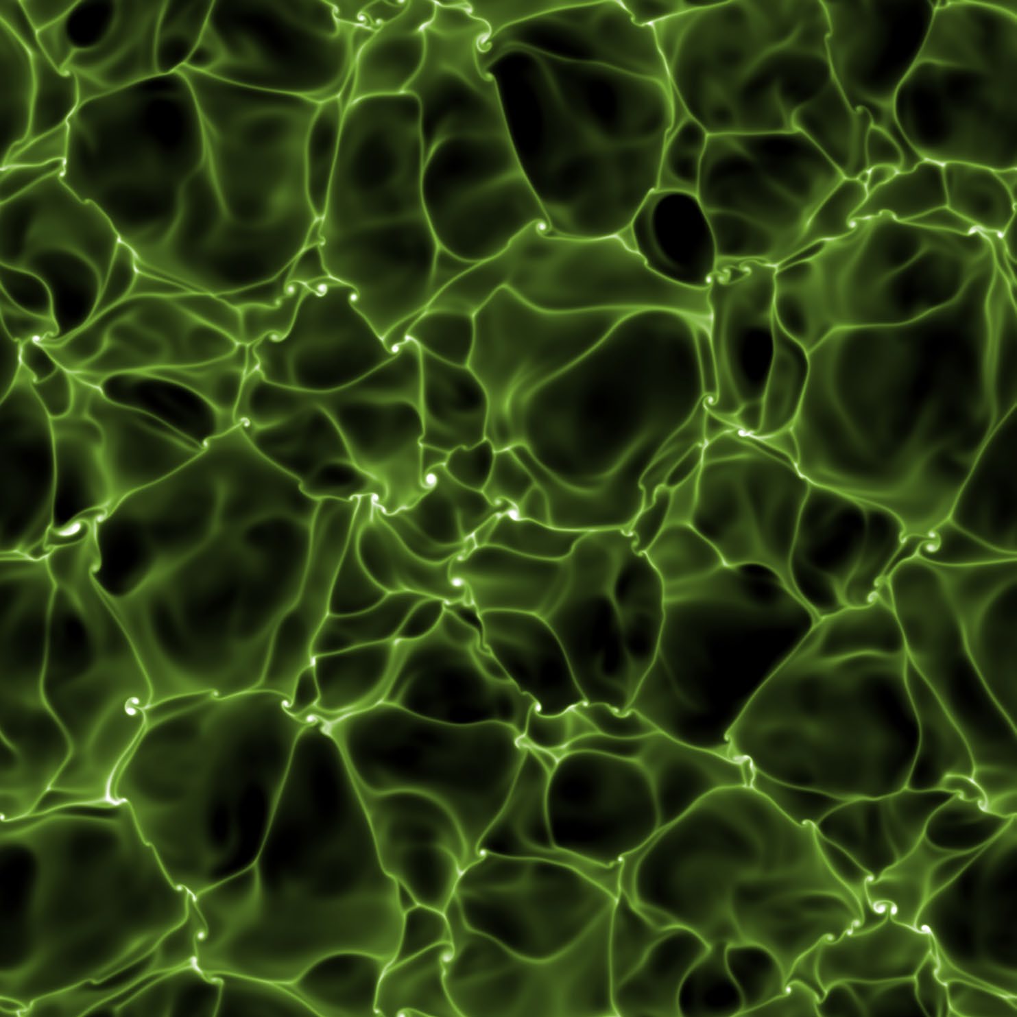
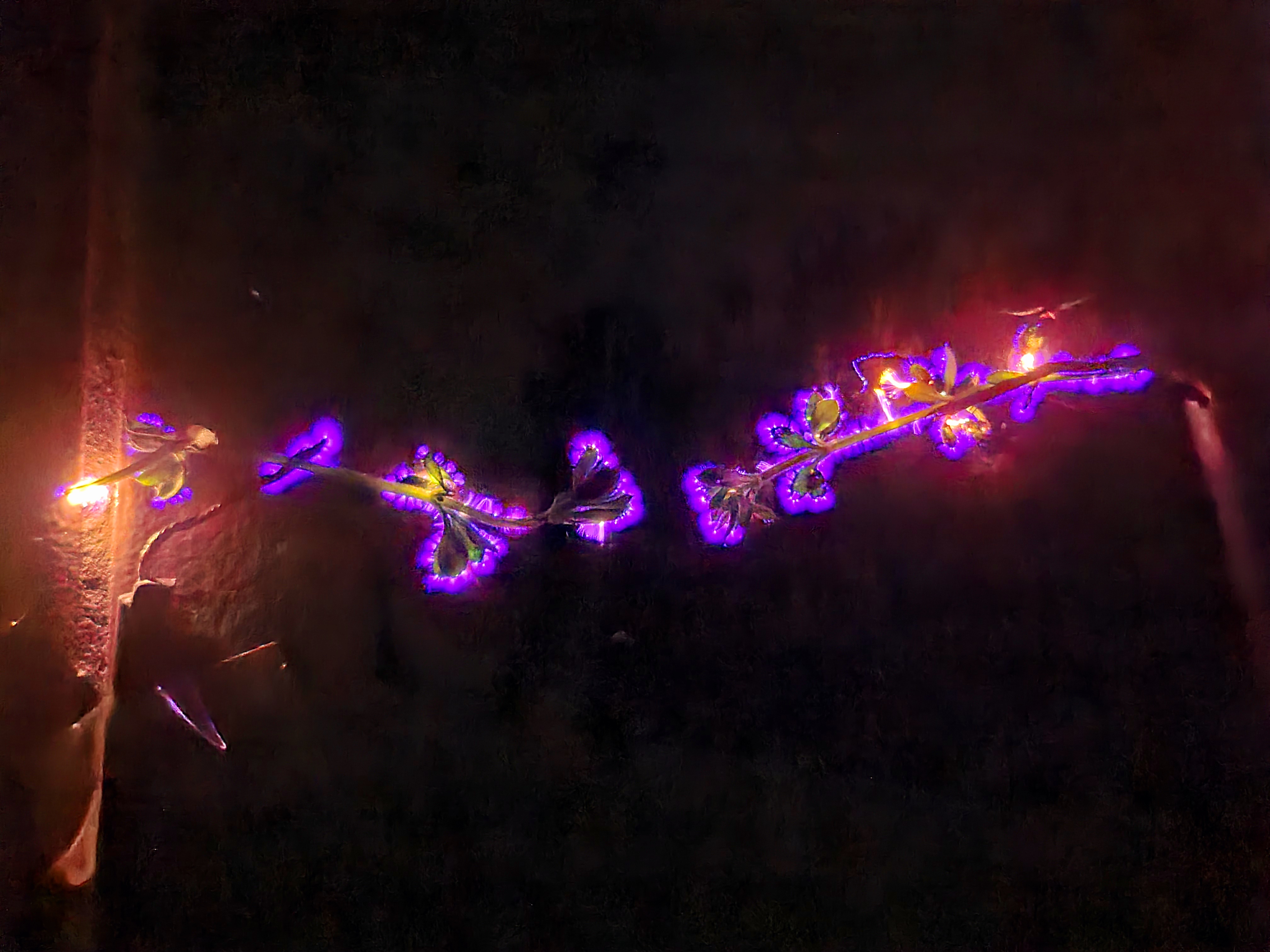
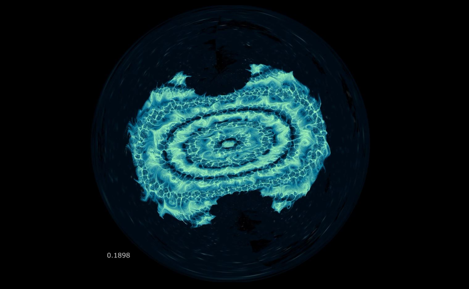
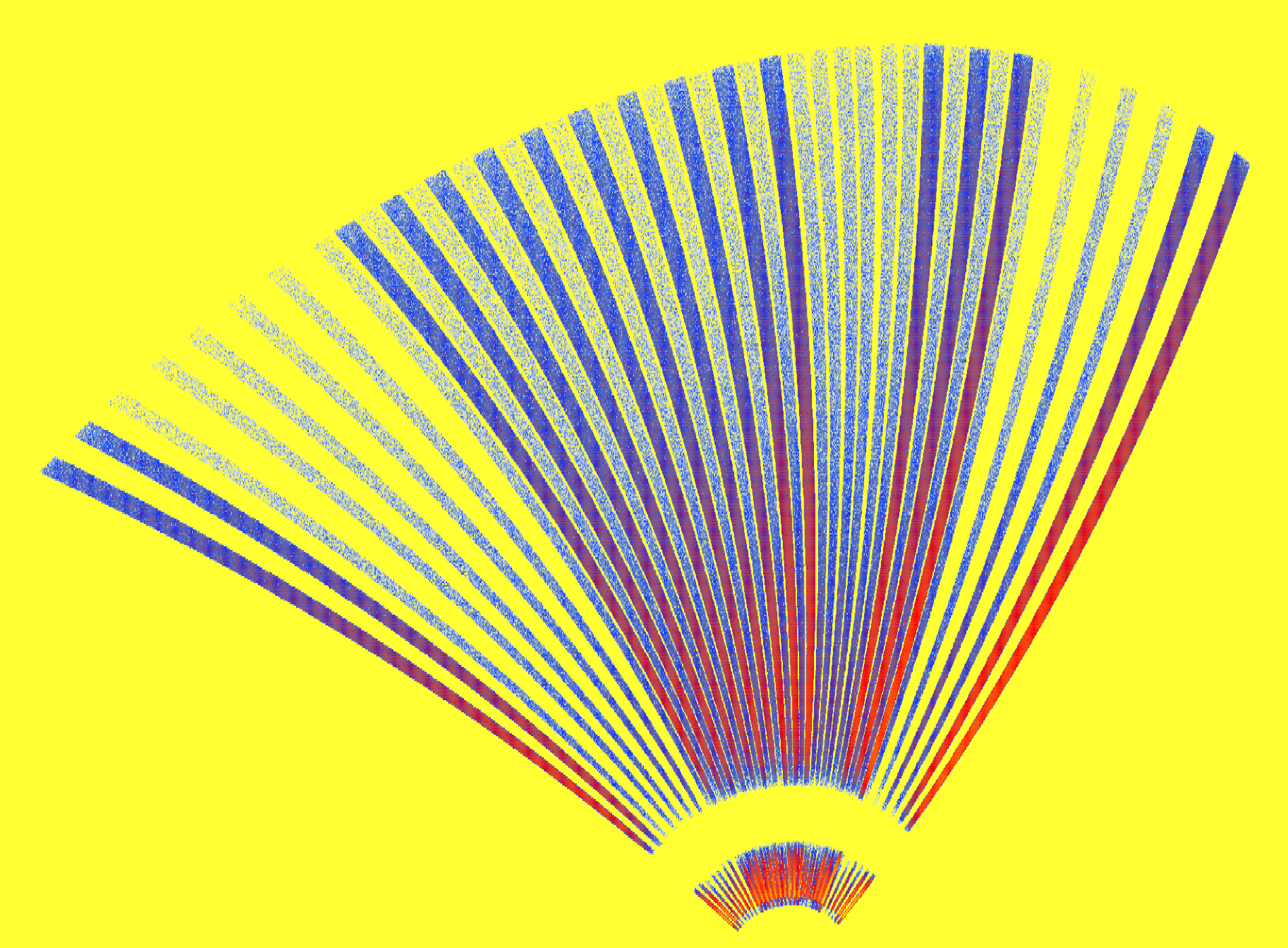
Fetal Movements of an Axion
by SungWoo Youn
IBS Dark Matter Axion Group
To search for the elusive dark matter believed to fill the universe, scientists examine faint electromagnetic traces inside specially designed resonant cavities. This image shows a series of simulated oscillation of the electric field in such a cavity. When viewed over time, the evolving patterns resemble the gentle movements of a fetus inside the womb. The axion, one of the most promising dark matter candidates, could one day be discovered through experiments like these—an achievement that may transform our understanding of the universe. Thus, this piece represents more than a simple simulation: it symbolizes the emergence of new physics from the darkness of the cosmos.
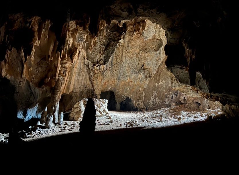
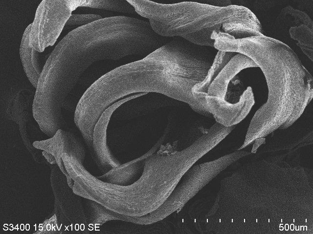
Memory of the Past Climate
by Nitesh Sinha
IBS Center for Climate Physics
This picture shows the entrance of a cave in Botswana, Southern Africa, along with the carbonate deposits formed inside it. These secondary cave formations— stalactites, stalagmites, and other speleothems—preserve climate records spanning hundreds of thousands of years and serve as valuable proxies for past climate change. Recent advances in speleothem research and dating techniques now allow us to reconstruct hydroclimate variations from the distant past, offering deeper insights into human historyand migration. Understanding past climate is essential for improving climate models and predicting future climate change. This image symbolizes “rocks with climate memory.” The sunlight passing through the cave represents the light of scientific research—illuminating the past hidden within these ancient stones.
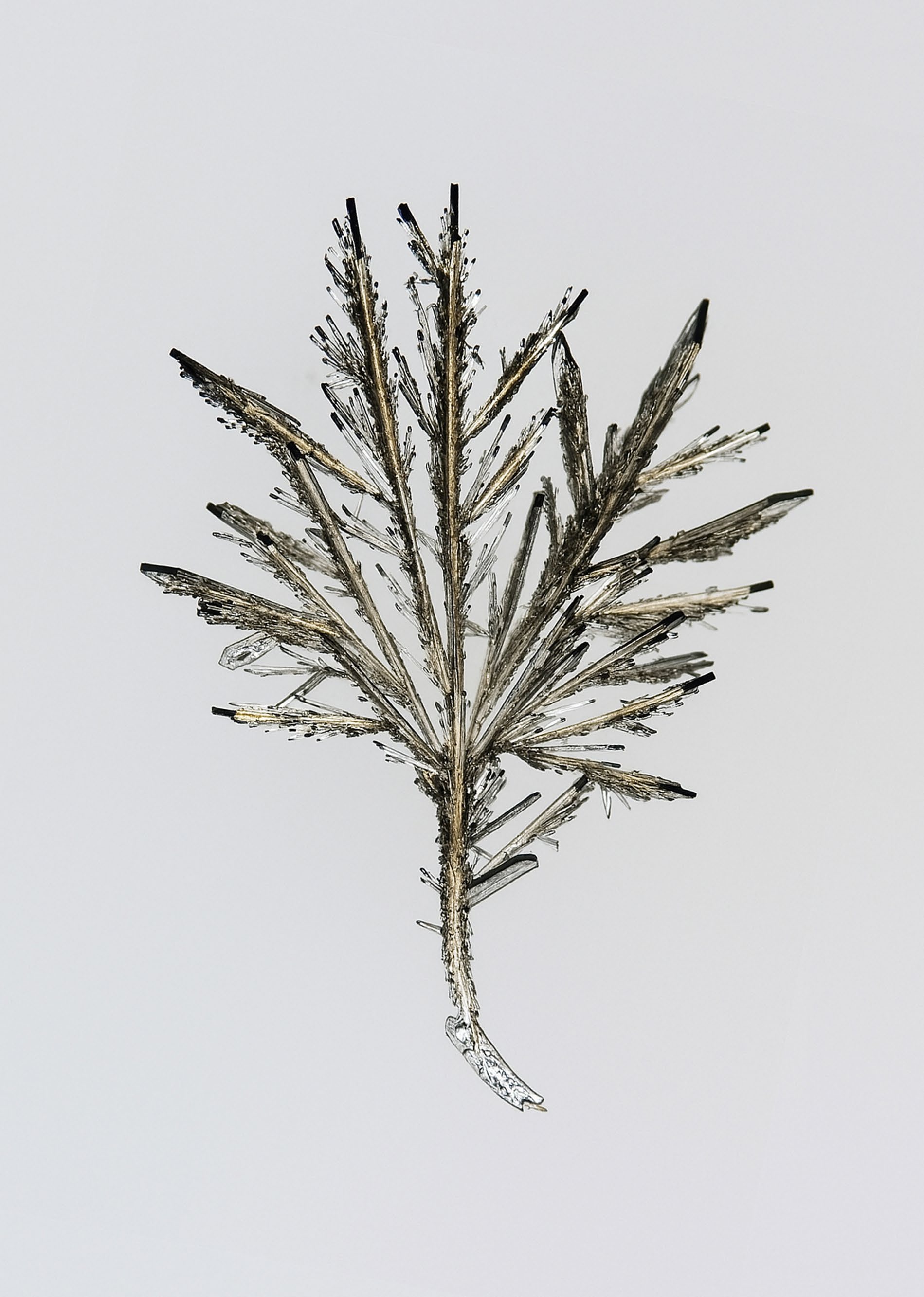
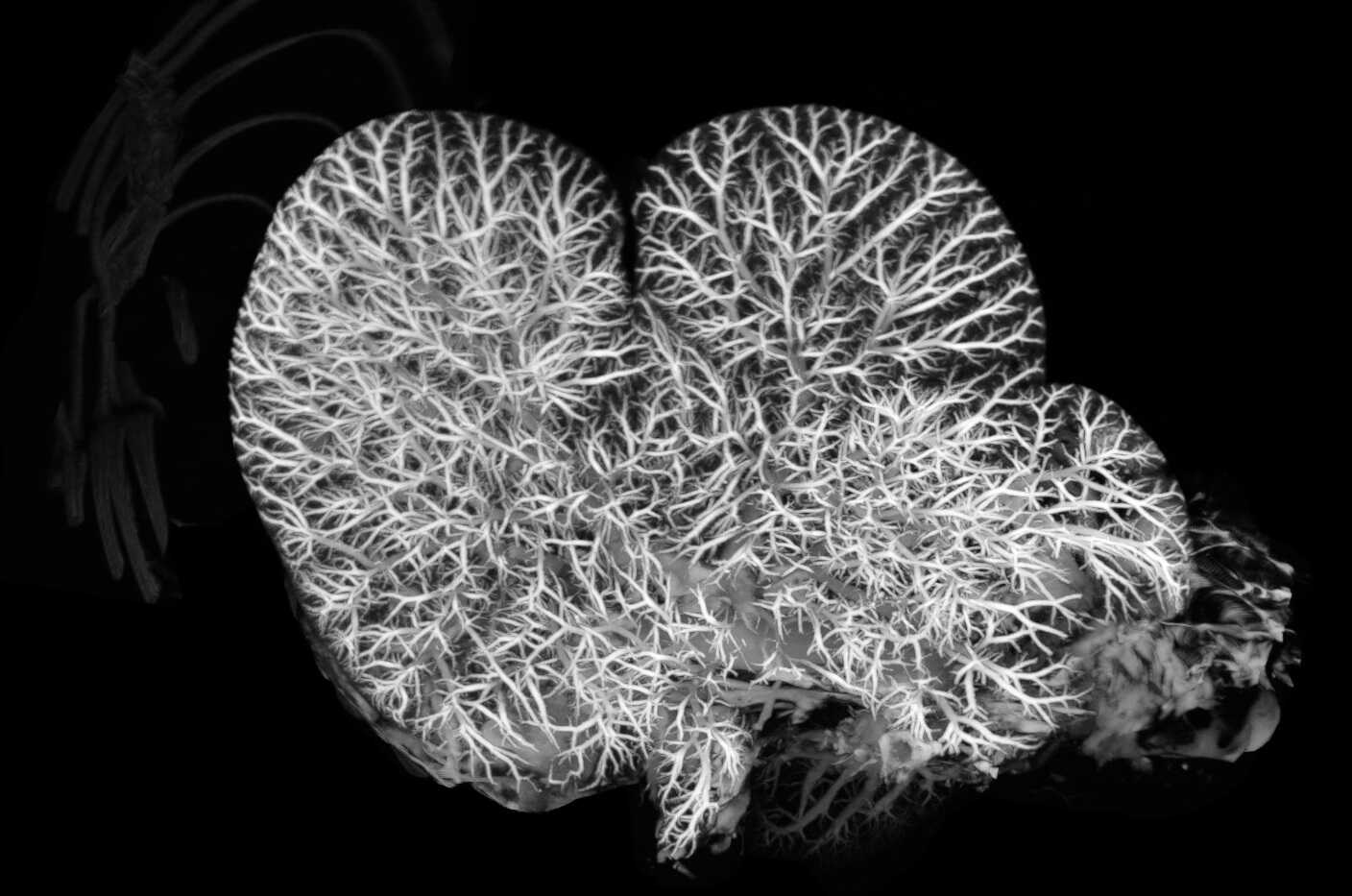
Patience and Hope
by Donghyeon Kim
IBS Center for Advanced Reaction Dynamics
One early spring day, while examining gold complex crystals, I unexpectedly came across a dendritic, tree-like structure. It was not the single crystal I had hoped for, and for a moment I felt a tinge of disappointment. Yet at the tip of its barren branches, there seemed to be a trace of hope. As the days grew warmer, and after many more attempts, I finally obtained the result I had long been seeking. That tree—once dismissed as a failure—was in fact time itself, quietly holding a hidden flower bud. And in the end, that long wait blossomed, passing beyond winter into spring. In an age when patience and hope feel increasingly rare, this crystal gently reminds us of something important: to endure quietly, to take root even beneath frozen ground, and to keep longing for the flowers that will one day bloom.
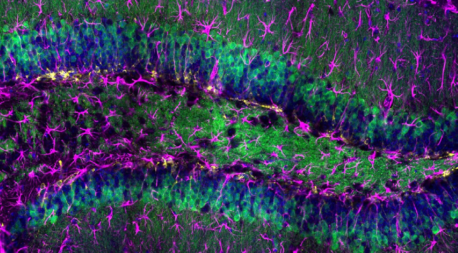
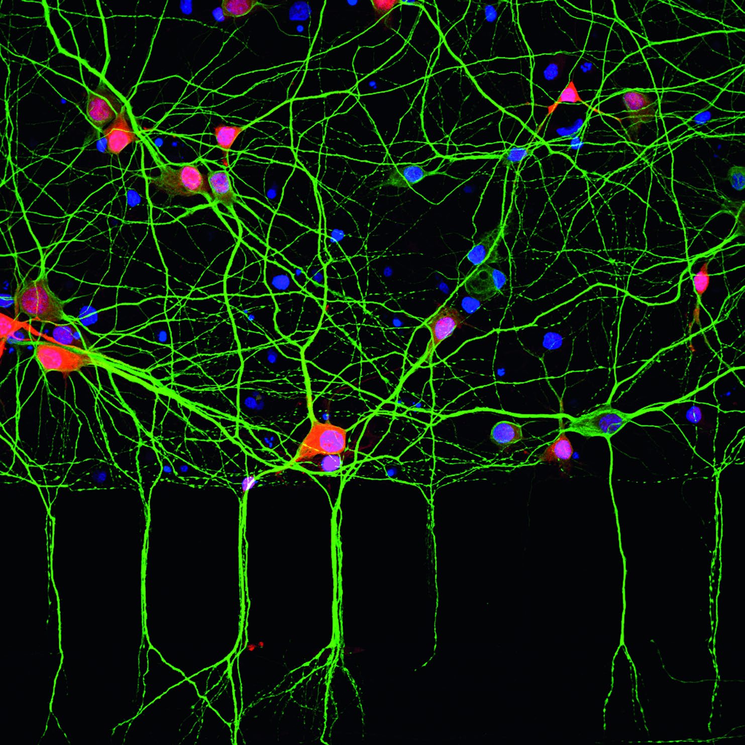
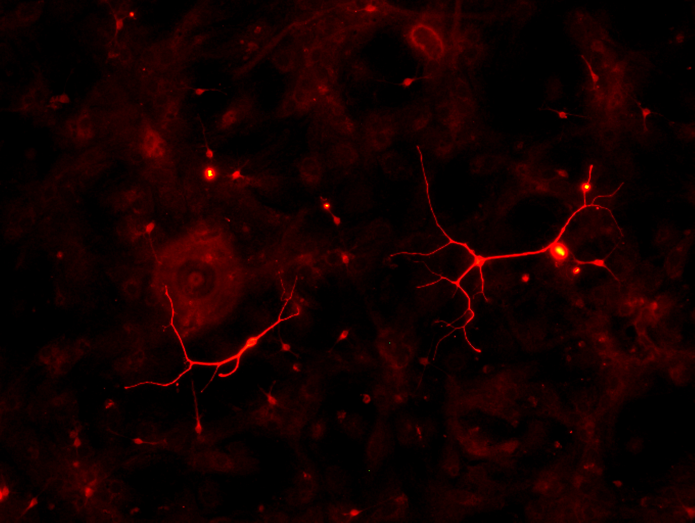
Cells ignite, life moves and brain keeps its own secret symphony
by Nishani Jayanika
IBS Center for Memory and Glioscience
This image shows the hippocampus activated by ultrasound stimulation designed to mimic natural brain waves. As the cells release calcium, they glow—sending signals to their neighbors in a motion that feels like a mysterious dance. Around them, countless other cells weave a dense network of interactions, each following its own internal dynamics while preserving numerous hidden regulatory mechanisms. Even under direct observation, the brain moves according to its own encrypted rhythm, guided by processes we still do not fully understand. This scene offers a glimpse into the profound and enigmatic complexity of the brain.
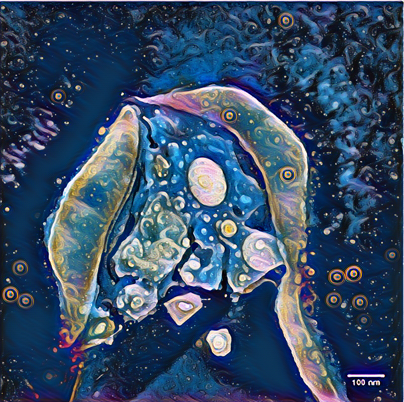
Quantum Star
by Caroline Hommel
IBS Center for Quantum Nanosciene
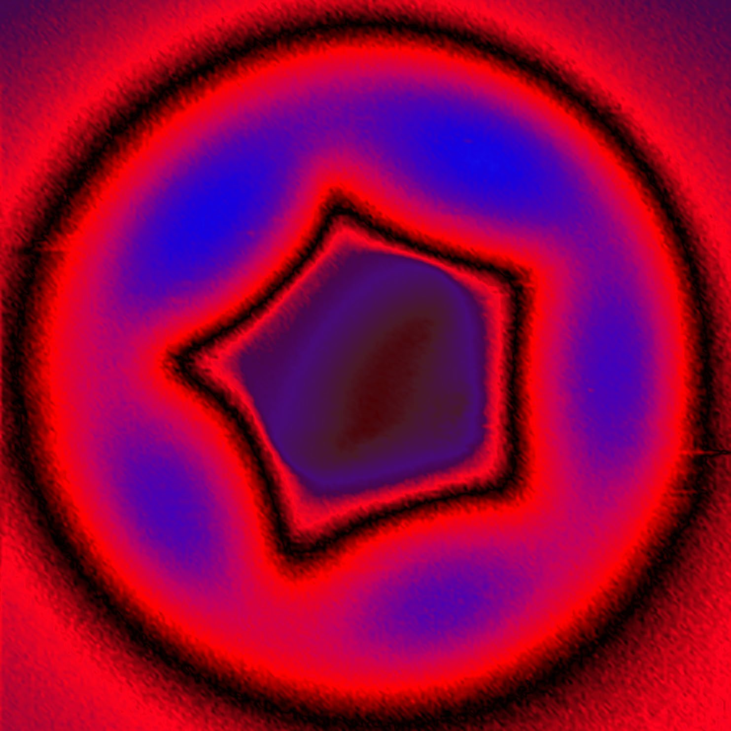
This AFM image shows a single pentagon within a fullerene molecule— the tiny, soccerball-shaped structure made of carbon atoms. Rendered in vivid shades of blue, black, and red, it looks almost like an emblem from a science fiction universe. Its sharp lines and glowing form evoke scenes of space travel, as if it were a gateway inviting us to explore distant galaxies. Yet what we see here does not come from the vast cosmos, but from atoms far smaller than the eye can perceive. At the nanoscale, symmetry creates patterns that feel otherworldly. Science reveals what has always been hidden around us. In that moment, the quantum world becomes as fascinating as the universe above. A reminder that great discoveries often begin in the smallest of worlds.
Atomic Moon
by Caroline Hommel
IBS Center for Quantum Nanosciene
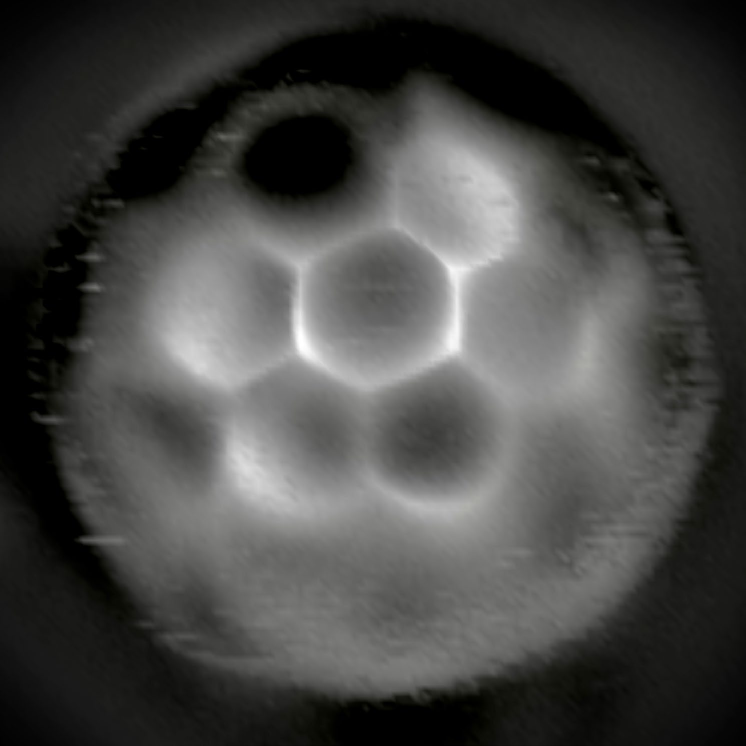
This STM/AFM image shows a fullerene—a molecule composed entirely of carbon atoms. Its spherical shape, built from hexagons and pentagons like a tiny soccer ball, also evokes the smooth, round form of the full moon that has inspired artists for centuries. Humans instinctively associate circular shapes with safety and harmony, a response rooted in our evolutionary past. In this nanoscale sphere, science an aesthetics come together in perfect balance. Beyond its visual beauty, the fullerene possesses remarkable scientific properties. It can trap atoms inside its carbon cage, forming bonds that cannot exist freely in nature. This hidden architecture reveals how entirely new forms of matter can emerge at the smallest scales. A molecular moon, shining not in the night sky, but in the quantum world.
Light of Heavy Ions, A Small Universe
by Kyongho Tshoo, Geonhee Oh, Jae Cheon Kim, Minsik Kwag
IBS IRIS Experimental System Divison & Experimental System Team
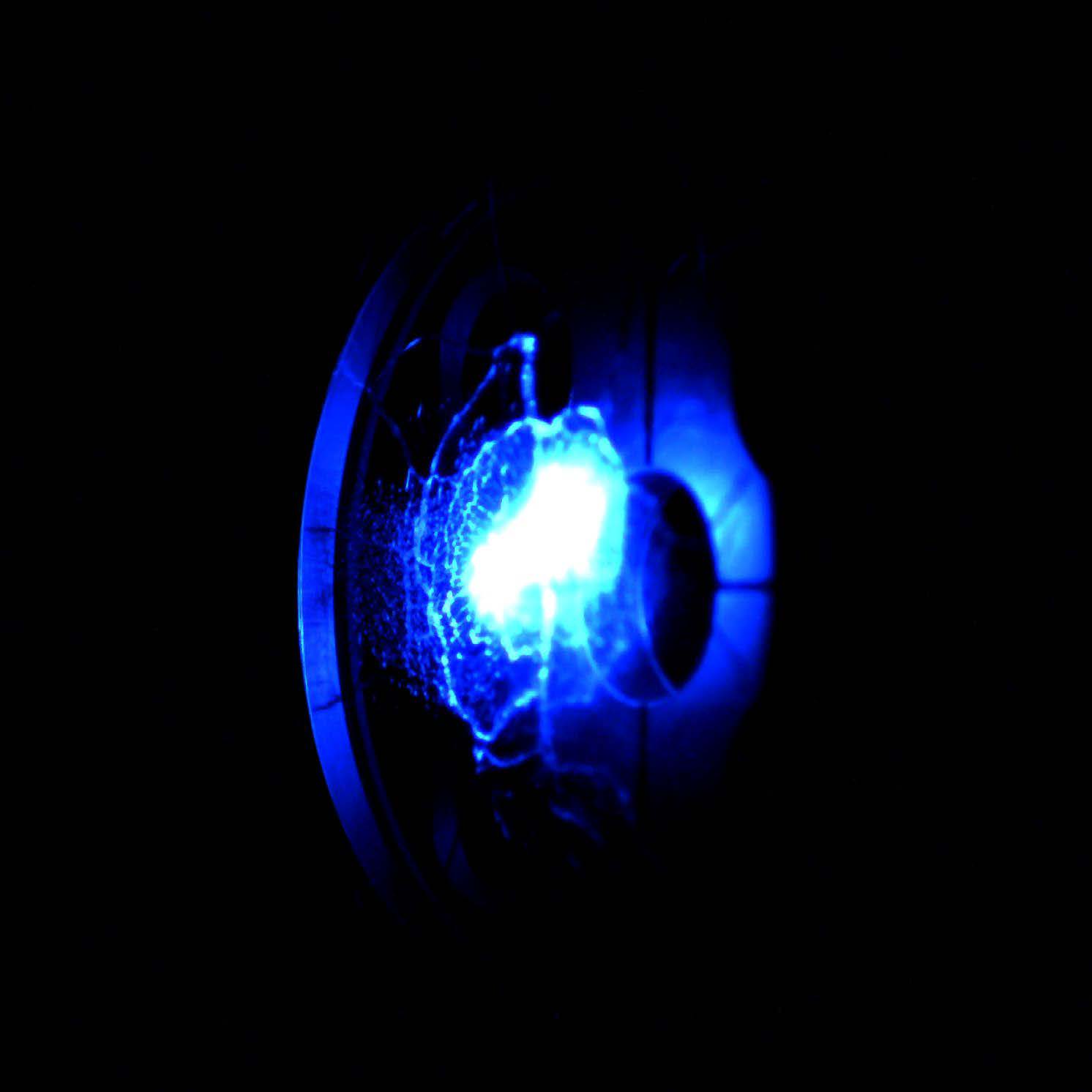
This photograph was taken in 2024 at the RAON heavy-ion accelerator in Korea. An accelerated argon heavy-ion beam strikes a quartz plate at the entrance of the KoBRA spectrometer, producing a flash of light. Repeated impacts generate fine cracks in the quartz, and each burst of blue light resembles interstellar matter scattered across space. Born from tiny particles, this light seems to bridge the microscopic world with the vast universe, evoking the same sense of wonder we feel when gazing up at the night sky. Within this single point of light lies the beginning of RAON’s journey to explore the origin of matter and the universe.
The Dance of Liquid Sheets in Light
by Gwangeun Ahn, Daehyeon Bang
IBS Center for Advanced Reaction Dynamics
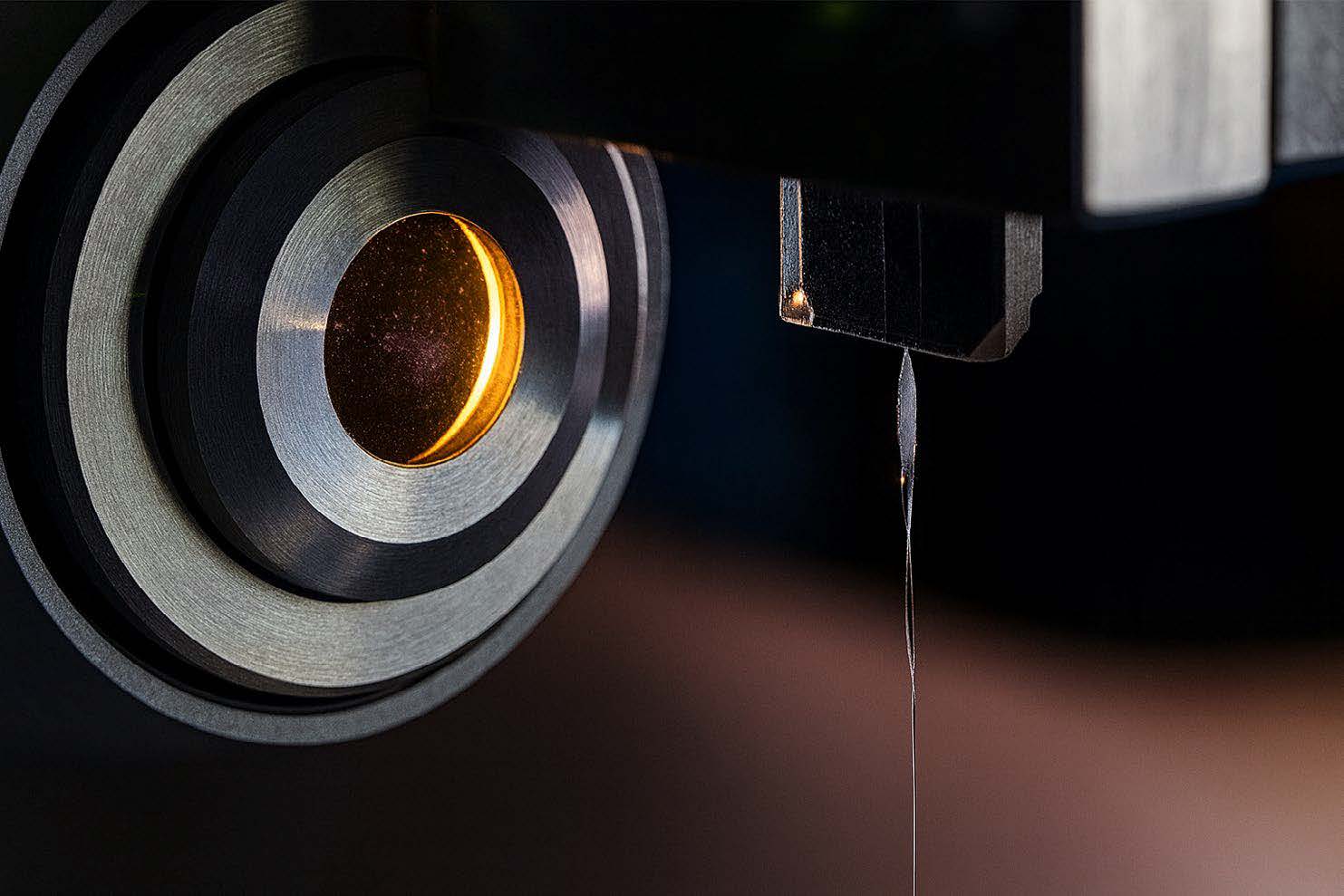
This image captures a liquid sheet jet formed by the collision of two ultra-fine streams of water. At the point of impact, a thin film forms and folds downward, developing into a chain-like sheet that alternates direction at right angles. Compared to a single liquid stream, this structure produces jets that are tens to hundreds of times thinner—an essential feature for ultrafast electron diffraction (UED) experiments, where only nanometerscale targets can unveil atomic motion. The glowing circular aperture on the left contrasts with the transparent, thread-like jet on the right, symbolizing the harmony between human-made precision and the delicate beauty of nature. Though the sheet jet appears as fragile as a strand of silk, it carries deep scientific significance by exposing the fundamental behavior of liquids at extreme scales. To scientists, it is an experimental target. To viewers, it becomes an artistic moment shaped by light and fluid. This image stands at the boundary where science and art converge, transforming the hidden dynamics of liquids into a visible expression of both knowledge and beauty.
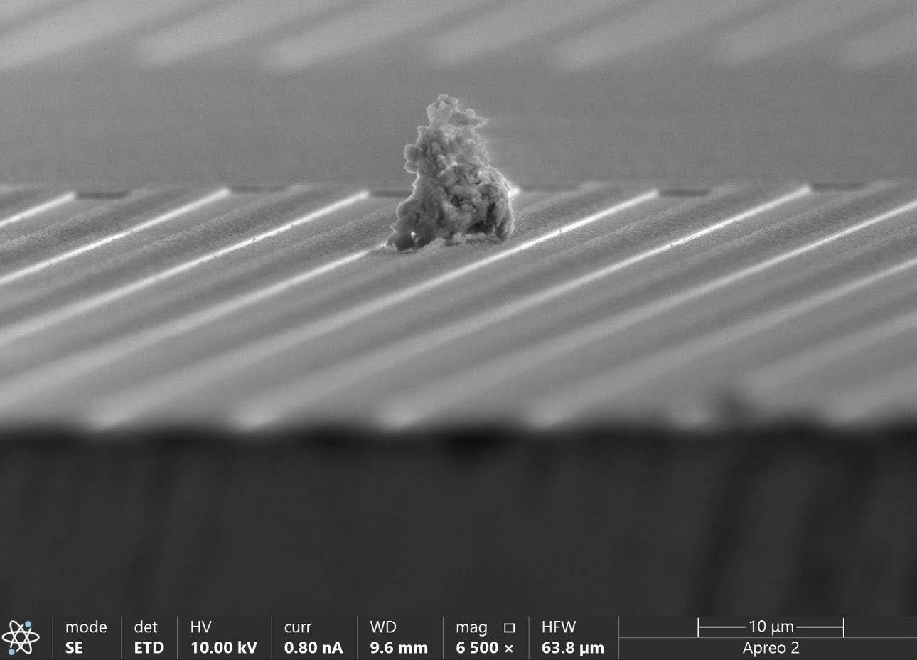
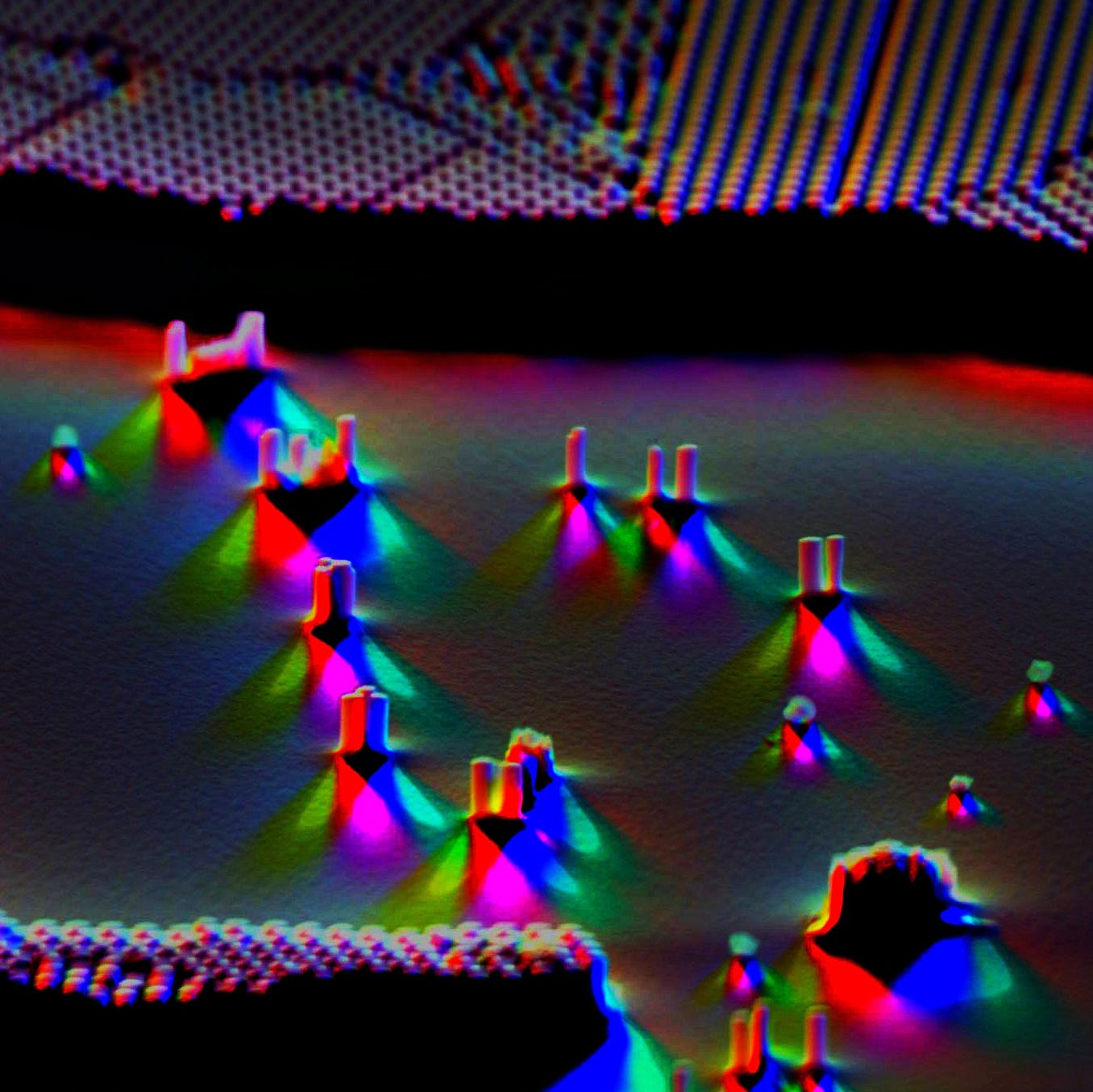
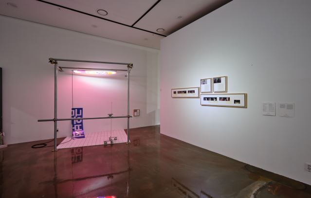
Unicorn Above the Superconducting Coil
by Jin-A Jeon
IBS Center for Underground Physics
This image shows a niobium coil, a key component of a superconducting detector, captured with a scanning electron microscope. The coil is only about 5 micrometers thick, roughly 1/20 the width of a human hair and is magnified 6,500 times. In a superconductor, electric current flows without resistance, a property that enables the extraordinary sensitivity of cryogenic particle detectors. Scientifically, the coil is nothing more than a pathway for current. Yet visually, it resembles a mysterious, enchanted track. Racing across it is an imagined “micro-unicorn”—a creature that does not exist, but whose presence lingers in the viewer’s imagination. In this way, a cold and precise scientific instrumentbecomes a stage for fairytalelike wonder—an experience we hope to share with everyone.
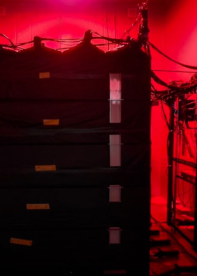
An Eye in the Deepest Darkness
by Lee In Soo
IBS Center for Underground Physics
This video captures the installation of the COSINE-100 detector deep underground. Heavy lead bricks, each equivalent to the weight of a small child, are placed one by one to build a shield that blocks external noise. At the center of this fortress rests the detector itself—nearly 100 kilograms—guided into position through careful coordination and teamwork. More than simple construction, the process feels like a solemn ritual of raising an eye toward the universe. Even amid heavy equipment and repetitive labor, the researchers continue with patience and focus. The interplay of dark structures and red lighting transforms the scene into something almost artistic, beyond science alone. Once installed, the detector begins its quiet vigil, waiting for invisible signals— the faint traces of dark matter. In the profound silence underground, humanity has opened an eye to the cosmos. That eye remains awake today, searchingfor secrets we have yet to discover.
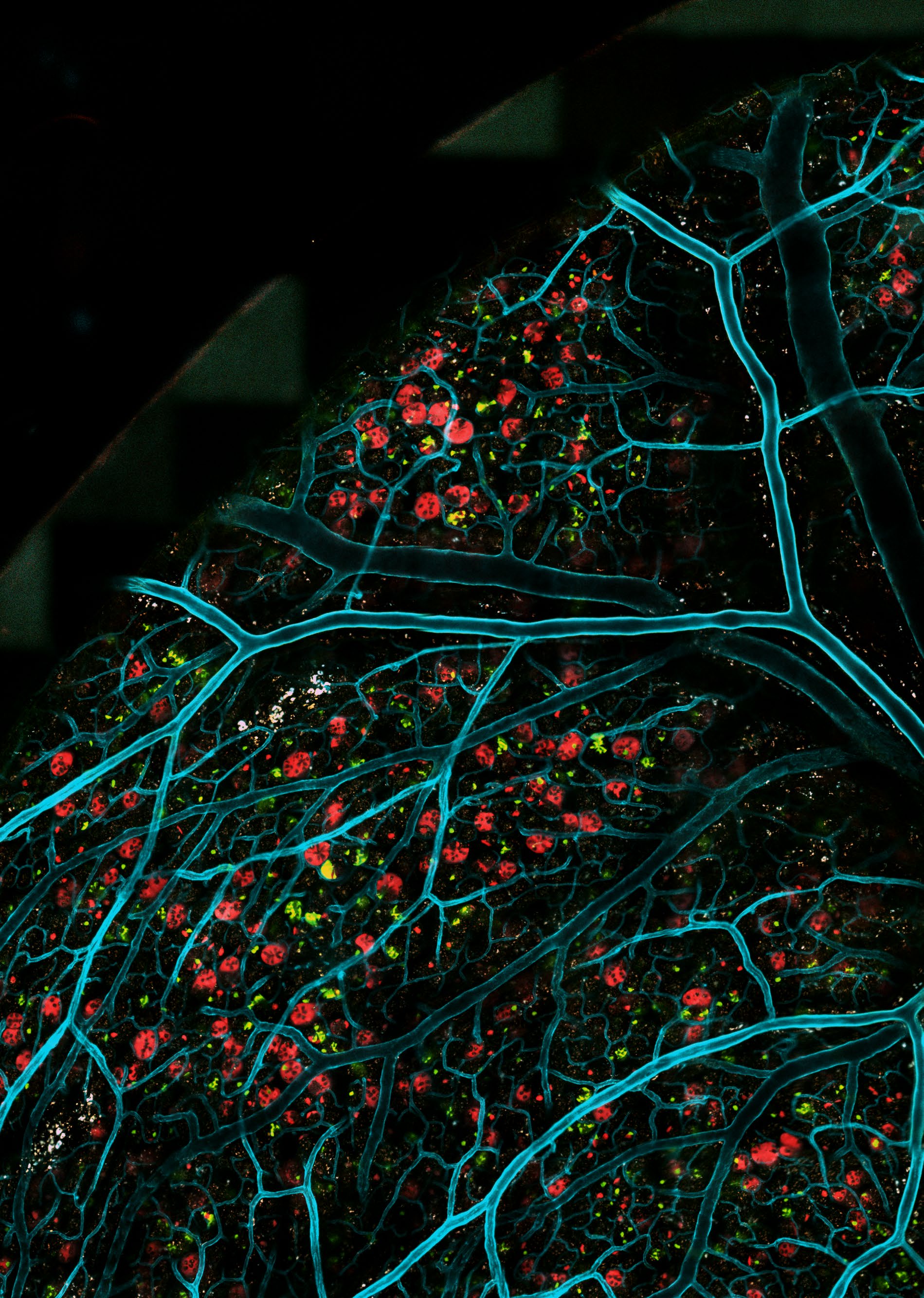
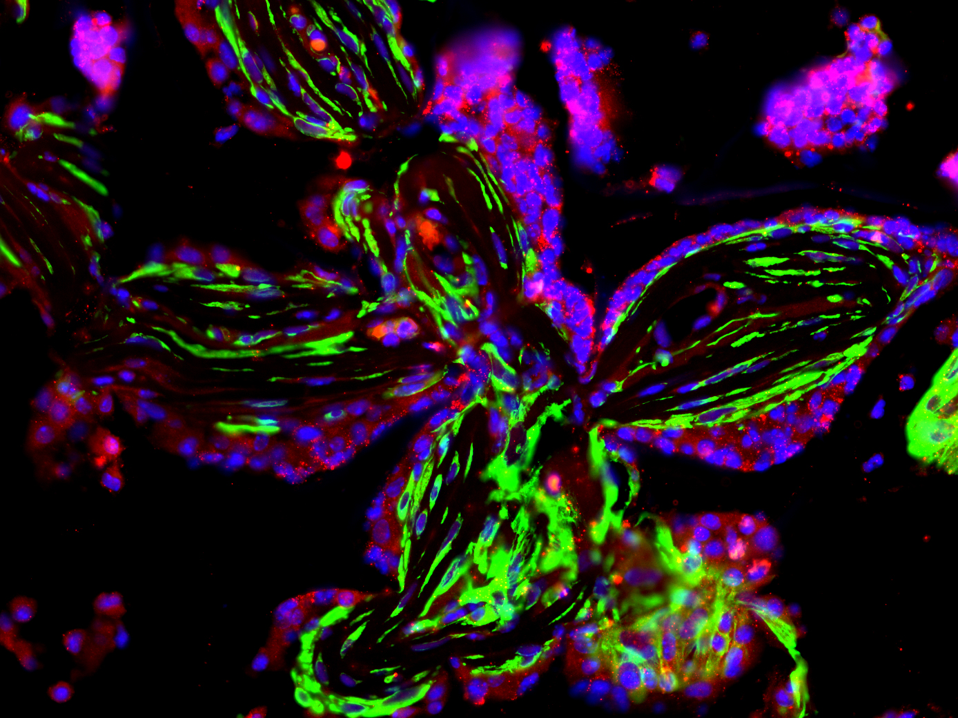
Tree of Good and Evil
by Prajwal Walke
IBS Center for Genome Engineering
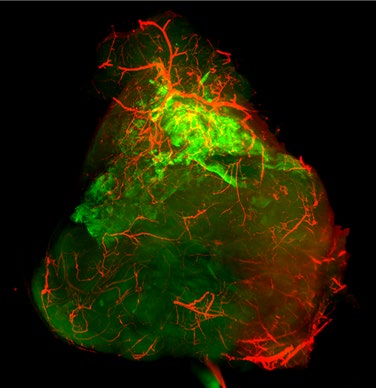
Like the Tree of the Knowledge of Good and Evil in the biblical Garden of Eden, this image captures the intestine of a live mouse. The oncogenic intestinal crypts glowing in red appear as the forbidden fruit, while the cyan branches represent blood vessels that support them. Each crypt resembles a fruit containing cellular “seeds” that ultimately give rise to cancer. These malignant red crypts aggressively outgrow and suppress the natural green crypts, steering the tissue toward an inevitable cancerous fate.
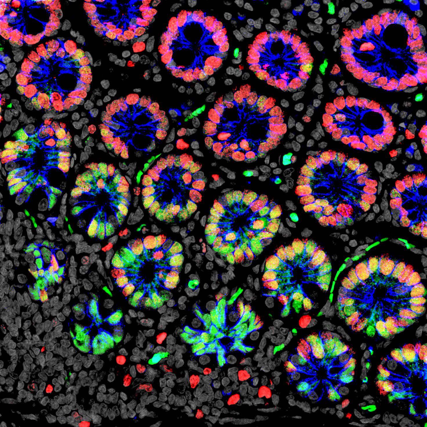
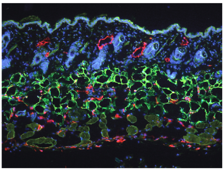
Harmony
by Young-Woong Kim
IBS Center for Genome Engineering
This confocal image shows small intestinal crypts from a monkey, stained with antibodies targeting three different proteins. The upper region is dominated by red, the lower region by green, and the boundary between them glows yellow where the two colors overlap. Looking at this gradient, I was reminded of Korea—the only divided nation in the world today. That is why I titled this work Harmony. Beyond the inter-Korean division, the image also brought to mind the many ideological, political, and social conflicts that split our society into opposing sides. A perfect yellow—a perfect 50/50 blend of red and green—may be impossible. Yet I still hope for a world where the extreme ends, the areas filled with 100% red or 100% green, gradually fade away. A lone figure unable to embrace diversity eventually collapses.
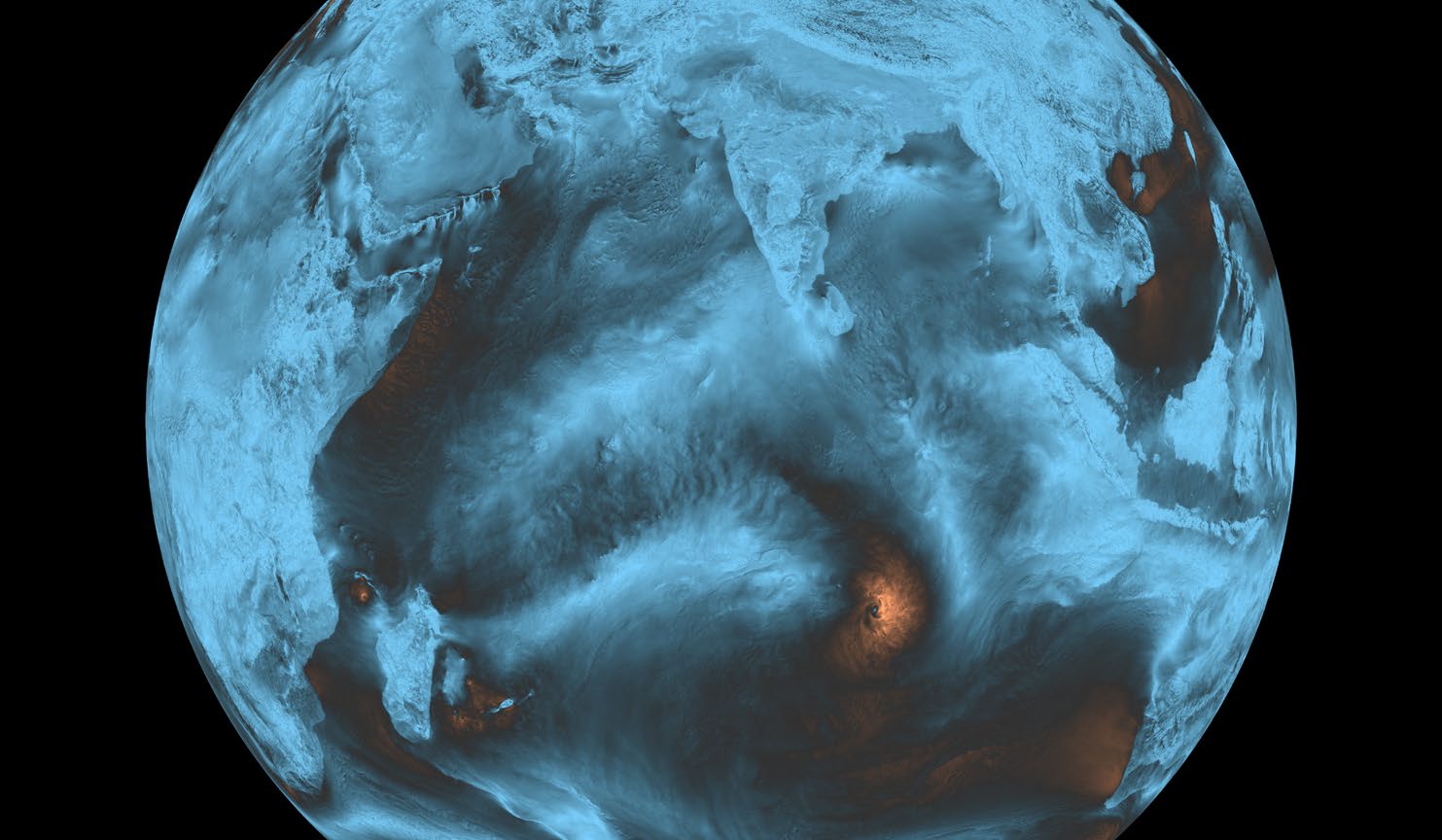
Winds of Change
by Axel Timmermann
IBS Center for Climate Physics
This image highlights the collaboration between the IBS Center for Climate Physics (ICCP) in South Korea and the Alfred Wegener Institute (AWI) for Polar and Marine Research in Germany. The simulation was carried out on the IBS supercomputer Aleph using an Earth System Model developed at AWI. Shown here is a 3-hourly snapshot of wind speed over the Indian Ocean from one of the highest-resolution global climate model experiments conducted to date. The simulation uses the OpenIFS–FESOM model, developed at AWI, with a global atmospheric resolution of about 4km and an ocean resolution varying between 4 and 25 km. Simulating just one model year requires approximately two weeks on the Aleph supercomputer. Colors in the image indicate wind speed: light blue represents weak winds and orange represents strong winds. The snapshot reveals the complex, multi-scale structure of atmospheric circulation— kilometer- scale features associated with convection, ring-shaped patterns marking the growth of gravity waves, and an orange swirl south of the equator corresponding to a tropical cyclone. The simulation also illustrates that winds are generally weaker over land than over the ocean due to surface friction. Additional features include mountain- gap winds (e.g., west of Malaysia and south of Yemen) and island wakes (e.g., south of La Reunion). This simulation is now being extended to quantify how global warming will influence regional- scale climate. Scientists from ICCP, AWI, and other international partners will jointly analyze the petabytes of data generated by this supercomputer experiment.
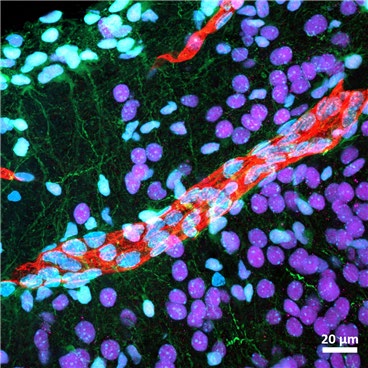
Rivers through a New Land
by Jong-Yeon Yoo
IBS Center for Synaptic Brain Dysfunctions
A river carves its path through unfamiliar land, carrying life wherever it flows. In this image, amouse blood vessel (red) extends deep into a transplanted human cortical organoid. This moment shows that the organoid can receive a blood supply from the host brain, a critical requirement for its survival and growth. Such vascular connections are essential for understanding how human neurons integrate into new circuits, and for developing models to study brain development and disease. What resembles a cosmic river under the microscope is, in truth, a glimpse of two species sharing life at the cellular scale.
CSF Conflux
by Jin-Hui Yoon
IBS Center for Vascular Research
Just as used water flows through drains and merges into a single channel on its way to a treatment plant, cerebrospinal fluid (CSF) follows a similar journey. This image of a mouse illustrates that pathway, showing how CSF travels from the nasopharyngeal lymphatic network toward the deep cervical lymph nodes after cleansing the brain. The green lymphatic vessels visible in the photo mark the very routes through which the CSF flows, carrying the brain’ “wastewater” to its next destination.
Flow of life
by Hokyung Jin
IBS Center for Vascular Research
This image captures the journey of cerebrospinal fluid as it flows from the brain to the lymph nodes in the neck. Tinged with red, the fluid carries the rhythm of life as it leaves the brain and circulates through the body—like a soft beam of light gently slipping through the darkness, revealing the most fundamental energy of life. The green lymph nodes that hold this light are calm and beautiful, reminiscent of auroras shimmering across a night sky. This place is more than a simple passageway—it is a space where life is cleansed and renewed. Through this single photograph, we glimpse the remarkable processes unfolding quietly within our bodies, normally hidden from sight, and the serene beauty that resides within them.
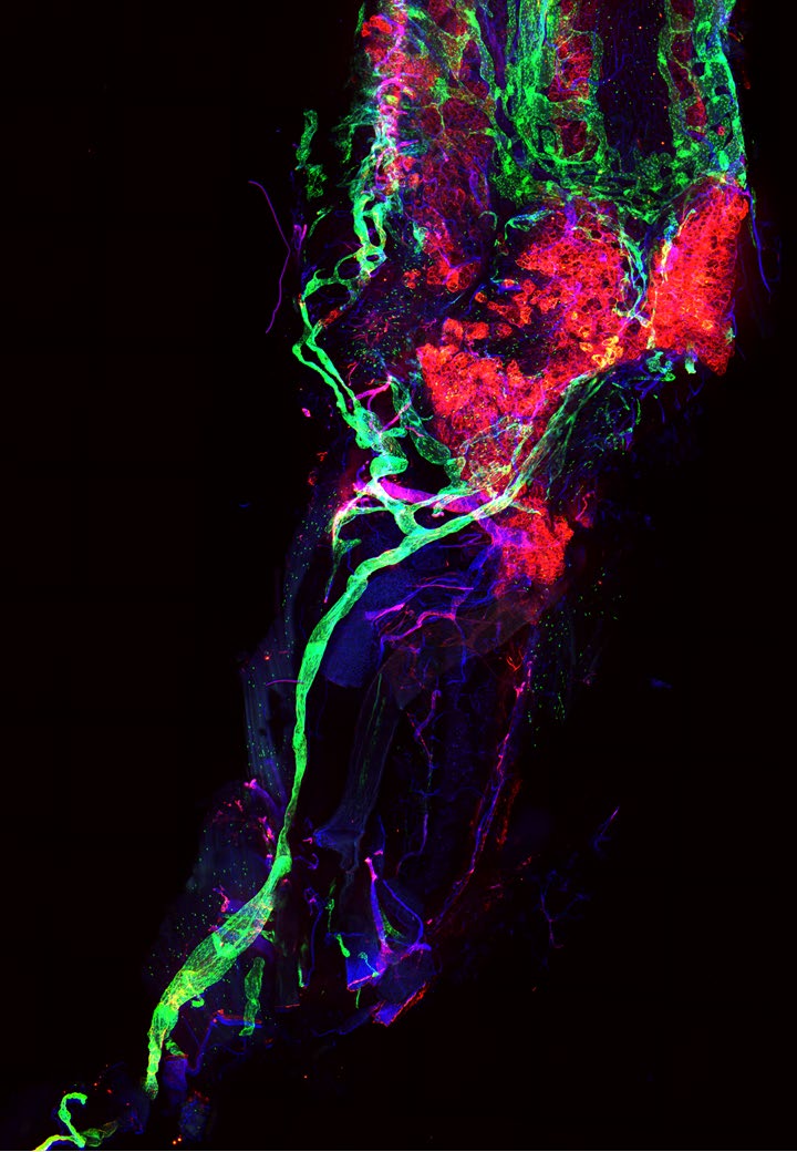
A Human Brain within a Mouse Brain
by Jong-Yeon Yoo
IBS Center for Synaptic Brain Dysfunctions
Inside the mouse brain, a fragment of human cortex begins to live and grow. The green region shows a transplanted human cortical organoid, while the red structures are mouse blood vessels infiltrating the tissue. This image captures a remarkable moment: two different species connecting at the most fundamental level of life. These vessels deliver not only blood, but also the possibility of survival, communication, and function. Through such grafts, we can explore how human neurons adapt to a new environment and integrate into host circuits, opening doors to understanding brain development and disease. A tiny human organoid inside a mouse brain may one day illuminate the profound complexity of the human mind.
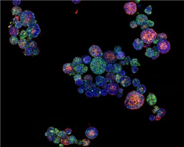
Fireworks of Life
by Heo Seo-Young
IBS Korea Virus Research Institute (Center for Study of Emerging and Re-emerging Viruses)
Fireworks are a spectacle born from countless fragments of light converging to illuminate the night sky. In this image, cells within a bat kidney organoid, revealed through multiplex immuno-fluorescence, glow in distinct colors like individual spark, scattering and gathering to form a single magnificent scene. This constellation of radiance is not mere decoration, but a symbol of the kidney’s vital functions—filtering waste and maintaining the body’s balance. Just as fireworks require the combustion of gunpowder to flare, each cell expends its own energy to fulfill its role. Though small, these luminous cells cooperate to preserve renal order, creating a grand festival of life. This image transcends a fleeting moment of beauty, embodying an everlasting fireworks of life, and a tribute to the devotion of the cells that sustain it.
The Spectrum of Love
by Hyun Jun Jang
IBS Center for Memory and Glioscience
Just as no two people in the world are exactly the same, love also exists in many sizes and forms. Some love burns brightly and intensely; some is faint and quiet. Some love is large and dazzling, while other love is small and simple. At times, love leaves us wounded, and sometimes it remains unfinished. This image was created by measuring various metabolites and lipid molecules in mouse brain tissue using mass spectrometry imaging. The cortex, hippocampus, and thalamus—each with its own distribution of biomolecules—together or individually resemble different heart shapes. As you search for these subtle hearts within the image, I hope you take a moment to reflect on the shape of love that lives within your own heart.
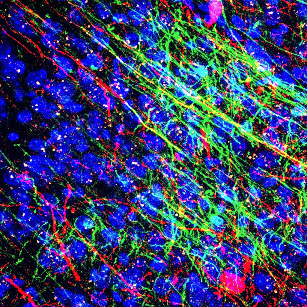
Hand in hand for us
by Lee Sangyeong
IBS Center for Memory and Glioscience
Where does the altruism to reach out to someone in pain truly come from? Empathy, the ability to feel another’s suffering, is the starting point of helping someone and the smallest building block of society. Yet, the capacity to help does not arise in isolation. It is shaped by society itself. Memories of receiving kindness, time spent with friends, and other social experiences strengthen oligodendrocytes and nurture empathy. In this image, the greenlabeled oligodendrocyte makes contact with the ACC–BLA neural circuit, a key hub of empathic processing. It looks as if one extended hand is gently met by another— a quiet gesture capturing the essence of empathy. In the end, the power to help others comes from within us, but it is society that teaches our hands how to reach.
The Azure Paradox
by Kwon SeongJin
IBS Center for Artificial Low Dimensional Electronic Systems
We anticipated perfect stillness, but the unexpected cracks we discovered within it offered a new source of inspiration. This image shows a cryogenic scanning tunneling microscopy topography of the quantum material Ta₂Pd₃Te₅. The exciton condensation formed by the union of electrons and holes in this material is expected to create a stable, perfectly ordered state, like a calm sea glowing red. Yet on the surface of this quiet order, paradoxical blue traces shine where they should not exist. An unforeseen quantum resonance locally disrupts the condensation, leaving these luminous marks behind. This work reminds us that the blue beauty blooming in the gaps of perfection is, in fact, the most convincing proof that such perfection exists.
*Encounters, Now, Here
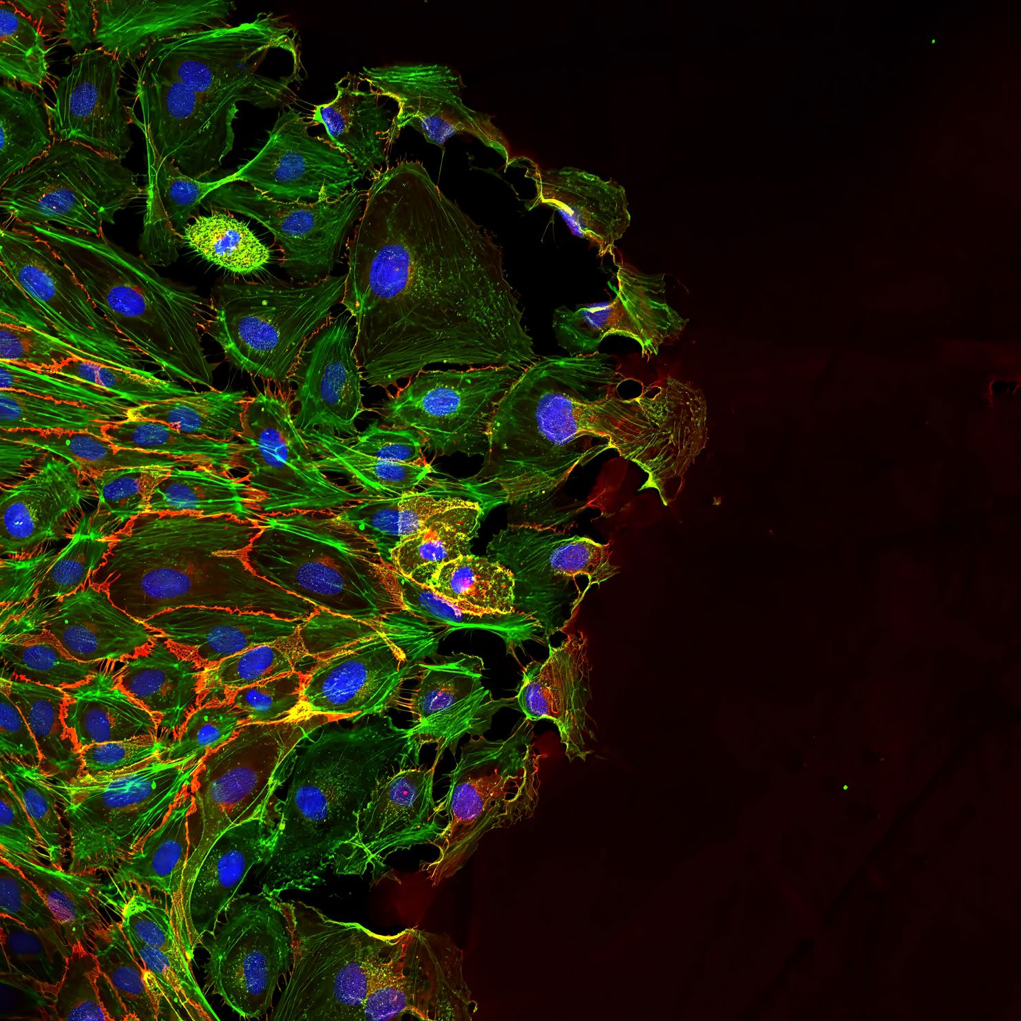
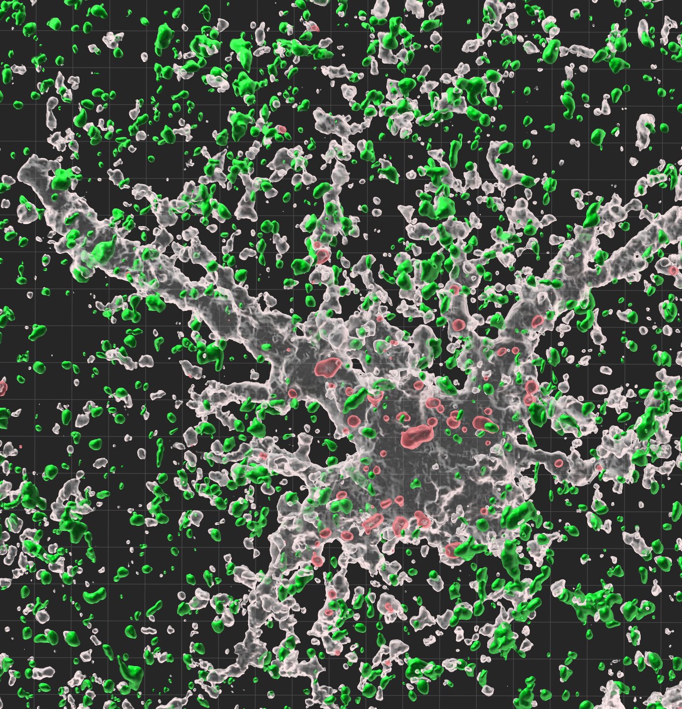
Collective Cell Migration
by Jinwoo Cheon, Minsuk Kwak
IBS Center for Nanomedicine
by Joachim Spatz, Senne Seneca, Harish Kumar Senapati, Ilia Platzman
Max Planck Institute for Medical Research
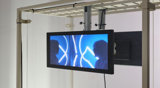
Epithelial cells move not as isolated units but as a connected, collective flow, coordinating their direction and speed through subtle mechanical cues. In July 2025, IBS and MPG established the Max Planck–Yonsei IBS Center for Nano Medicine to jointly develop next-generation nanoscale biomedical technologies. Captured at the Max Planck Institute for Medical Research, this image marks an early point of that collaboration and hints at the new scientific journeys the partnership will make possible.
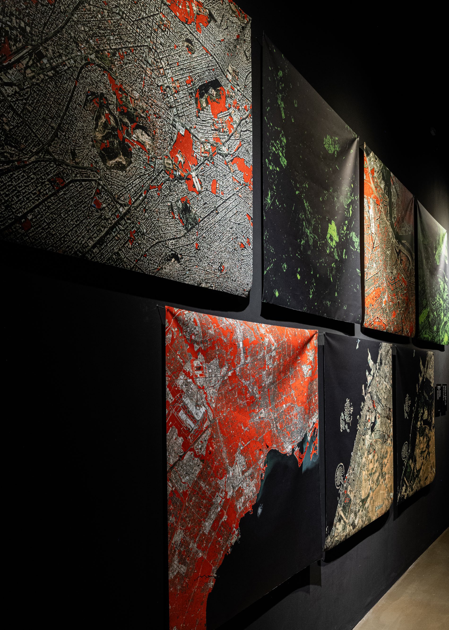
Urban green space and happiness in developed countries
by Meeyoung Cha(Former) CI, (Current) Director of the Max Planck Institute for Security and Privacy
BS Center for Mathematical and Computational Sciences Data Science Group
by Jeasurk Yang
Max Planck Institute for Security and Privacy
by Inho Hong
Max Planck Institute for Human Development
by Woo-Sung Jung Chair of the Korea Foundation for Science and Creativity(KOSAC)
University of Science and Technology(POSTECH)
by Donghee Y.Wohn
New Jersey Institute of Technology
by Oh-Hyun Kwon
Korea Advanced Institute of Science and Technology(KAIST)
This image visualizes an IBS–MPG study mapping urban green space worldwide at 10-meter resolution using satellite-based NDVI. By measuring per-capita greenery in major cities, the research shows that access to green space is closely tied to happiness, especially in wealthier nations. The work underscores how insights into global urban challenges emerge through international scientific collaboration.
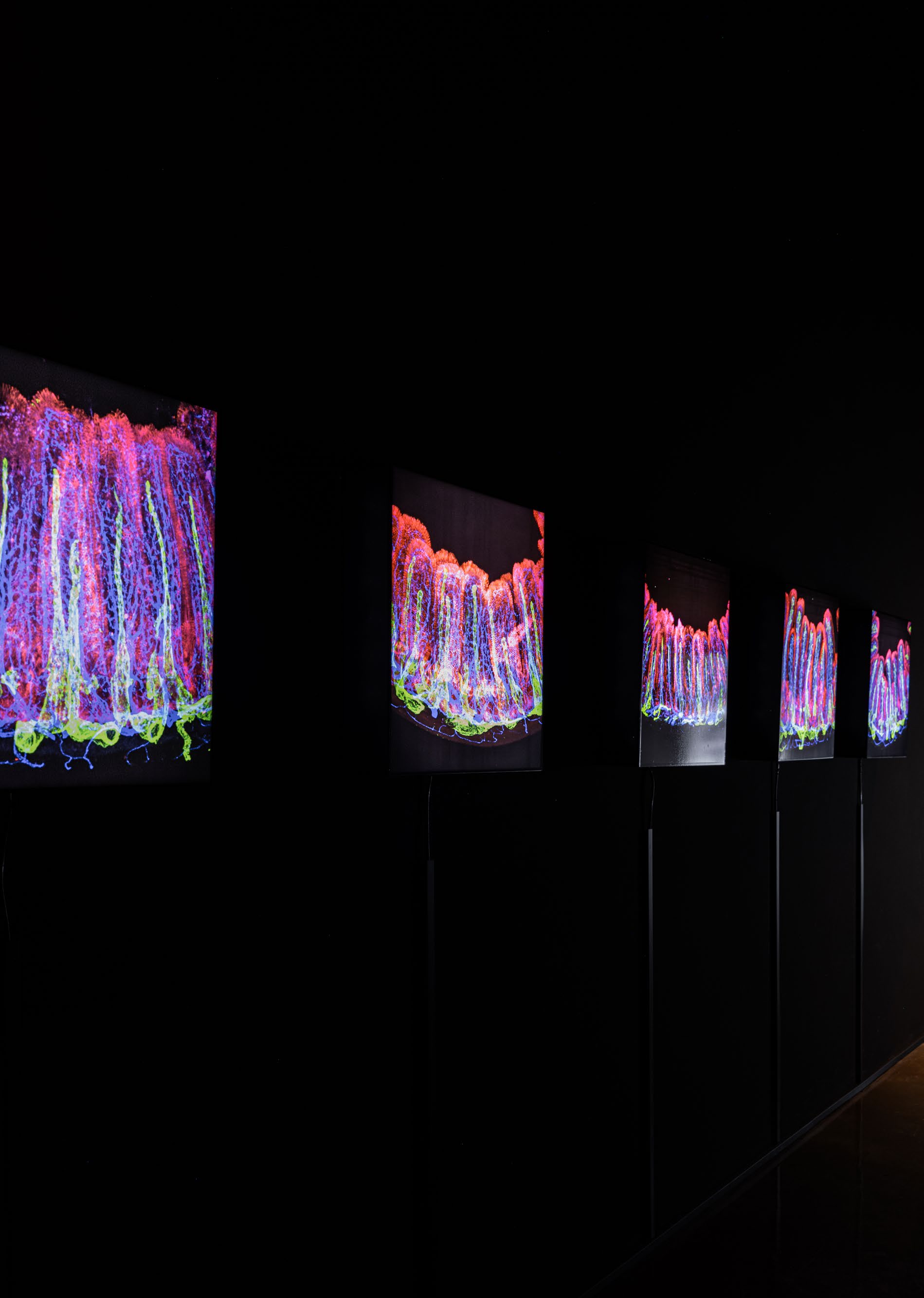
Distinct fibroblast subsets regulate lacteal integrity through YAPTAZ-induced VEGF-C in intestinal villi
by Gou Young Koh (External Scientific Member of the Max Planck Institute for Molecular Biomedicine), Seon Pyo Hong, Hyunsoo Cho, Intae Park, Hosung Bae, Sang Heon Suh
IBS Center for Vascular Research
by Ralf H. Adams
Max Planck Institute for Molecular Biomedicine
The lacteal within the intestinal villi is a delicate channel for absorbing fats. This image contrasts normal mice with those showing YAP/TAZ overactivation, revealing how subtle shifts in cellular signals can reshape this structure and disrupt organ function. Collaboration with MPG deepened the understanding of these fine-scale interactions.
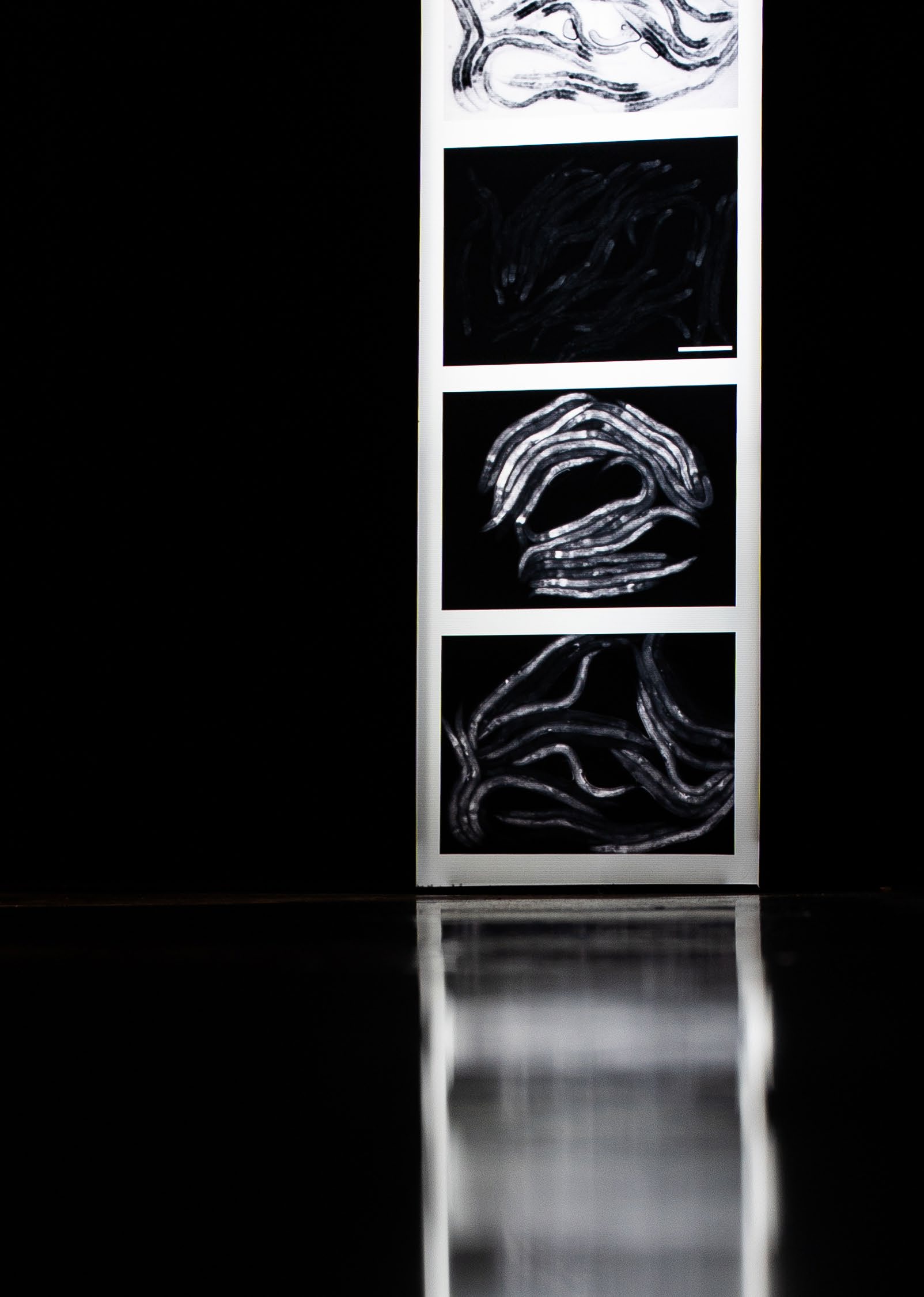
Genome-wide RNAi screen for regulators of UPRmt in Caenorhabditis elegans mutants with defects in mitochondrial fusion
by Stephane G. Rolland, Barbara Conradt
IBS Center for Genomic Integrity
by Assa Yeroslaviz
Max Planck Institute for Biochemistry
This image captures the stress signal that lights up in C. elegans when mitochondria malfunction. Disrupting certain genes triggers a brighter fluorescence, seen here as shifts in grayscale. Through joint work, IBS and MPG revealed how this hidden alarm system is sensed and regulated inside living cells.
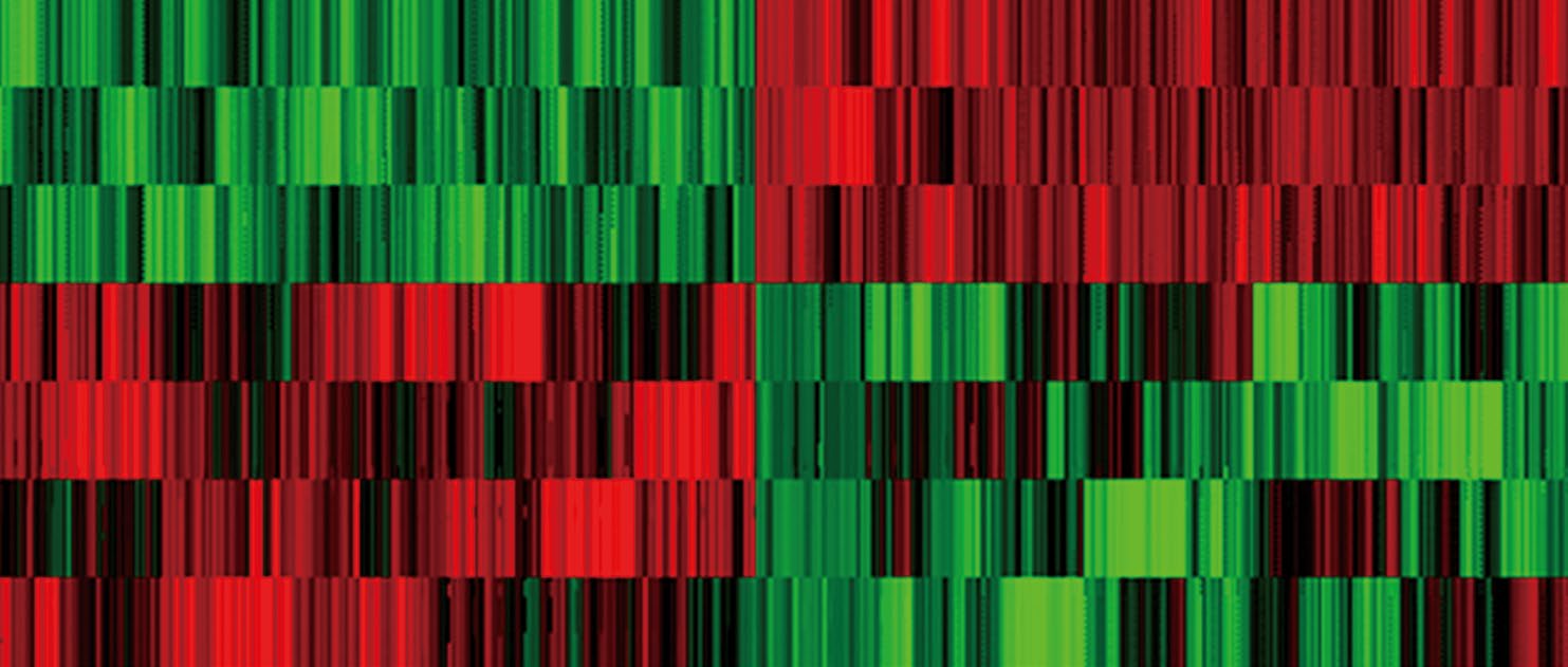
YAP TAZ direct commitment and maturation of lymph node fibroblastic reticular cells
by Gou Young Koh (External Scientific Member of the Max Planck Institute for Molecular Biomedicine), Seon Pyo Hong, Hyunsoo Cho, Intae Park, Hosung Bae, Sang Heon Suh
IBS Center for Vascular Research
by Ralf H. Adams
Max Planck Institute for Molecular Biomedicine
This image is a color-coded “map” showing how proteins inside lymphnode stromal cells change when the YAP/TAZ proteins become abnormally overactive. Compared with normal cells, some proteins rise sharply while others drop, creating completely different expression patterns. It reveals how small molecular shifts inside a single cell can ultimately reshape lymph-node structure and immune function.
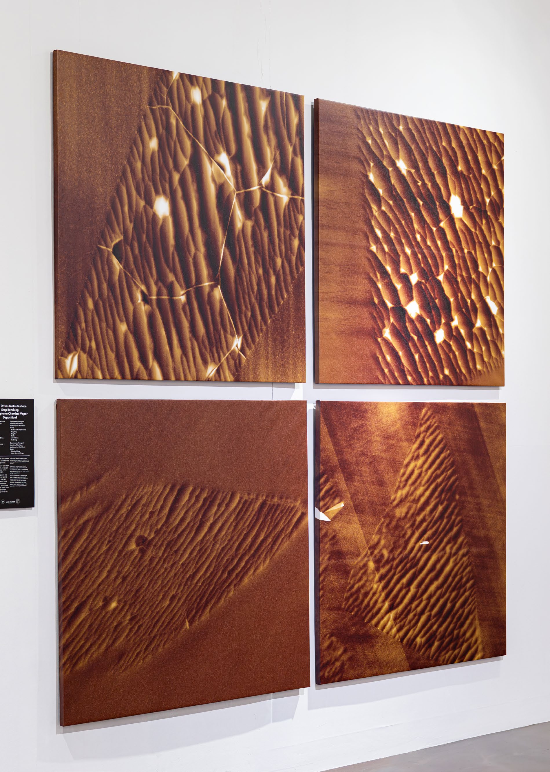
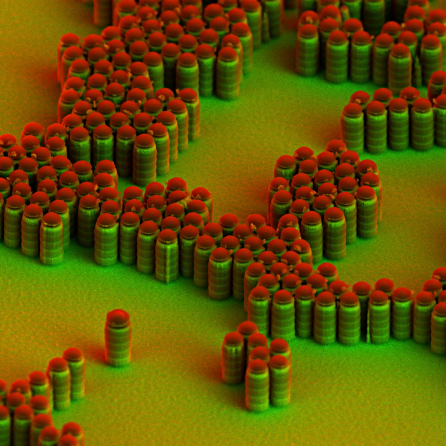
What Drives Metal-Surface Step Bunching in Graphene Chemical Vapor Deposition?
by Rodney S. Ruoff, Feng Ding, Ding Yi, Da Luo, Jichen Dong, Xu Zhang
IBS Center for Multidimensional Carbon Materials
by Zhu-Jun Wang, Marc-Georg Willinger
Department of inorganic chemistry, Fritz Haber Institute of the Max Planck Society
This image captures how the copper surface beneath graphene begins to rise and ripple like small desert dunes when heated. Contrary to previous assumptions, these features do not form because of compressive strain in graphene but emerge naturally from the metal surface at high temperatures. The study overturns a longstanding view of how surfaces interact with two-dimensional materials, and close collaboration with the MPG — renowned for real-time, atomic-scale surface imaging — was essential in revealing this behavior.

Synaptic adhesion molecule IgSF11 regulates synaptic transmission and plasticity
by Eunjoon Kim, Yeunkum Lee, Doyoun Kim, Sun Gyun Kim, Junyeop Daniel Roh
IBS Center for Synaptic Brain Dysfunctions
by Christoph van Riesen, Jeong-Seop Rhee
Max Planck Institute for Experimental Medicine
Three key proteins come together at a synapse to form the core structure that enables neurons to communicate, shaping how we learn and remember. Through joint research, IBS and the MPG revealed how this delicate assembly is organized and how its disruption can lead to disorders such as Alzheimer’s or autism. Their collaboration sheds light on this hidden “architectural site” of the brain and lays groundwork for future therapeutic advances.
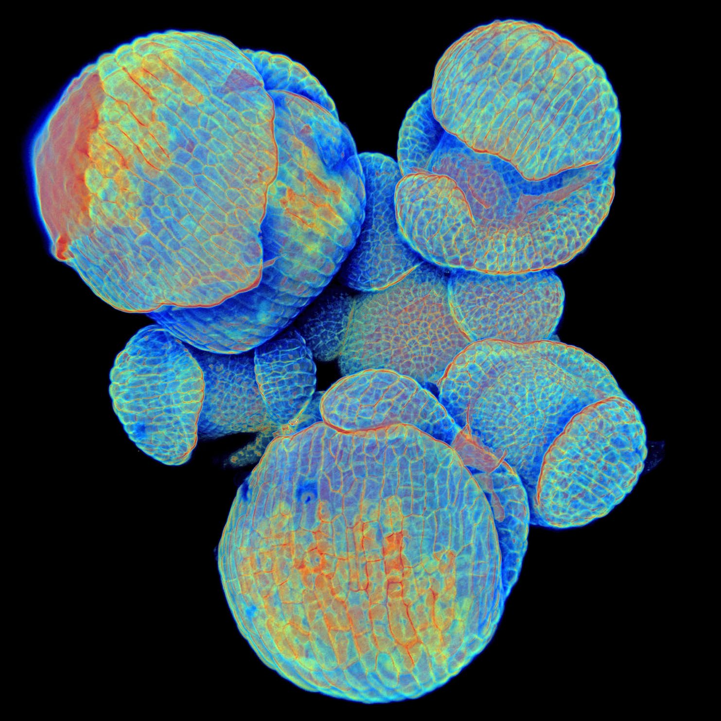
Just at the right time
by Ritika Kulshreshtha
Max Planck Institute for Molecular Plant Physiology, Potsdam-Golm
In the life of a plant, too, much depends on the right timing – from seed germination to growth and flowering. So plants can measure the length of the day or the temperature in order to switch from the growth phase to the reproduction phase at a suitable time – in other words, to flower. The image shows how the flowers of a thale cress, botanically called Arabidopsis thaliana, develop from the dome-shaped tissue of the shoot tip. The thale cress is a so-called long-day plant that blooms in spring. The plant measures the length of the day via its leaves. If the light period is long enough, messenger substances migrate from the leaves into the shoot tips and transmit the flowering signal. In this way, the plant ensures that the flowers are formed at the right time to ensure fertilisation, seed formation and species conservation.
Rainbow in the Nanoshade
by Bjorn Hoffmann, Silke Christiansen
Max Planck Institute for the Science of Light, Erlangen
Solar cells provide a climate-friendly energy supply. To ensure that solar cells increase their efficiency in converting sunlight into electricity and require smaller amounts of silicon in the future, scientists are researching photovoltaic elements that do not consist of a closed silicon layer, but a thin yet dense ‘carpet’ of nanowires. In this way, the light is trapped between the columns, hall-of-mirrors style; and in addition, the nanowires have special optical properties that allow them to absorb more light than a smooth layer. Aesthetically, however, isolated nanowires are far more pleasing - especially when they are scanned with an electron beam that is captured by three diodes which show the scattered electrons in red, green or blue, respectively. This colour display is created when one or two of the diodes are in the shade, cast by the electron beam on a nanowire.
Summer in the City
by Chiel van Heerwaarden
Max Planck Institute for Meteorology, Hamburg
It forms clouds, causes storms – and presents considerable challenges for climate researchers: turbulence forms in the atmosphere when warm and cold air come together. For example, if a city heats up considerably more than its surroundings in summer, the warm air rises rapidly, like in a chimney. Along the edges, it mixes with the colder ambient air in numerous large and small vortices. Particularly interesting situations arise when the rising plumes of heat from two or more sources of heat interact. The likelihood then increases that they will churn up the atmospheric layering of an entire region and eventually influence the climate. Scientists use computer simulations to vary the magnitude of the heat sources and their distance from each other.
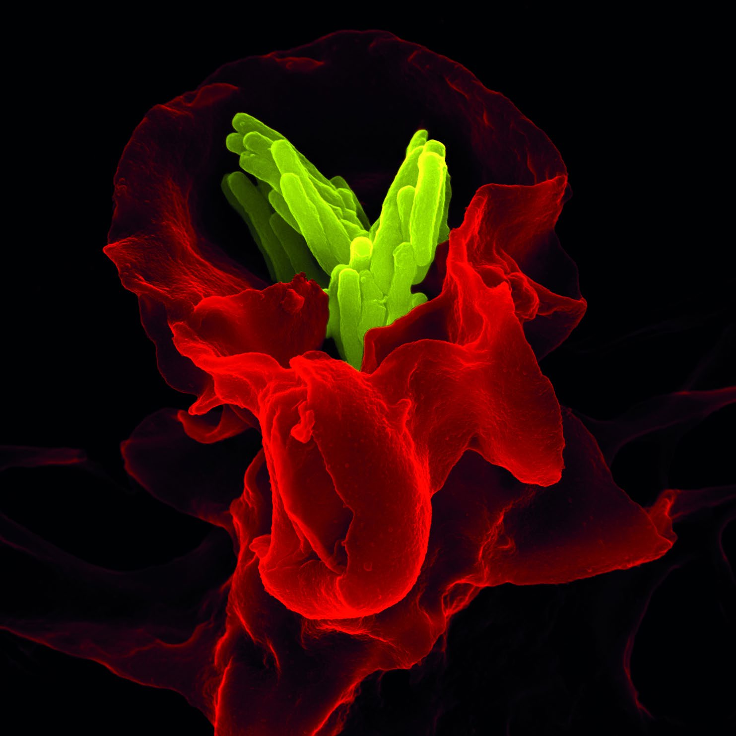
Immune System at Work
by Volker Brinkmann
Max Planck Institute for Infection Biology, Berlin
What looks like an exotic flower at first glance, is, in fact, the human immune system at work: a white blood corpuscle (shown here in red) is in the process of disarming tuberculosis bacteria (yellow). The pathogens are encircled by the scavenger cell membrane, pulled into the interior of the cell and locked in there–ideally forever. However, Mycobacterium tuberculosis is an extremely tough customer: thanks to a particularly resistant membrane, the bacteria can survive for many years inside the scavenger cells and may be released again if the host immune system is weakened, for example through diseases like AIDS or the effects of ageing.
Towers of silicon
by Bjorn Hoffmann, Silke Christiansen
Max Planck Institute for the Science of Light, Erlangen
Researchers hope for higher efficiency in future solar cells. To achieve this, they form the silicon for photovoltaic elements not into today’s conventionally smooth layers, but into ‘carpets’ of nanowires which absorb a lot more light. Since the wires are just 100 nanometres thick and two micrometres long, they resemble tiny towers. MPG researchers produce these structures by initially coating a thin layer of silicon with polystyrene spheres. They then remove the silicon, which is not protected by the beads, by etching it with plasma, i.e. strongly ionized gas. Normally, the silicon towers stand close together. Here, researchers have left gaps between the polystyrene spheres to be able to inspect the wires from the side as well. The electron microscope, with which the picture was taken, distinguishes between different materials because it uses two different detectors. It shows polystyrene in red and silicon in green.
Turbulent Exchange
by Juan Pedro Mellado
Max Planck Institute for Meteorology, Hamburg
Turbulent currents play an important role in climate events, for example in cloud formation or – as calculated and visualised here – in the exchange processes that occur on the surface of water bodies. When the water on the boundary with the air cools down, through convection and uplift, a typical cell-like pattern of the heat distribution in the water arises in the layer underneath. The dark zones in the image are relatively warm areas, which move upwards while cooler areas, often just a few millimetres wide, move down – the light-coloured edges of the ‘cells’ here. Tiny whirlpools arise at the network nodes, sometimes even double vortices with opposite directions of rotation.
Microcosmic Transport System
by Karen Kohler
Max Planck Institute of Colloids and Interfaces, Potsdam
Pharmaceutical substances are most effective and cause fewest side effects when they are released directly in the diseased area of the body. MPG Planck scientists are working on the development of a drug delivery system that only releases drugs when it recognises the target cells: microcapsules with special recognition molecules dock directly onto diseased cells, e.g. cancer cells. The drugs can escape through the capsule walls as a result of changes in the temperature, pH value or salt content. The image shows different types of such capsules that were exposed to different temperatures: some shrivelled to form solid balls (yellow) and others melted to form bigger capsules (green), which collapsed when they dried out.
Neurons on Track
by Ina Bartnik, Erin Schuman
Max Planck Institute for Brain Research, Frankfurt am Main
Learning and memory are based on the constant modification, dismantling and re-establishment of the connections between cells in the brain. Simplified models are needed to enable scientists to study and understand these complex processes. Researchers at the Max Planck Institute for Brain Research grow neurons in fine microchannels on plates with a photolithographic structure. In this way, the complex threedimensional network of neurons in the brain is reduced to two dimensions in the cell culture. The researchers can thus analyse how the synapses between the cells form or dissolve, and examine the role played by substances like neurotransmitters in these processes. This cell culture technology is also of great interest for the development of new active pharmaceutical substances.
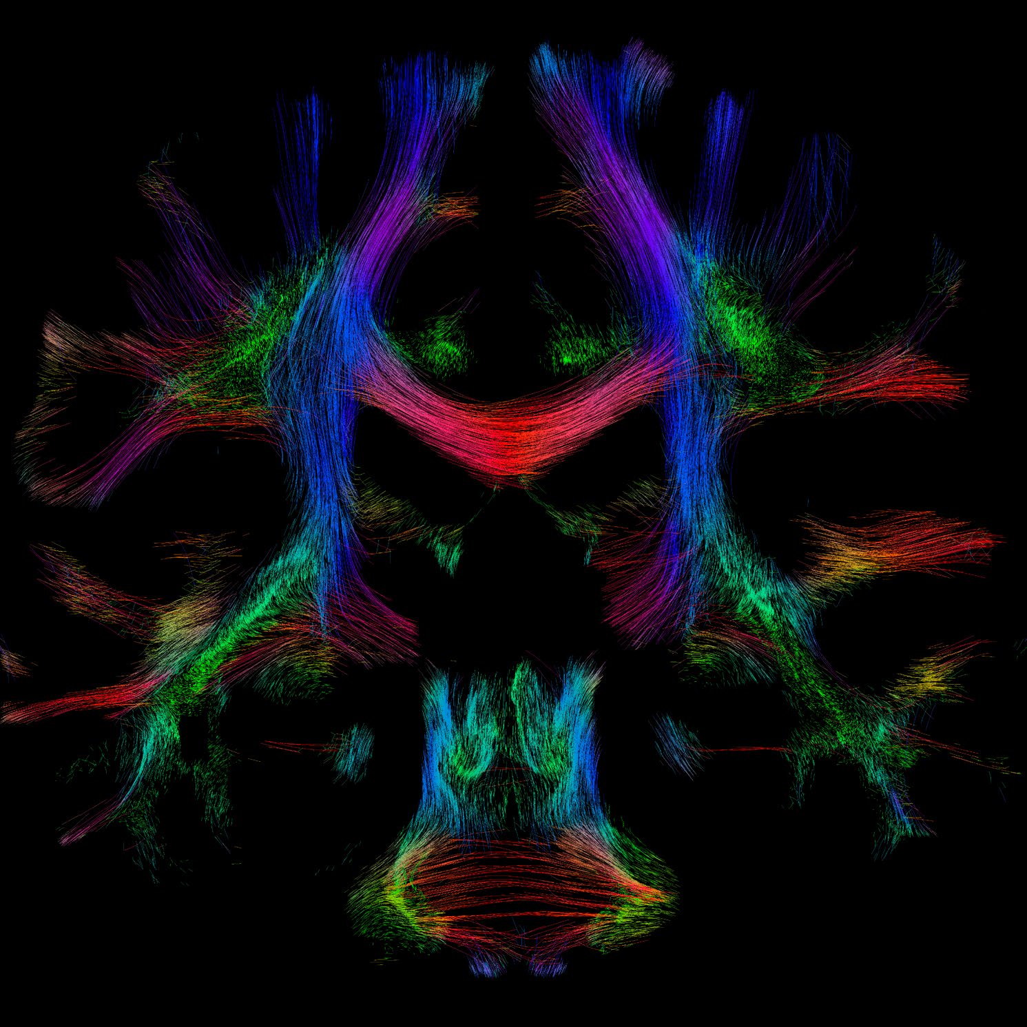
Highways for Thoughts
by Ralph Schurade, Alfred Anwander
Max Planck Institute for Human Cognitive and Brain Sciences, Leipzig
What is 17 multiplied by 146? Or 111 plus 97? Complex cognitive skills such as calculation wouldn’t be possible without complicated connections of neuronal circuits in various brain regions. With the help of diffusion-weighted magnetic resonance imaging (MRI), neuroscientists are able to uncover how these nerve fibre bundles connect different regions of the brain. To this end, the scientists use the natural magnetism of the particles in the brain in order to measure the diffusion movement of water molecules in the tissue. This enables them to draw conclusions on the pathways and signal orientation of the large nerve fibre bundles. The researchers translate the measured diffusion gradients into bright colour patterns, with the colours corresponding to the direction of the fibres (red: left-right; green: front-back; blue: top-bottom).
Structure is Key
by Hugo Sandim, Katja Angenendt
Max Planck Institute for Sustainable Materials
Steel is one of the most versatile construction materials ever. Since its properties can be influenced over a wide range by forming, heat treatments, and not least by alloying – i.e. adding different quantities of other elements – tailor-made grades are now available for a wide variety of applications. This image shows the crystallographic structure of a specific version of a stainless steel very often used in the food industry due to its chemical resistance. The material was manufactured by 3D-printing. The colours illustrate the crystal orientations, which have a decisive influence on the properties of the material. A closer look reveals that our image was created by duplication: The original result was mirrored on the longitudinal axis.
The 7th Art in Science
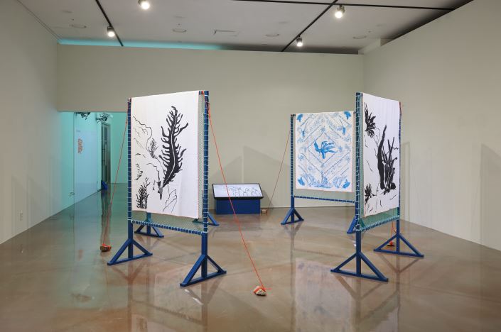
<ea Plants, Bare Hands, Entangled Gaetbawi>, 2022
by Rice Brewing Sisters Club
Rice Brewing Sisters Club’s Sea Plants, Bare Hands, Entangled Gaetbawi is a work that focuses on seaweed, bare-handed fishing, and coastal rocks(gaetbawi), consisting of four installations and one video. This work delves into how sea plants coexist with the ocean, as well as the wisdom of bare-handed fishers who physically experience the sea within a mechanized fishing industry. While this kind of knowledge appears to oppose objective, empirical science, the work suggests that both knowledge and wisdom are necessary in life. Emphasizing the importance of learning from nature, this work marks the first step of the exhibition that starts the collaboration between science and art.
<Catnap>, 2024
by Yoon Seo Yeong
Yoon Seo Yeong explores the connection between bodily sensations and mental states through her work. The sporadic drawing series Catnap illustrates how the fragmented sleep patterns of contemporary individuals transform our ways of thinking. During her visit to the Center for Cognition and Sociality, the artist could have an idea on her own interrupted sleep patterns and the effects of insufficient sleep on the body, mind, brain and their operation. This idea influenced the installation’s formation, allowing viewers to appreciate both the external and internal aspects of the work.
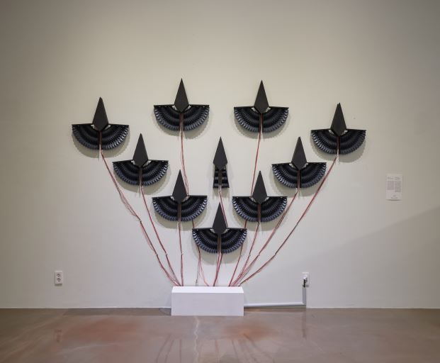
<Community>, 2024
by Jang Young Rok
Jang Young Rok focuses on the phenomena and movement of community formed by individual entities through kinetic art. His new work, Community, is drawing migratory birds, inspired by numerical precision obtained from large-scale data analyzed by the Center for Climate Physics. For example, the V-shaped flight formation of migratory birds reduces wingbeat effort by 14%. This insight led the artist to closely observe and simplify the movements of machine wingbeats. By isolating only the motion in each simplified individual element, the work captures the unique dynamism of groups and species.
<Falling Asleep Radio>, 2024
by Lim Se Eun
Lim Se Eun’s Falling Asleep Radio begins with the sudden start of youtuber Eresh’s live show, addressing themes such as youth isolation, workplace bullying, traumatic memories, and PTSD. Eresh’s effort to connect with viewers and overcome past wounds is similar to the artist’s effort to make an artwork. This work embodies the mechanisms of memory formation and regulation, as well as human affect and cognitive resilience against past memories, reflecting the creator’s unique perspective.
<O – Republic of Korea – Baekban: The Taste of Home – PULL!>, 2024
by KIMCHEEERS!
KIMCHEEERS! researches the relationship between human food culture and art. In this exhibition, she presents the new work O – Republic of Korea – Baekban: The Taste of Home – PULL!. When viewers pull back the blinds, the food covers rise, revealing an image underneath that visualizes taste at a cellular level. This work shows the complex process of taste perception, redefining gastronomy as an act rooted in cultural and social contexts beyond sensory experience.
<Patches>, 2024
by Park Eun Seo
Park Eun Seo contemplates ways to represent objects as they are. The work Patches employs flexible materials like PVC, EVA, and leather to reveal the memory and cognitive flexibility involved in adapting to and understanding unfamiliar objects through repeated thinking. During her research related with the Center for Cognition and Sociality, the artist encountered the concept of cognitive flexibility. She integrates this idea into the structure of the work, reflecting the process by which glial cells modulate the signaling of surrounding synapses to create malleable and flexible memories.
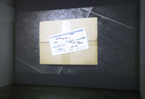
<Memory of the Box>, 2024
by Min Jihun
Min Jihun’s work Memory of the Box follows a journey by placing a recording device inside a delivery box to trace its travel route. This video captures the infrastructure of the transportation industry from the perspective of a box—one that is not accessible to us, the customer. By borrowing the viewpoint of the box and its recording device, the work reflects the artist’s ongoing interest in how humans perceive mechanical movements and the perspective of machines, delving into considerations of human cognitive structure.
<Set Extracted from “pee pee”>, 2024
by Choi Jinho
Choi Jinho’s work emerges from his long standing interest in film as a medium and is deepened by his recent engagement with climate issues through a visit to the Center for Climate Physics. Based on the fictional film pee pee, these works show the special techniques such as how film fakes climate within their narratives and screen with computer graphic and set design. The artist investigates how cinema evokes emotions in the audience, overlaying this exploration with considerations of climate.
<ound of Body_M1>, <Round of Body_B #3, #5>, 2024
by Min Sung Hong
Min Sung Hong creates installation works by collecting and recombining objects discarded by migrating humans. He captures the memories and traces of life embedded in things often deemed useless. These works displayed in this exhibition are also composed of items commonly found in traditional Korean families, highlighting the relationships between societal values, humans, space, and the environment in the context of migration.
<Transformative Intervention>, <Additional Intervention>, <Deconstructive Intervention>, 2024
by Park Ji Young
Park Ji Young discovered moss growing on the floor while exploring the underground space of the Center for Underground Physics. The Intervention series, inspired by the wonder of plant life, arises from this unexpected emergence in an artificially created environment for research. The three works, Transformative Intervention, Additional Intervention, and Deconstructive Intervention, study the themes of transformation, addition, and deconstruction, showing morphological variations that result from these interventions.
<Floating in Empty Air>, 2024
by Jung So Jin
Jung So Jin’s work Floating in Empty Air is an installation that continues the act of knitting with mohair yarn and wire, creating a formal connection to climbing plants. During her visit to the Center for Underground Physics, the artist discovered the intriguing forms of the underground space in the Yemi Lab. The paths made by scientists, who descend underground to study dark matter while avoiding background noise made by space ships, appeared meandering yet had a distinct direction for each. This spatial character inspired the artist and has been reflected in her current work.
<The Pallid Life Form>, 2024
by Kim Ri Ha
Kim Ri Ha’s new work, The Pallid Life Form, is an exploration centered on the K-pop idols and industry, infused with the artist’s unique imaginative perspective. The work questions how we can recover the essence of life in a contemporary society filled with endless replication driven by capitalism algorithms. Through this work, the artist seeks to reflect on the fundamental meaning of change as a human being and as a singular existence.
<NOV01-1400>, <Existences Outside Language>, 2024
by Jeoung Jae Hoon
Jeoung Jae Hoon is currently researching the relationship between names and reality arising from personal experiences. He presents new works NOV01-1400, Nomenclature, and Existences Outside Language in this exhibition, based on his visit to the facility of the Center for Underground Physics in Jeongseon, Gangwon-do. His work starts with the methods scientists use to infer the existence of intangible concepts like dark matter and focuses on the process and the elements of assigning names to entities that have yet to take form.
2022 IBS Art in Science

A Ray of Light
by 정송문(정재용, 송지천, 문혜람)
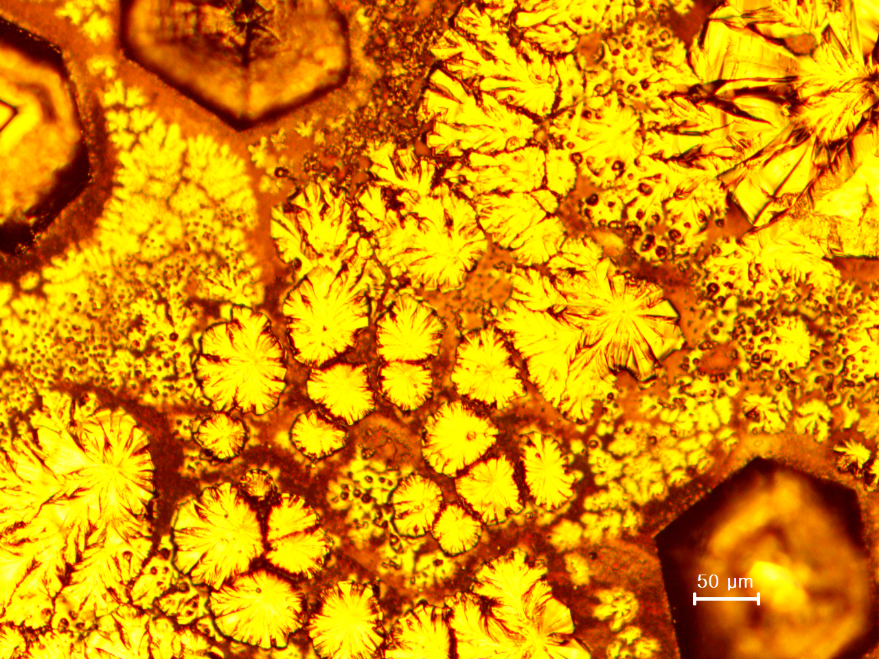
This image, resembling a ray of light illuminating the dark cave, was captured during the measurement of a specific crystal using MALDI-TOF Mass Spectrometry. The researchers created the crystal by mixing the CHCA (α-Cyano-4-hydroxycinnamic acid) matrix with SiO2 aerogel for the MALDI-TOF mass analysis, which is widely used for measuring the molecular weight of proteins or DNA fragments. When this crystal was examined under a scanning electron microscope at a magnification of 50,000 times, a fantastic scene was created.
The Beauty in the Pain
by Phan Thuy Tien
Born and raised in Vietnam, a country with a long history of war and colonial rule, I have witnessed the pain and loss of soldiers who were affected by Agent Orange in their lives during the American war, both mentally and physically. In the early 20th century, the "Gate control theory of pain" was proposed, which suggests the role of neural mechanisms in the dorsal horn of the spinal cord on the flow of nerve impulses and the development of therapeutics targeting it. The picture was taken by confocal microscopy showing the star-shaped glial cells called astrocytes (in green) during the stress, which induces them to change shape and swell up. It is reminiscent of the soldiers in the war who sacrificed themselves to protect their country, transforming their own pain into the most beautiful flowers for their country and future generations.

From a distance, it looks like a comedy, Up close, it's a tragedy.
by 고지훈
The researchers observed the process of angiogenesis (the formation of blood vessels) in pancreatic cancer patients to study ways to block cancer cells from creating blood vessels to fuel their growth. In the artwork, the blue represents cell nuclei, the red represents cancer cells (EpCAM), and the green represents vascular cells (CD31), combined which appear like vibrant flowers blooming on a verdant stem. This artwork, born from the sufferings of cancer patients, is given its title based on its determination to develop new treatments.

The Aesthetics of Emptiness
by 김영웅
This is an image created created out of vascular cells, which are usually neglected in colon research. The distribution of blood vessels in the mice colon, where vascular cells are expressing a distinctive red fluorescence, was observed through a confocal microscope. The commonly studied epithelial cells are represented in black, forming the background, while the vascular cells, which had been marginalized until now, are revealed in this mystical scene.
Mystery of Hearing
by 이재훈
The cochlea is a structure in the ear responsible for hearing. Depending on the frequency and intensity of the sound, it is selectively stimulated from its basal end to the very tip, detecting sound through hair cells. The cochlea has a systematic structure where each region, like the keys of a piano, represents the characteristics of sound. To understand this structure, extensive research is being conducted worldwide. The researchers captured the Corti organ located on the spiral cochlear membrane to depict the wonder of hearing.
We are like two poles of a magnet
by 이건희
When we captured the flow of current with 15,000 volts of voltage between the two electrodes, a series of sparks emerged in the midst of gases. When the voltage between the two electrodes is increased, spark discharge occurs in the gas. If the electric field between the electrodes is not uniform, the glow discharge occurs in the areas with a higher electric field on the electrode surface. This is known as corona discharge. In the case of an arc discharge, intense light is emitted when the electrodes are heated and hot electrons are emitted.
Poodle's Electron Cloud Stroll
by 유석열
The atoms that make up our world can form bonds using electrons when they are close to each other, and they can break these bonds when they move apart. This is known as a chemical reaction. During a [3.3] sigmatropic rearrangement, researchers simulated changes in electron distribution using a quantum mechanics program called Jaguar 9.1 ibo view. This program describes the position and shape of electrons as quantum mechanical wave functions, and it produced an image that resembles a poodle made of balloons taking a stroll.

A Tiny Mars Inside My Body
by 허숙경
This image, obtained from a study examining the effects of the LIGHT protein (HVEM-L, TNFSF14, or CD258) on osteogenesis in human bone marrow-derived mesenchymal stem cells (hBM-MSCs), evokes the surface of Mars and its valleys. During the experiment, researchers observed the results of calcium and phosphate deposition staining under a microscope and discovered that the LIGHT protein plays a crucial role in enhancing osteogenesis and stem cell therapy.
Flight
by 진강태
When tungsten oxide powder is placed on a sapphire substrate and heated to 900 degrees in intense heat, it creates an image resembling a phoenix in flight. The presence of crystalline structure on the sapphire substrate causes the tungsten oxide to become thermodynamically relatively oriented (microcrystals or polymer chains aligning in a specific direction). The flaming wings display some directionality while the surrounding area depicts disorderly flames.
The Birth of Light
by 심혜원
In an experiment testing the antioxidant properties of vitamin C, researchers discovered the crystallization of ascorbic acid and observed it through a polarizing microscope. It appears as if a scene from Georges Lemaître's "Big Bang Theory" is being recreated, with numerous rays of light emanating from a single point. At the beginning of spacetime, a tremendous explosion is heard, and everything in the universe is born from a single point. This image offers a glimpse of the beginning of all light, the essence of the Big Bang.
The Decomposer of Life
by 진주 in 진주 (이나윤, 이바다, 노경래, 석진주)
On the surface of moss collected from beneath a stone wall, researchers discovered a previously unknown aspect of the moss surface. Moss, an organism, that thrives in dark, damp places, appears rough on the surface. But when they examined it using a scanning electron microscope, they observed a pattern that resembled vines or seaweed swirling and being drawn in, akin to the shape of a bird's nest. This image provides a fresh perspective on moss, which acts as a decomposer that breaks down the remains and waste of other organisms, transforming organic matter into inorganic matter.

Waves of Hatred
by 팀 심심이(송현호, 신민기, 진효진, 차미영, 최정회)
Over the course of 365 days, researchers created a visual representation of the types and quantities of hate speech used by people toward a chatbot. The color visualization and motion in the image resemble the waveform of some music. The motion in the image demonstrates changes in color and undulating movements, much like the ebb and flow of waves. Hate speech are poured onto the chatbot in various forms and quantities every day. Through visualization, the researchers aim to bring objectivity and concreteness to the seriousness of hate speech usage, with the hope of promoting self-reflection and change in the users.
We both met and grow together
by synapses(Zafira Puan Adelin, Hazeta Rahmani Wafda)
The story behind every encounter is unique and it feels like it just happens to be like that. Similar to the neuron in the picture, they were just a single cell that differentiates into neurons, and they grow together day by day, expanding their axon until finally being able to function as a signal transmitter. The neuron is differentiated from the Mouse cell in the subventricular zone, by using immunocytochemistry (ICC), we stained the cell with DAPI to identify the neuron. Then, we looked at the slides through a fluorescence microscope and get the image.
2021 IBS Art in Science
Beautiful lives of the stars
by JUNG JUN HYUK.
Students majoring in CT at Seoul National
University
of
Arts
The work started out as a curiosity about what would happen when we gather and visualize the luminance data that shows the birth and death of stars. A star is another name for self-illuminating objects that provides mystery to the night sky. The stars are formed from interstellar matter such as gas and dust when these matters coalesce in the denser regions of interstellar space. Stars born in this way express their existence and mystery by emitting light, as they live through the pre-main sequence, main sequence, and post-main sequence stages, ending in death. In the work, the luminosity data of stars repeatedly appear and disappear as waves. If you observe the luminance data over several hours, you can see that they maintain a similar pattern and change regularly. As such it is possible to express the beauty of the stars’ lifetime through data visualization. *Luminosity: The brightness of a star according to the intensity of light emitted over a certain period of time. This brightness can be obtained from the radius and the surface temperature.View video on YouTube
The origin of the earth
by The debate about the origin of life on Earth has been going on for a long time. To support the panspermia hypothesis that life came from other celestial bodies, a research team from the Tokyo University of Medicine observed how microorganisms can survive in outer space for three years from 2015 on the International Space Station. As a result of the experiment, it was verified that microorganisms can survive for a long time in outer space. This means that the origin of life on the Earth possibly began when microorganisms migrated between planets by sticking on meteorites. Microorganisms have been the only living organisms for half of Earth's history and are fundamental to the birth and survival of all living organisms. However, we have identified less than 1% of the microorganisms with a light microscope. Unraveling the mysteries of microbes may therefore be one way to approach the origins of life on the Earth.
A Mother's Hands
by CLARISSA ELIZABETH MARIA
intern at CSLM (Center for Soft and
Living
Matter)
This image was taken using the transmission electron microscope (TEM), in the process of optimizing liposomes sample preparation. Liposomes will typically dry up and form circular structures. However, in some rare cases, they might break and form unique amorphous structures. If one were to look closely, this structure looks like hands. Not just any hands, but a mother's embracing hands. A mother's hands are gentle yet powerful at the same time. They are more than powerful to protect us from dangers, and they are also the gentlest hands where we run to, they are the hands that are always wishing for our safety and happiness, no matter how many miles apart. As an international student staying thousands of miles away from home, it feels reassuring to know that even the nanoscale world is trying to remind me that my mother will always wish for my safety and happiness.

This is not a tree.
by Kang Seok
Ph.D. students at KAIST Graduate School of Medicine /
researcher at IBS Vascular Research Group
The metaphors using trees have been widely used in most cultures across time and place. In Norse mythology, Yggdrasil was the source of the universe and life. In the Garden of Eden in the Bible, there were the Tree of Life and the forbidden fruit. There were also aging trees of Seonangdang in the old village of Korea. A tree is rooted in the ground and extends its branches and meets the sky. Since the sky begins at the end of the tree, mankind dreamed of the sky through the tree. In a horizontal world dominated by gravity, the vertical axis of the tree was a passage connecting the different dimensions of the earth and the sky. It's like Jack climbing the beanstalk to the giant's house. The inside of the donut is the same as the outside. The human digestive system starts from the mouth, passes through the esophagus, stomach, small intestine, large intestine, and then goes to the anus in the form of a single tube. The essence of the digestive system is nothing more than a cylinder with a hole in the middle, with donuts hanging down. (In other words, donuts and the digestive system are isomorphic.) So the inside of the mouth is the same as the outside of the body. Food consumed by humans is broken down into its components through the process of digestion. However, it is still located outside the body. The outer universe is distinct from the inner universe. This work was obtained using a confocal fluorescence microscope after staining blood vessels and lymphatic vessels of the mesentery (a thin membrane connecting the intestines and our body) of a 5-day-old mouse. Blood vessels supply oxygen and nutrients to the intestine, and conversely, lymphatic vessels absorb the digested nutrients as they reach the intestine. Substances from outside the body move into the body through lymphatic vessels. In other words, the lymphatic vessel is a passage that connects the different universes (phases), inside and outside. Through this, the inner and outer universe becomes one connected universe as the boundaries disappear. Just as a tree connects the sky and the earth, lymphatic vessels connect the ‘inside’ and ‘outside’. Whether anyone who sees this work associates it with a tree or perceives it directly as the anatomical structure of lymphatic vessels, the essence of this work is neither the tree nor lymphatic vessels. This is the cosmic gate that connects the different dimensions.
Blossom of snow
by Park Junho
Professor Kim Cheol-joo of Pohang University of
Science
and
Technology, Ph.D. in the lab
This image expresses the scene where the pine leaves that endured the cold winter meeting again with the new spring flowers that are flying under the warm spring sunlight. The pine leaves and the color of the cold air on the left represent winter, while the yellow flowers and bright colors represent the early spring. How these yellow flowers blend in with the image of a pine tree appears to represent how the pine tree overcame a cold difficult winter and gets to see a warm spring once again. This scene is a photomicrograph that I accidentally obtained while synthesizing organic semiconductors. It was taken back during a personally difficult time period at the beginning of my doctoral program, due to repeatedly failing experiments. At the time, the goal was to make a clean film, but after it was confirmed that the film grew in a wire-like formation due to the bonding anisotropy of the organic layer, I felt despondent due to failing another experiment. However, because the picture was so beautiful, I took a picture and stored it deep in the data file. Two years have passed since then, and now after successfully completing the research and writing my paper, I was very happy. When I encountered this picture again, I was reminded of the difficult and hopeless times in the past. I realized that I am now enjoying the spring, and I wanted to show this image to the people to other people who may be living through a hard time right now. What is unusual is that the leaves of these pine trees are not broken, but are all connected as one. While each leaf boasts its own individuality, they are all connected and appear like a single big flower. The first thing you may notice in the photo may be the shape of the yellow spring flower, but if you keep looking, you will notice the huge pine leaves displaying their beauty in their strength. If you think about it, these seasonal changes are similar to our lives. Due to the coronavirus, our lives may have changed for the worse over the past year. However, as spring always comes after winter passes, I hope that through this photo, we hope that our daily lives will be able to return to normal in the near future.
smell of the sea
by Lee Junhyuk
Department of Bioscience and Neurology at the Korea
Institute of Science and Technology
The brain works hard even when we perform simple tasks such as smelling an object. In particular, the role of the olfactory cortex is important for 'recognition' beyond 'sensing' odors. This image was obtained while expressing synaptophysin mCherry-eGFP in the olfactory cortex via fluorescence staining to perform an observational study of the astrocytes in the region. This image, taken at the beginning of summer, is reminiscent of the beach. I think this is an image that represents my desire to go on vacation to the beach while being tired of repeated labwork this year. Even at this moment, I can already imagine the smell of the sea, so I named this work the scent of the ocean. I hope I can go to the beach soon.
hippocampal cleanup
by Lee Se-young
graduate student at the Korea Institute of Science
and
Technology
In the hippocampal synapse, necessary synapses are created as memories are formed, while unnecessary synapses disappear. However, it is still not well known whether this process is important for memory formation and through which mechanism it occurs. To study this question, the researchers hypothesized that astrocytes, one of the glial cells, would "consume" unnecessary hippocampal synapses to eliminate them. In this image, the normal synapses show green color (red mCherry and green eGFP), while the consumed synapses show red color (green eGFP is degraded and only red mCherry remains), and astrocytes display white fluorescence. This research revealed that in order to form new memories, the astrocytes must eat and destroy unnecessary synapses. This knowledge suggests that sometimes, we should take a break rather than just move forward.
Lost in the forest of love
by Choi Cham, Park Sooah, Kwon Yoonhee
Optosken Technical
Support/Team
members belong to UNIST Research Support Headquarters
A contrast agent is essential for conducting a CT scan of soft tissues. Contrast agents need to be administered to the tissue without clogging blood vessels. By administering a contrast agent to the mouse liver, which has the size of a fingernail, it became possible to observe even the microscopic blood vessels inside the liver. The liver capillaries show up as the shape of a heart from a certain angle. This inspired us to submit this image as artwork. This work expresses the snowy forest of love, which is easy to get lost in. An additional touch that adds a woman who is sitting and crying on one side and a man who is wandering in search of such a woman on the other side could allow for a more in-depth expression.
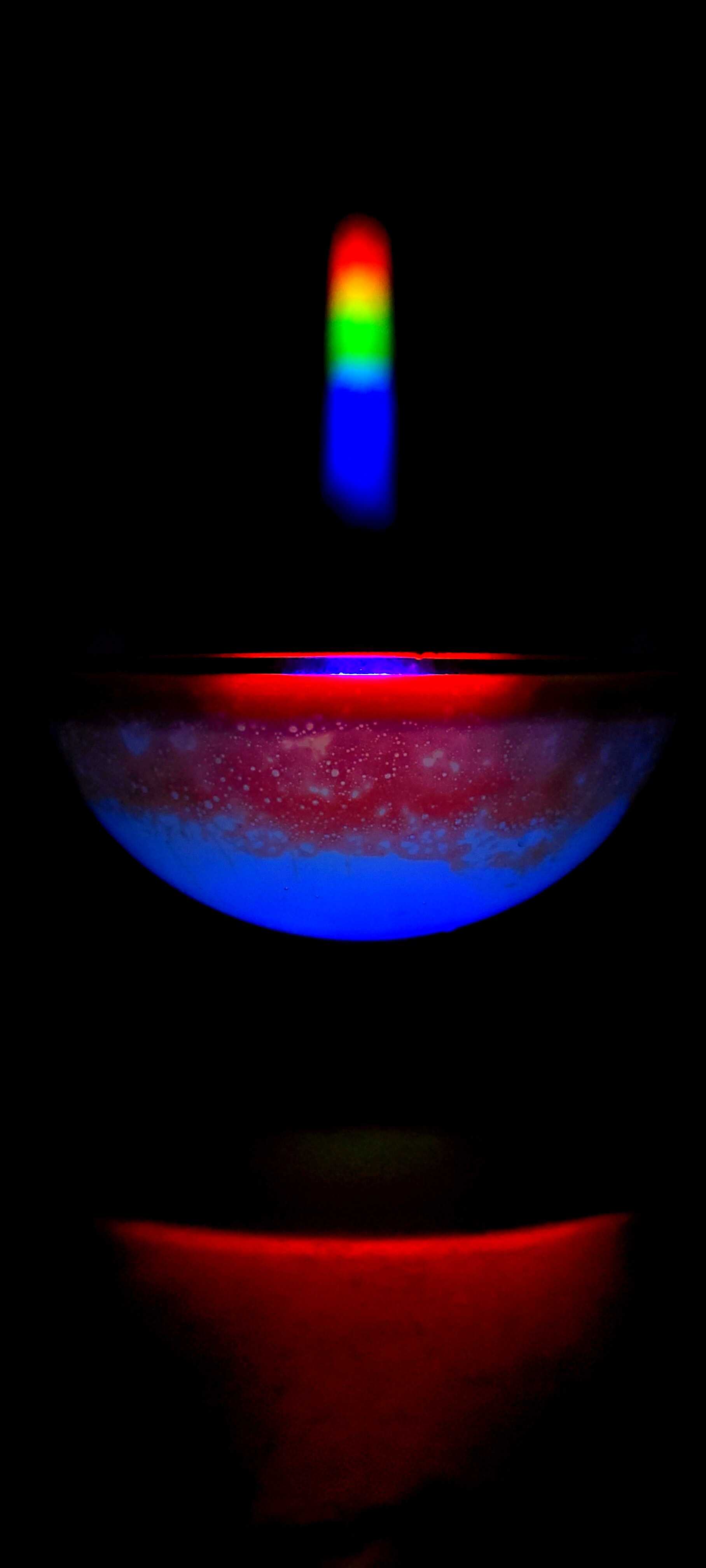
end of extraterrestrial planets
by Kang Juwan, Seo Seongmin, Lee Sang Eon, and Kim Seowi
Gyeongnam
Science
High School student.
The light that illuminates our daily lives is made up of many different wavelengths. Of these, visible light, which we can see and feel, is the most beautiful gift that nature has given us. The white contains light red, yellow, and green lights which have different characteristics such as different diffraction angles. The three primary colors of light pass through a narrow gap and deflect each other slightly, creating a variety of new colors. On the backside of the CD, which is a representative diffraction grating plate, the light is reflected by numerous thin gratings and causes interference, showing colorful patterns. Similar to CD, the grating film also consists of many thin gratings. The visible light with a wavelength of 400 nm to 700 nm spreads widely on the diffraction grating films with several thousand thin grooves per 1 cm on a transparent plate. To observe the pattern more easily, a hemisphere bowl of water was filled with water and oil bubbles so that vivid colors can be displayed on the surface. The red light passing through the white oil bubbles mixed in the water looks like a gas giant planet with active convection at its surface.
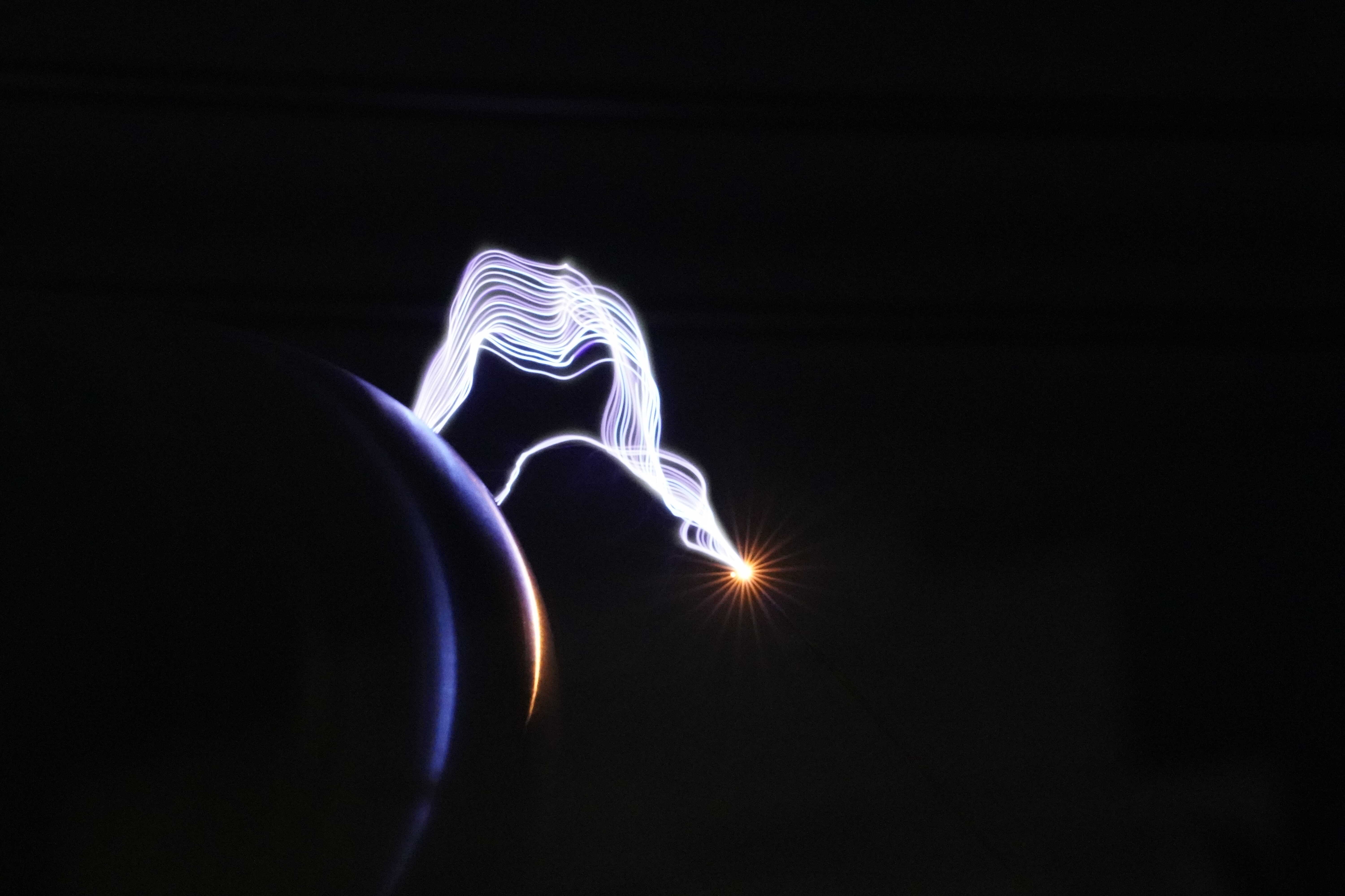
electric noodles
by Ahn Youngjun
Daejeon Metropolitan Office of Education Daejeon
Doan
Elementary School
What comes to mind when you hear the word 'Tesla'? Many of you are probably thinking of an American electric vehicle company. In fact, Tesla is named after Nikola Tesla, who invented the 'Tesla coil'. A Tesla coil is a device that converts low voltage into high voltage. This device allows you to create voltages as high as tens to millions of volts. By comparison, the voltage we use at home is 220V. The high-voltage electricity in the Tesla coil produces a loud noise and light when it is discharged. This principle is the same as the thunder and lightning we experience in nature. The picture above is a picture of electricity being discharged from the Tesla coil and heading towards the discharge sphere. Its principle is the same as lightning, but the discharge occurs so quickly that it is difficult to observe its appearance. Therefore I decided to take a picture to show my students. The above Tesla Tower is a photo taken at the Daejeon Science Experience Center operated within the Daejeon Educational Science Research Institute under the Daejeon Metropolitan Office of Education. (This is the institution I worked for from 2019 to 2020.) The Tesla Tower (called the Tesla Tower because two coils stand like twin towers) located at the entrance of the Basic Science Museum is built to showcase the principle of the Tesla coil. It is also possible to play music by controlling the discharge sound. In order to take a good picture of the discharged light, it was necessary to turn off all the surrounding lights. The photo was taken using Sony A9, 90mm macro (close-up) lens, F16, with a 1/20 second shot. At first, I shot the picture with a higher shutter speed, but it was difficult to see the trajectory, so I had to incrementally lower the shutter speed. The result was a picture that resembled the noodles at a Chinese restaurant. With the 'electric noodles' above, we hope many kids will be more interested in science!
chrysanthemum garden
by Kim Kwangbae, Song Ohsung, Kim Hojun
Kim Kwang-bae, Information
Materials Research Center, Department of Materials Engineering, Seoul National
University
This image was obtained while developing a perovskite solar cell during the crystallization process of the perovskite layer on the carbon layer. The magnified image is reminiscent of a garden full of chrysanthemum flowers. A solar cell is a material that uses solar energy and converts it into electrical energy, and it is being widely studied as a potential renewable source of energy. In particular, perovskite solar cells are classified as third-generation solar cells, since they have superior price competitiveness compared to conventional silicon solar cells and have a simpler manufacturing process. In addition, perovskite has a three-dimensional crystal structure composed of organic cations, metal cations, and metal anions. In this study, a perovskite solar cell device with a glass/FTO/TiO2/ZrO2/perovskite/carbon electrode structure was fabricated. Perovskite was produced by preparing a solution of inorganic PbI2 and organic MAI and heat-treatment at 100℃ for 30 minutes after dropping the solution. This image captures a phenomenon where the crystal growth rate of PbI2 and MAI are different during the perovskite crystal formation process, resulting in the excess MAI layer. When PbI2 and MAI are grown in a 1:1 ratio, a perovskite crystal structure called MAPbI3 is formed. The image of a chrysanthemum garden, captured via optical microscopy, arose due to the difference in the growth rate and ratio of crystal within a perovskite solar cell, and it is considered a work of art. The modern research environment is increasingly focusing on incremental improvement in efficiency by testing a minor change in the composition within a given range using a known process. This perovskite chrysanthemum garden is also an image that was obtained by chance during the process of finding the optimal perovskite layer formation conditions, which was then submitted as an artwork.

distortion of time and space
by Lee gunhee
Busan Science High School
This photograph shows a model of a black hole was floated on the water, which bends the background under the water due to surface tension. Normally, the pen should not be visible due to being hidden behind the black hole model, but the surface tension created by the black hole model causes the light to bend so that the pen underneath is refracted and revealed. This shows the same effect as gravitational lensing where light is bent under the influence of gravity due to a large object such as a black hole. Gravitational lensing is a phenomenon in which light from a very distant celestial body is bent by strong gravity from a large celestial body during its travels along the curved space-time. The gravitational lensing effect has been presented as strong evidence to support Einstein's general theory of relativity. In the photo above, the black hole model represents a celestial body with a large mass, a curved surface of the water is a curved space-time, and the pen is a distant celestial body.

A glorious day in solar cells
by Jo daehyeong
Korea Electronics and Telecommunications Research
Institute's Nano-New Materials Application Laboratory
The sun rises up, and we wake up. We hurry up, and the sun is at noon. We move fast and the sun goes down. We become tired and the day end. Do solar cells experience such a daily cycle? This photo shows the solar cells as if they were expressing the various emotions of the day using color. The vivid and brilliant color that the cells displayed was reflected in the title of the work. Why are solar cells are usually dark in color? This is because it needs to absorb as much light as possible to generate electricity. But is it possible to create more beautiful and colorful solar cells? This was the starting point of our study. We developed a solar cell whose color changes according to the angle of incidence of sunlight. This was done by taking advantage of the fact that different wavelengths of light are diffracted at different angles by the grating structure. By forming 300 nm wide and 770 nm long grids composed of zinc oxide on a thin-film solar cell, it was possible to produce various colors depending on the angle. At the same time, high solar power generation was possible thanks to the transparent property of zinc oxide. (Reference: Nano Energy 2021 Vol. 80 p.105550). The brilliant color of a solar cell is not the sunlight nor the pattern on the solar cell, but what is perceived through our minds. Perhaps creating such a phenomenon that can move people's minds is also a 'science'.
Korean Fan - "Boo Chae"
by Stuhl Laszlo
ibs 소속 / Center for Exotic Nuclear
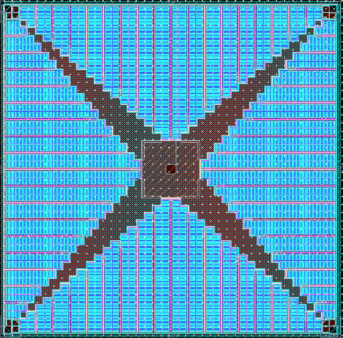
In this study we measured the Gamow-Teller Giant Resonance in 11Li, the nucleus with the largest neutron-proton asymmetry factor. The figure shows the excitation energy of the reaction products of the experiment as a function of the center-of-mass system. The bar like structures are the individual neutron detectors, and their bending comes from the transformation from laboratory coordinate system to the center-of-mass coordinate system, resulting a perfect Koran Fan at IBS. This result will largely contribute to understand the neutron stars and Equation of state in connection to pion exchange.
four-leaf clover power semiconductor
by Kim Myungjun
Professor Cho Sung-hwan's laboratory at KAIST's
Department
of Electrical and Electronic Engineering (https://ccs.kaist.ac.kr/) PhD program (to
be
graduated in February 2022)
Today, semiconductors are being used in all areas of life, and therefore power management of semiconductor circuits is very important. The (system) semiconductor responsible for this power management is called a power semiconductor. In other words, power semiconductors supply and distribute current to almost all semiconductor circuits. The problem is that when many currents flow in a power semiconductor, the currents bump into each other and generate resistance. This means that the efficiency is reduced and the heat generation is increased. Because of this, the design shape of power semiconductors is an important research topic; Power semiconductors must operate extremely efficiently without generating heat. While doing this, our research team was inspired by the shape of plants. Plants need to absorb as much sunlight as possible for photosynthesis. In particular, the "four-leaf clover" stretches each leaf widely as possible in a symmetrical manner; This is to maximize contact with sunlight by maximizing the surface area. As a result, the plant can maximize the efficiency of photosynthesis. We thought that if we imitate this design inspired by nature, it would be possible to evenly distribute the current flowing in the power semiconductor so that it can operate efficiently with minimum heat generation. Our research team commissioned TSMC Foundry from Taiwan to actually produce a prototype of this "Four Leaf Clover Power Semiconductor". For this contest, we would like to submit not only the computer design drawings (left) but also micrographs of the manufactured chip (right). After successful testing, our research team could learn how nature can produce such efficient and beautiful designs.
Seoul, the glowing city
by Boitet Maylis
PhD student at Institut Pasteur Korea - campus of
University of Science and Technology
Our laboratory is focused on understanding the basic biology of Pathogenic bacteria, and more specifically Mycobacterium tuberculosis, the bacterium responsible for tuberculosis (TB) in humans. Using unique technologies cell-based phenotypic screening technology platform for drug discovery, our laboratory uses bioluminescent and fluorescent proteins to track pathogens. Bioluminescence occurs through a chemical reaction that produces light energy within an organism's body. Bioluminescence can be found in many organisms: bacteria, algae, jellyfish, worms, crustaceans, sea stars, fish, and sharks to name just a few. Taking advantage of bioluminescence genes key in the process to generate light, bacteria at the Institut Pasteur Korea were engineered to glow in the dark. Using a matrix pattern on which glowing bacteria freely grow, we chose to represent Seoul with light. Controlling pathogens in a given pattern resonates with South Korea health capability to have kept COVID-19 outbreak under control.

shining wings
by Kim Jung-soo
Professor Joo Cheol-min of the Department of
Mechanical
Engineering at Yonsei University
This work is an image of the birefringence characteristics of vitamin C crystals which was taken using a polarizing microscope. Birefringence is a phenomenon that occurs when light is incident on an optically anisotropic medium, the light is split into two refracted lights due to the refractive index is different depending on the polarization direction of the light. The birefringence property provides a variety of information about the composition and structure of a material, such as the shapes and arrangement of molecular crystals inside the material. Since the technology for imaging the birefringence properties of materials have a wide range of applications, such as in bio, medical, and food industries as well as materials sciences, we were able to take this image while conducting related research. This birefringence image of vitamin C can make us all say “is this really vitamin C?” This shows how we can find beauty in the small parts of everyday life that we don’t know about. “The real voyage of discovery consists not in seeking new landscapes, but in having new eyes.” - Marcel Proust
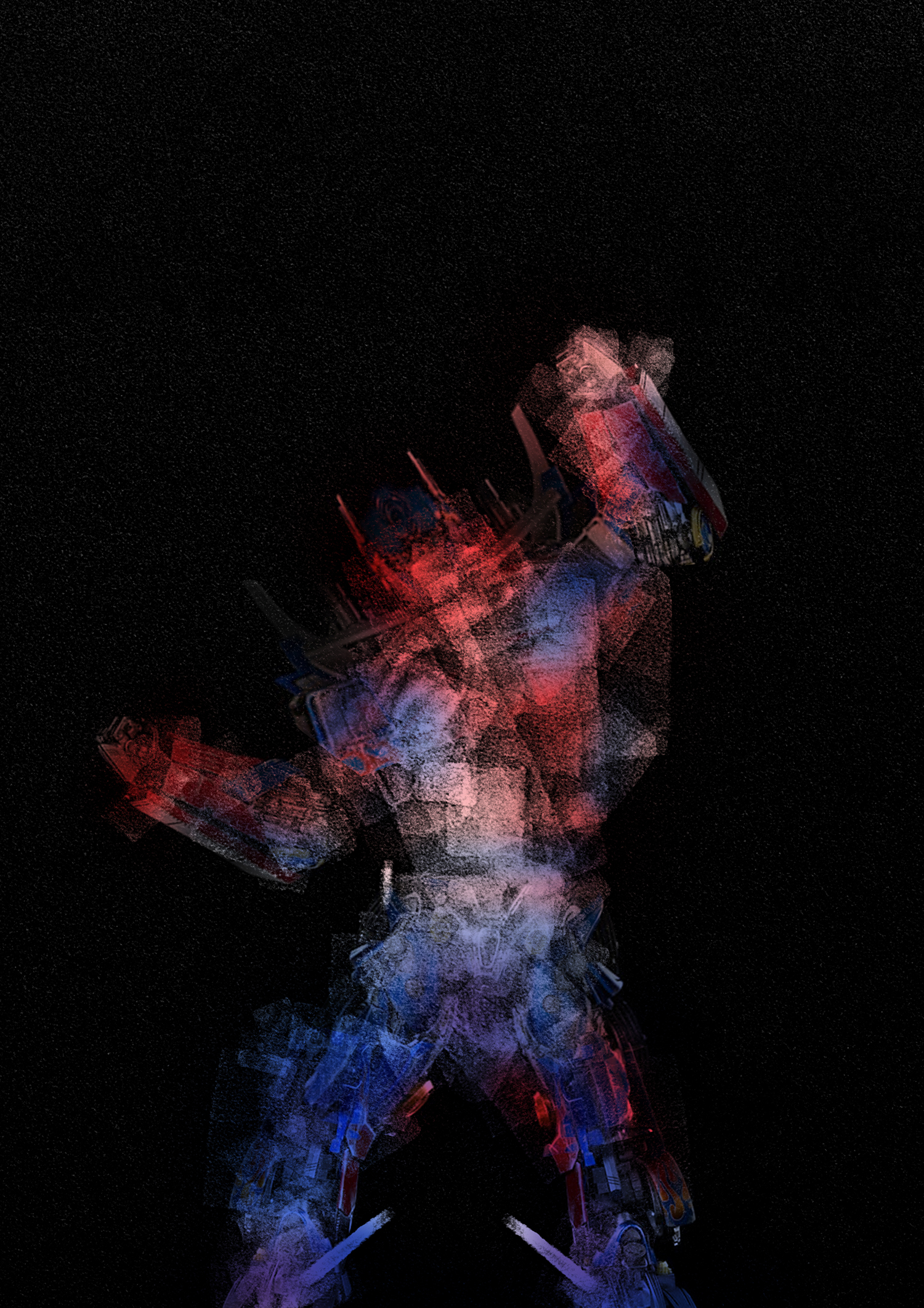
Nano-transformers: driving a sustainable future
by Sampathkumar Jeevanandham
graduate student (PhD) from
Department of
Chemistry, POSTECH
“Imagination is more powerful than knowledge” - Albert Einstein. I envisaged the protagonist - Optimus prime from the movie “Transformers” with the Transmission Electron Microscopy (TEM) image of TiO2 nanostructures. Can you imagine a pandemic partner of coronavirus? Yes, it is nothing but the everlasting climate change due to global pollution. Enormous growth of industrialization accompanied by excessive CO2 emissions are the root cause for global warming where the demand for sustainable energy generation remains inevitable. Major energy crisis in this fast moving world desperately needs some rapid “Transformers” to drive our future in a sustainable way. Emergence of TiO2 nanostructures as a promising photocatalyst accelerates the potential of energy conversion in next-generation technologies where the global carbon emissions can be drastically reduced and transformed into value added fuels. Herein, the exfoliated ultrathin 2D nanosheets (60 x 40 nm) of TiO2 resembles the assembly of animated “Transformers” with ultimate motto to protect the mother-nature from harmful activities. The unique characteristics of these ‘nano-transformers’ possess in-built tunability on their properties, acting as a core catalytic platform and driving force for clean energy production. This illustration bridges the ideology between science and technology insisting that the power of imagination is the key to solve complex problems and develop advanced futuristic applications.
four-leaf clover in Gologanoid
by Kim Hwan
Assistant Professor of Nanochemistry Materials
Engineering
at
the National Transportation University
Three-dimensional bone organoids are produced using chondroitin sulfate-based biomaterials and mesenchymal stem cells and vascular cells. After culturing for about 6 weeks in mice, this work shows the ossification formation center, where ossification is starting to take place. It appears that the shape of the ossification process was similar to that of a four-leaf clover. Cell nucleus was stained with DAPI, mesenchymal stem cells and human cells were immunostained with h Vimentin 9, and osteoblasts undergoing ossification were immunostained with OCN (osteocalcin).
Microcosm
by Yoon Sun-young
Faculty of Cosmetic Science at Chow Women's
University
This image was obtained while studying the relationship between lymphatic vessels and hair growth. In the skin tissues of mice, the nucleus (blue), lymphatic vessels (red), and adipocytes (green) were stained and images were obtained using a fluorescence microscope. There are various cells in the skin, which is like a huge universe. Individually, cells are arranged chaotically and appear disordered, but they end up discovering a unique order and form the skin tissue. The presented image shows the magnificent order established by diverse types of cells to the extent that it can be called a microcosm. All living things, not just the mouse used in the experiment, have their own microcosms that are like cosmos of chaos.
Who changed this picture?(The color of the famous painting that we know will change?)
by Kim Donghyuk, Kang Namsoo
Kang Nam-soo, a teacher at Chungbuk
Science
High School / Kim Dong-hyuk, a sophomore at Chungbuk Science High School
The light around us is an electromagnetic wave, which oscillates in all directions while traveling in a straight line. However, there is a specific case in which it oscillates in only one direction, and that is called polarization. When a polarizing film is used, only the light that oscillates parallel to the polarization direction of the film can pass through. But can you believe that this light 'rotates' when it passes through starch syrup? This rotation of the polarization direction of light is called optical activity. The rotation angle of light depends on the wavelength of the light and the distance the light travels through the starch syrup. Therefore, when white light passes through the polarizing film (polarizer 1) and then passes through the starch syrup, each wavelength within the light will rotate differently according to the path length through the starch syrup. When this light is viewed through another polarizing film (polarizer 2), a particularly large amount of the light that has a polarization direction parallel to that of the film (polarizer 2) passes through, which means that only light of a specific color can be seen. When the polarizing film (polarizer 2) is rotated, the color that passes through is changed accordingly. While studying the optical activity phenomenon according to the temperature of starch syrup, we attempted to make a work of art with the intense color as shown here. In particular, we reproduced Mondrian’s art style using the optical activity phenomenon. While Mondrian’s painting stays the same color, our version can change dynamically. People think that works of art, especially masterpieces, should always be timeless. On the other hand, our work provides a new artistic paradigm that challenges this rigid view.View video on YouTube
2020 IBS Art in Science
Ancient Future
by KANG Seok
Center for Vascular Research
The sperm combines the egg to become a fertilized egg. A single fertilized
egg
cell
then undergoes cell division and divides into a myriad of cells. The process in which
these
cells divide, differentiate, and move to develop into an organism is called
'morphogenesis'.
The
cell's DNA sequence contains a schematic of this development process. All cells start
from
one
fertilized egg, which means they all start with an identical blueprint. Although they
all
have
the same origin, each cell ultimately becomes a different cell with its own shape and
function.
Individual cells gather at a predetermined time and location to form different tissues
and
organs within an individual. If you look at a cell early in its development, it will be
difficult to determine the fate of that cell. However, all cells undergo development as
if
their
future had been predetermined from long ago. In that sense, it is natural to look at an
egg
and
envision a chicken that it will become in the future. Likewise, all fertilized eggs have
are
potentially destined to become an individual. In other words, the “the ancient future”
of a
fertilized egg is the “individual”. We developmental biologists explore the processes
and
mechanisms behind this predetermined future.
To observe the development of blood
vessels,
CD31 and PDGFb proteins in 10.5 days old mouse embryos after were stained with
fluorescent
antibodies, and images were taken with a confocal fluorescence microscope.
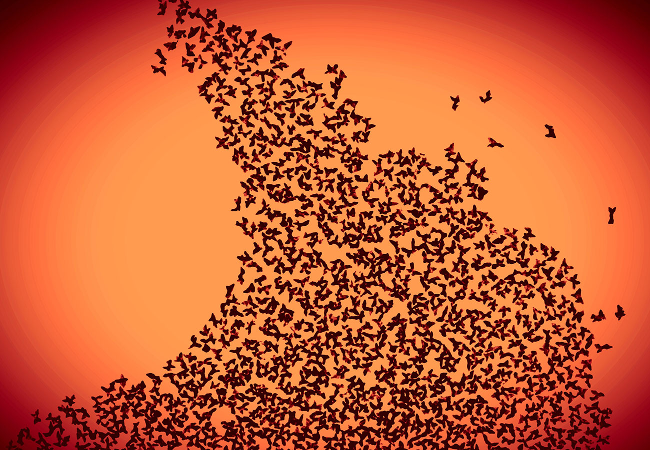
A new dawn!
by Anubhab Acharya
POSTECH
“And, when you want something, all the universe conspires in helping you
to
achieve it”- Paulo Coelho. The flock of birds flying high in the dawn sky,
chasing
the
dreams in the light of hope. We do not taste success all the time, but the hope of
accomplishing
dreams should not be rested.
The idea of “never giving up” can be visually imagined
in
the
Transmission Electron Microscopy (TEM) image of very tiny asymmetric gold nanoparticles
(average
size: 90 nm). The stimulating characteristics of uniquely shaped gold nanoparticles and
their
interaction at molecular level with genes, viruses and proteins, etc. open the door for
advanced
futuristic biomedical applications. This enlightening picture signifies a new journey
both
for
the birds and science; the incredible potential for new discoveries to be made!
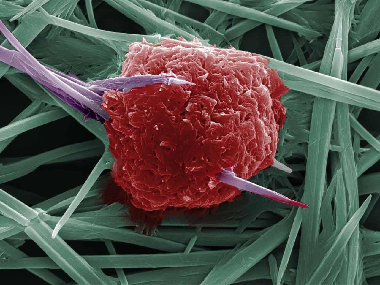
Invisible sacrifice
by JEONG Jae Yong
Yonsei University
When foreign antigens (viruses, microorganisms, etc.) invade our body, our
body
launches a strong immune defense system to protect itself. The red cells in the photo
are
macrophages, which are one of the immune cells in the body. This is a picture of
macrophages
consuming monosodium urate crystal (MSU), which is a substance that induces gout, and
displaying
the antigens to lymphocytes. The image was taken using a scanning electron microscope
(SEM).
Although it is just a cell, watching these cells risk their lives to fight these
intruders
to
protect our bodies makes us feel grateful.
At a time when the world is struggling to
eradicate COVID-19, I hope this work will serve as an opportunity to rekindle our
appreciation
to the medical staff, scientists, and our own immune cells for working hard from
invisible
locations to protect us against the disease.
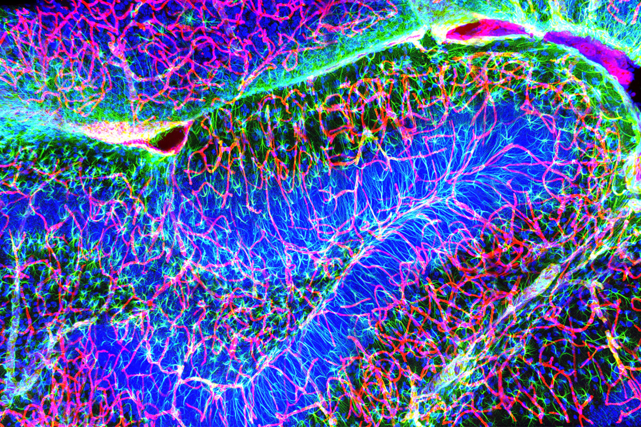
The clown's smile
by IM Hyeon Gyeong, HAN Ga Yeong
Center for Neuroscience Imaging
Research
The curved structure of the hippocampus of the brain in this image appears to resemble a smile. The cell nucleus (blue), blood vessels (pink), and star glia (yellow-green) were colorized to create a gorgeous image with three colors. The overall structure and color schemes seem to look like a clown’s smile. However, in the media, clowns are often portrayed as people who laugh superficially but are actually not that happy in reality. Like a clown's smile, the hippocampus of our brain stores many memories that evoke various emotions. Although the image itself is bright and colorful, it seems to evoke a strange feeling reminiscent of the Joker.
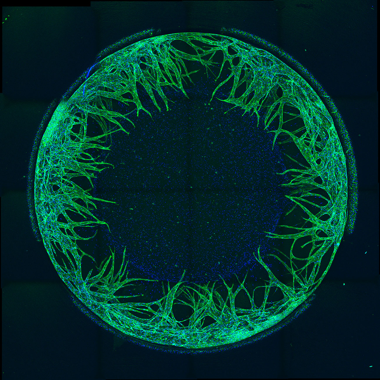
Bloody Eyes
by KOH Ji Hun
Seoul National University
Organ-on-a-chip is a microchip that can reconstruct human cells in a
three-dimensional environment in vitro. Based on an understanding of microfluidics, this
microchip is capable of patterning a hydrogel containing cells in a designated area.
With
the
ability to co-culture two or more cells, these microchips are capable of simulating a
variety of
organs and closely simulate their microenvironments.
The above work is a fluorescence
image
of a model that mimics the mechanism of angiogenesis, which plays an essential role in
cancer
growth and metastasis. Vascular endothelial cells patterned around a circle show
angiogenesis by
fibroblasts that emit vascular endothelial growth factors in the center. As the
concentration
gradient of the growth factor is formed in a radial shape, vascular endothelial cells
have a
tendency to be generated toward the center.
As implied in the title “Beautiful
Bloodshot
Eyes,” we depicted the phenomenon where a human eyeball became bloodshot. These models
can
be
presented as an excellent alternative to cosmetic toxicity experiments which currently
use
rabbit eyes. As animal testing is becoming gradually prohibited, this model can be used
in a
wider field of drug testing, thanks to the reliability and the efficiency of this in
vitro
model
compared to the existing experiments.
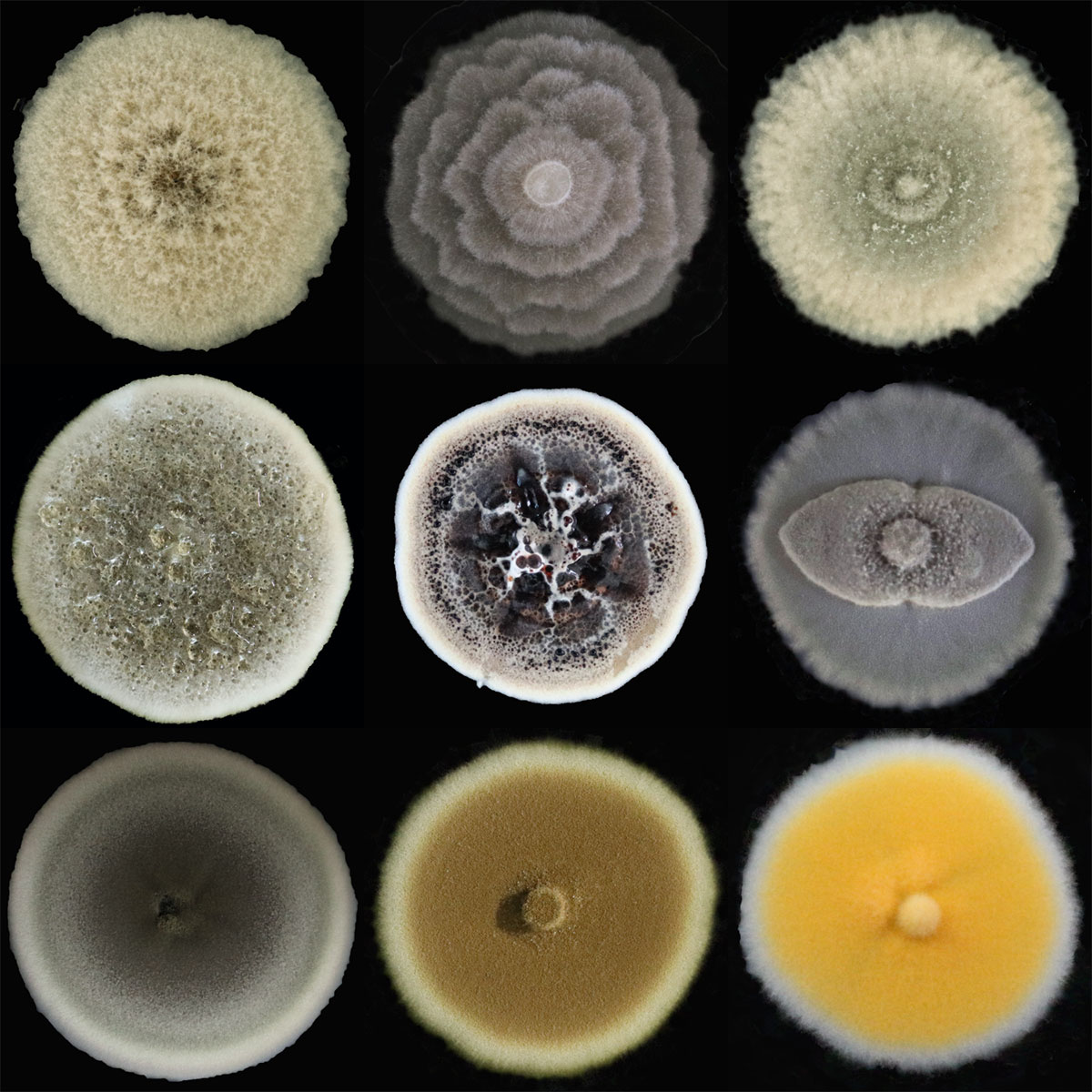
The presence of those we don't know about
by SUH Hui Ju
Dankook University
There are many organisms on Earth that we do not know about. In fact, there
are
many
small and insignificant organisms that constantly live next to us. What are they? They
are
fungi. Fungi act as decomposers that break down the material within the dead organism.
Even
though these are creatures that faithfully carry out their duties from nature’s point of
view,
people look upon them with disgust and believe they are dirty and causes of infectious
diseases.
But not all fungi are seen in such negative light. Some are highly valued
as a
producer of valuable pharmaceuticals, such as penicillin, while mushrooms are seen as
delicacies
or healthy functional foods in many cultures. In addition, fungi have been spotlighted
in
the
agriculture industry in recent years due to their soil-enriching properties. In many
ways,
whether we know it or not, our lives and fungi are intertwined with each other.
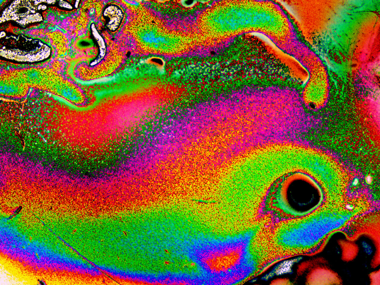
Sorrowful Rainbow Fish
by JEONG Ju Hyeon
POSTECH
This image is the surface of a germanium substrate that was deformed due to
contamination caused by a tweezer. This occurred while cleaning the substrate in order
to
grow
graphene on the germanium substrate, and the substrate had to be discarded due to this
incident.
This is an image I got while analyzing the surface with an optical microscope in order
to
figure
out what happened so that I do not repeat this mistake.
The name of the work,
Sorrowful
Rainbow Fish, is intended to reflect the state of my mind at the time of this incident.
Failing
an experiment or experiencing a setback inevitably leads to stress and pressure on most
graduate
students. The work was named as such since I was sad about not even being able to
conduct
the
experiment that I had planned.
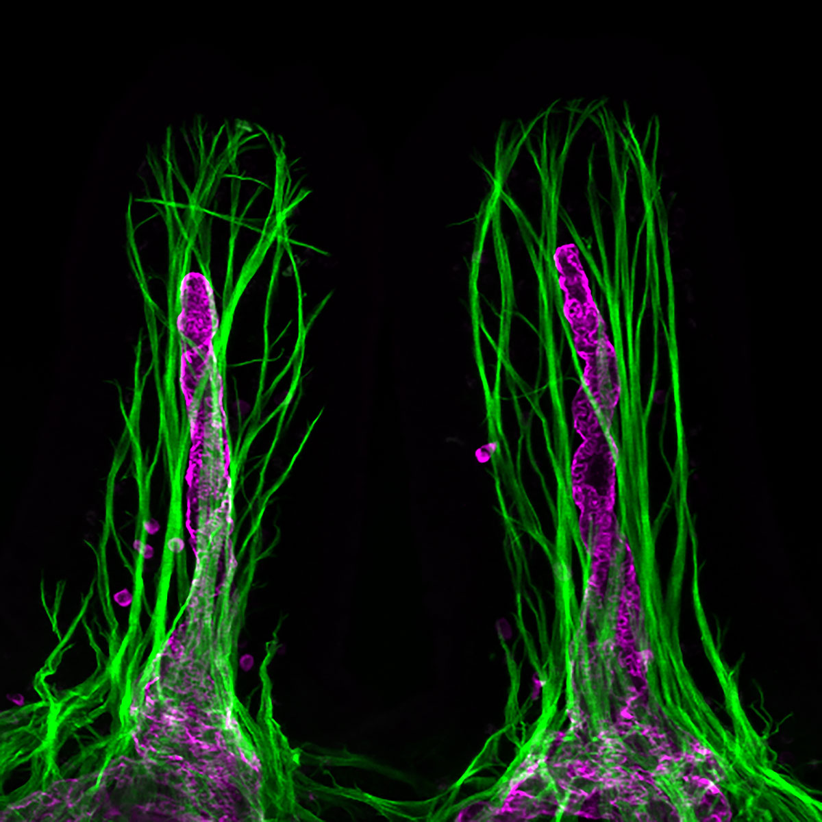
Flowers covered with grass
by HONG Seon Pyo
Center for Vascular Research
Inside the villi of the small intestine, there are special lymphatic vessels
called
chyle ducts, which are composed of endothelial cells in the form of elongated tubular
shapes.
Chyle ducts play an important role in absorbing fat-soluble nutrients such as fatty
acids
and
vitamins that enter the intestine. Our research team has investigated and researched the
shape
and function of these chyle ducts. In order to to do so, a deeper detailed observation
through
various experimental techniques was required. The further observation allowed us to
learn
about
the structure and function of the chyle ducts as well as their importance. The above
picture
was
taken in the process of studying the interaction between the chyle ducts (purple) and
the
surrounding smooth muscle cells (green).
The process of discovering and researching
the
chyle
ducts reminds me of a poem called “The Flower of Grass” by poet Na Tae-ju. 'It's pretty
if
you
look closely, it's lovely if you look at it for a long time, so are you.'
The
photograph
also
closely resembles an image of flowers that are hidden behind the grass.
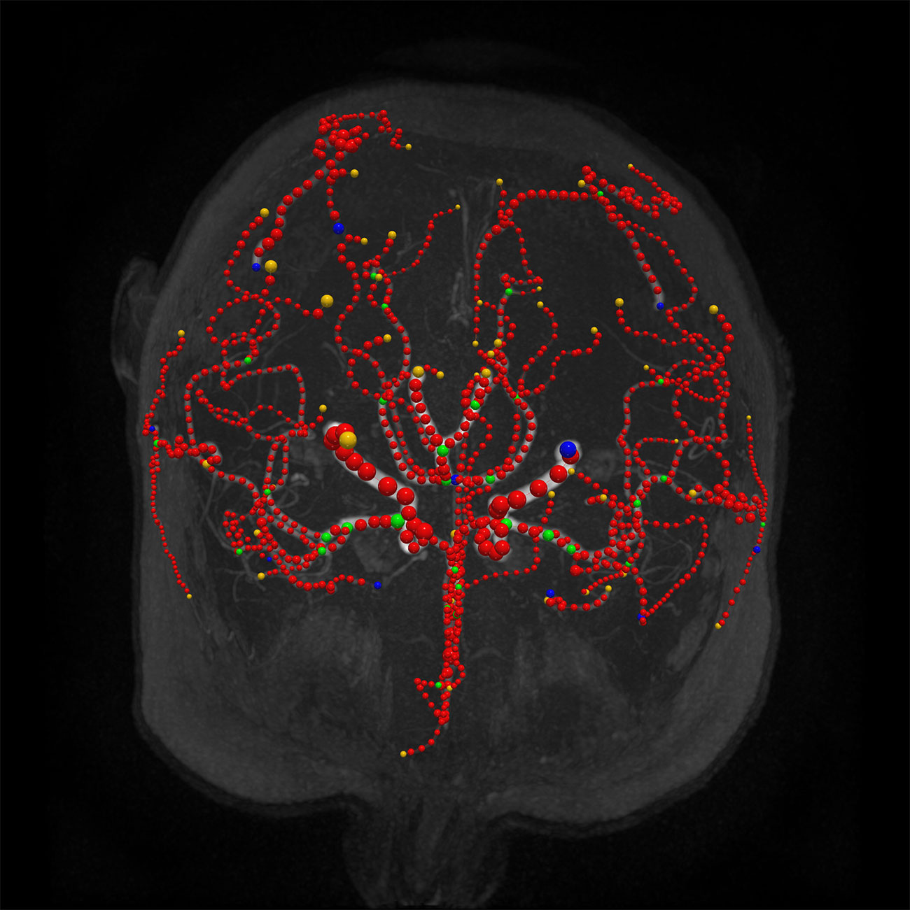
My Memory of Us
by NA Yu Ri
Korea Institute of Science and Technology(KIST)
We all age.
When I was little, I wanted to quickly become an adult. But
now
as an
adult, I am sitting here counting my age every year. My grandmother and grandfather are
no
longer with me as an adult. My aging parents are now in that vacant place. When I was
young,
my
grandmother, who was my best friend, suddenly became a different person. I even felt
shocked
and
betrayed by my grandmother, when she didn't remember me.
I wanted to know what
happened
to my
grandmother, why she had suddenly changed, and why she had to become a different person.
So,
as
a child, I decided to become a neuroscientist to study dementia.
This image was
obtained
while conducting a study on the brain through auto-detection for Cerebral Small Vessel
Disease
(CSVD) using the computational imaging informatics method. Our memories of us are
eternal,
beautiful, and precious. In order to keep these precious memories, I want to unlock the
mysteries of the brain, one step at a time.
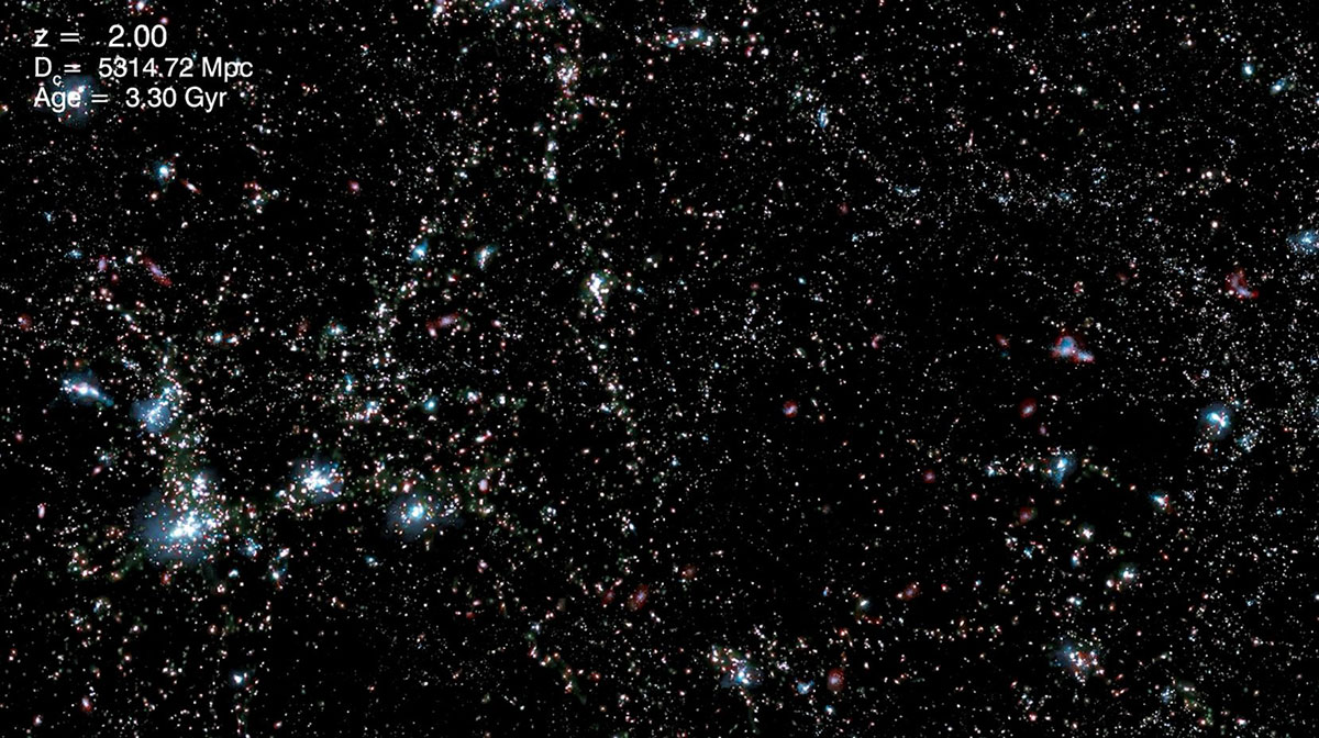
Time travel in the Universe
by LEE Jae Hyun, PARK Chang Beom, KIM Joo Han, SHIN Ji Hye, KIM Yong
Hwi
Korea Institute for Advanced Study(KIAS)
Horizon Run 5 is a large-scale project that performs cosmological fluid
dynamics
simulations in a huge virtual universe with a side length of about 3.1 billion
light-years.
Using a third of the supercomputer's computational resources exclusively for more than 3
months,
the formation of cosmic giant structures and the formation and evolution of galaxies
were
calculated with a resolution of up to 3140 light-years.
The submitted video dates
back to
about 1 million to 7.8 billion years ago after the creation of the universe, and it
shows
the
cosmic giant structure and the changes to the galaxies over time. As we go back to the
younger
and more distant universes, we see bluer, smaller, more irregular galaxies, and the
cosmic
giant
structures that contain galaxies become progressively weaker and sparser. This is an
inspirational video of what our universe would have looked like when it was younger.
Starting
from an image of a galaxy's stars, gases, temperatures, and metals visualized in
different
colors, it shows how the universe changes in terms of the composition of its stars, gas
density,
temperature, metal content, and dark matter.
View video on
YouTube
Jewelry Fish
by MIN Jae Myeong, YANG Soo Yeon
Bloomwork,Inc.
Can artificial intelligence serve as an art tool to become the new normal of
creation?
The long-held belief that art is a human-specific domain is collapsing.
Human
role
in the future may no longer be about creating a work, but involve making decisions on
how to
train artificial intelligence.
When the artificial intelligence was tested on the
public
work
<Fish>, the AI did not only reproduce the surface texture in detail but also
recreated
the
work using a new artistic style.
This means that artificial intelligence is capable
of
both
imitation and creation. This spawns a debate on whether artificial intelligence should
be
relegated to mostly assistance role, or be recognized as a new creator. One thing is
certain
that artificial intelligence may become the new normal for creativity. Do you agree?
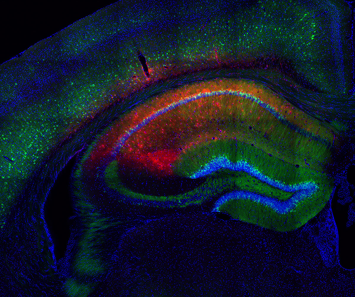
Star, Flower, River of memory
by LEE Gyu Hui
Center for Cognition and Sociality
The hippocampus is known as the part of the brain responsible for memory. In
order
to form and maintain memory in the brain, the roles of not only neurons but also glial
cells
are
important. However, there are still many things to be studied about the interaction
between
neurons and glial cells. This experiment determined how the neurons and the star-shaped
glial
cells called astrocytes differ in their activation and how interact with each other when
mice
performing certain behaviors in experiments. We expressed the green calcium indicator
protein in
the neurons and red calcium indicator protein in the astrocytes. The cell nucleus was
stained
using blue color, and the image was taken using a confocal microscope.
This image is
reminiscent of a work of an impressionist painter, which uses vivid natural colors to
illustrate
green grass and red poppy flowers under the starry night sky, against a river in the
background
flowing around the flower garden.
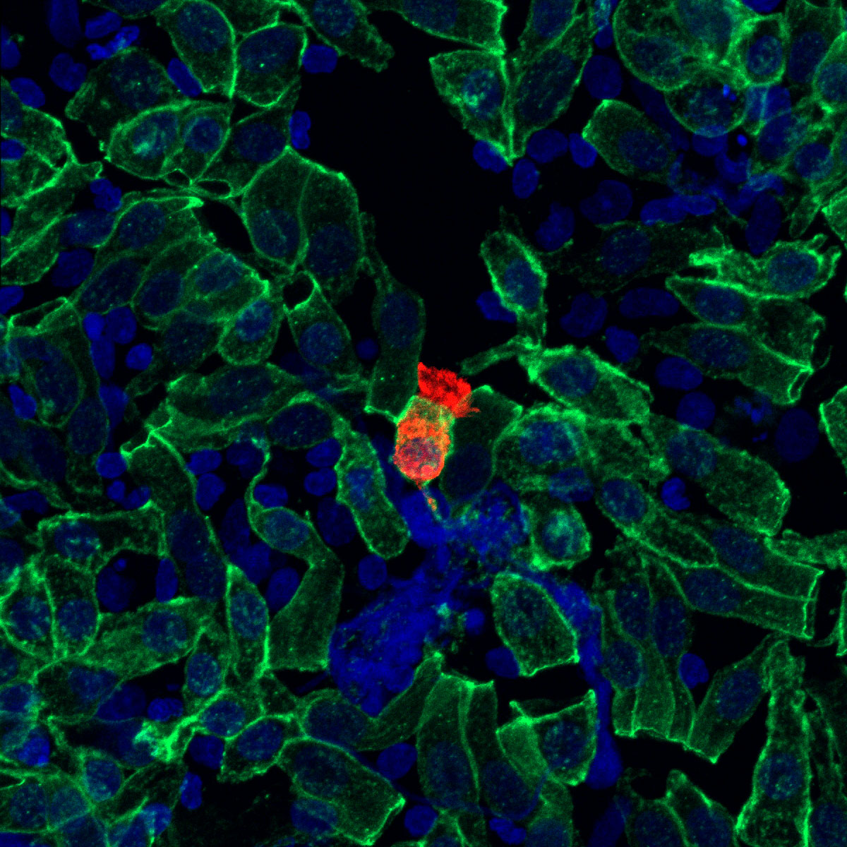
Red alert: COVID-19
by AHN Ji Hun
Center for Vascular Research
At the time COVID-19 showed signs of becoming a potential pandemic, humanity
may
have been underestimating the virus. The Sars-CoV-2 virus enters the respiratory tract
upon
inhalation, multiplies within our cells, and gets expelled within our breaths to rapidly
infect
the people around us. This work is a fluorescence microscope photograph that was taken
after
the
cells were collected from the nose of an asymptomatic COVID-19 patient and were stained
with
a
virus-specific protein antibody.
In this image, viruses (red) are actively
proliferating
within an infected nasal epithelial cell, which is in the middle of many uninfected
nasal
epithelial cells (green). These cells appear to be the personification of ourselves who
lived in
blissful ignorance in the early days of the pandemic without realizing the seriousness
of
the
situation. If you imagine that each of the cells in the picture is an individual, there
seems to
be a good argument about the importance of social distancing and early diagnosis.
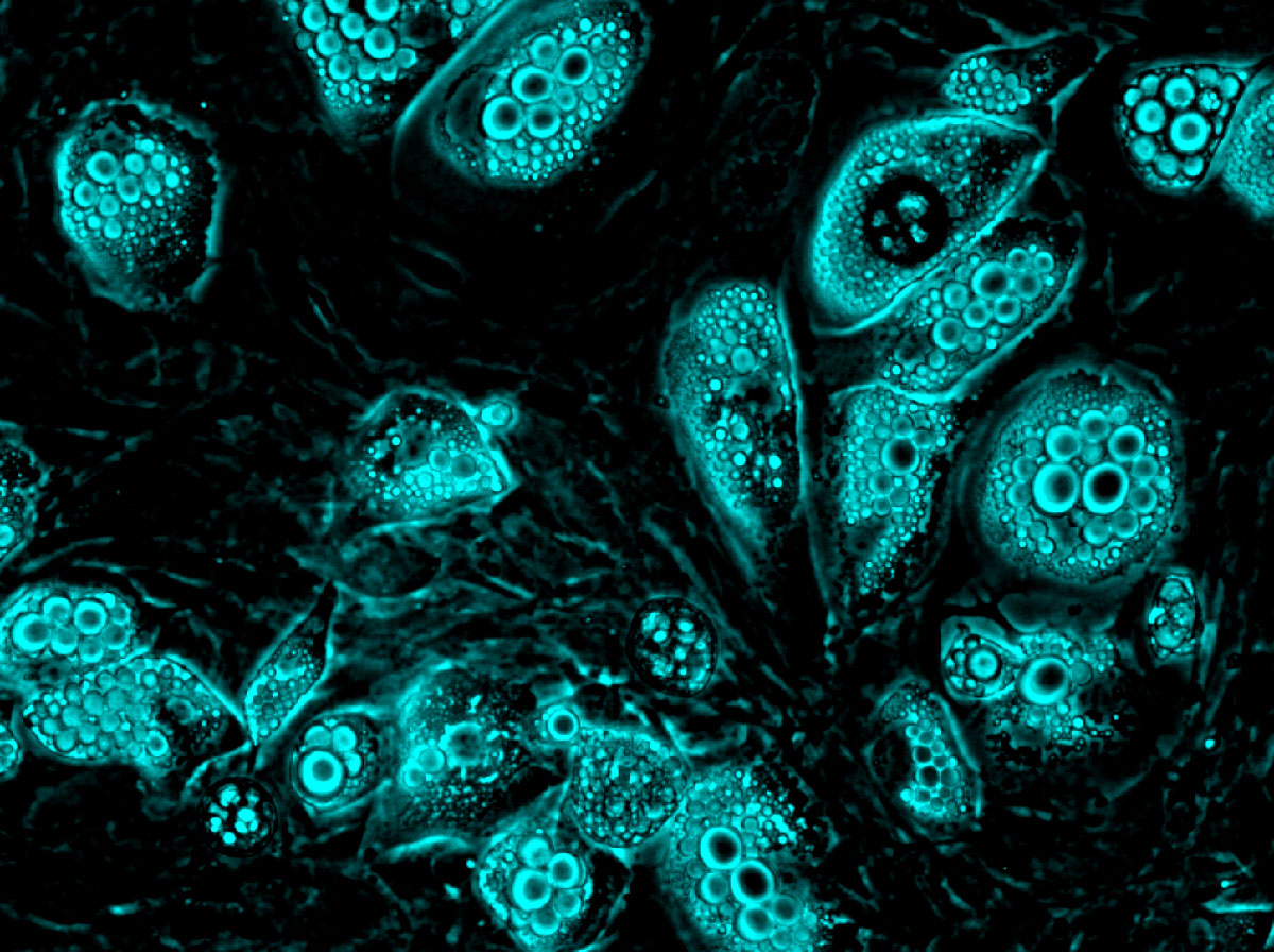
Pearly shells
by KANG Nam Yeong
Center for Self-assembly and Complexity
It is said that natural pearls usually require 2 to 4 years to be created. The probability of obtaining a natural pearl in the wild is very slim at approximately one out of 10,000 shells. Even among this, the probability that the pearl will have a large enough size and spherical shape for it to have a commodity value is even lower. Just as rare as these pearls are the brown fat cells in our body. While they are more plentiful in newborn infants, they gradually disappear as the individual grows into an adult, leaving behind only unhealthy white fat cells in the body. This study involved the induction of brown adipocytes from white fat for therapeutic purposes. The appearance of brown adipocytes in this image was reminiscent of the appearance of a shell containing pearls, and hence this is why the work was named Pearly Shells. It is said that the probability of successfully differentiating brown adipose tissue obtained from baby mice (Usually 10 to 14 days of differentiation time and complex culture conditions are required. Normally, the cells fail to undergo differentiation and are discarded) is said to be based on random chance. Therefore, it can be said that the brown fat cells obtained from this study are comparable to natural pearls. A cyan color was applied to the obtained image to give them a color similar to that of the natural pearl.
An Inner Struggle In The Body
by SEO Hyun Ji
Kyungil University
We consume numerous tablets throughout our lives. These range from
prescription
medication that you take when you are sick to various supplements, vitamins, etc. that
you
take
daily. One day, I was curious about what happens to a pill when it enters the
body.
Because
the pill is in a form of compacted powder, it melts when it comes into contact with a
liquid
such as water. Therefore, I dissolved the pill with hot water and filmed the process
using a
macro lens. During this operation, I could see that individual pill's shapes changed in
various
ways while melting. Some pills melted like a firecracker while some became spread like a
wave,
while the others had an appearance of melting little by little while maintaining the
original
shape. The appearances of these pills melting seemed like they were fighting viruses
inside
the
body, which led to the name of the work "Inner Struggle in the Body."
*Macro Lens: High-resolution lens made for close-up
photography
View video on YouTube
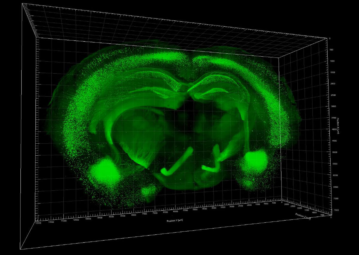
Steering wheel
by HONG Sung-guk
Bainary,Inc.
The steering wheel is an important part of a car that is required to change
the
direction of the vehicle. So how do all animals, including humans, decide their
direction?
The
brain contains about 100 billion neurons and 900 billion glial cells, and together with
the
spinal cord, they form the central nervous system. It evaluates environmental stimuli,
responds
appropriately to maintain the body's homeostasis, allows for movement, and controls
muscles.
This work is an image of a 2mm-thick section of a mouse brain that was
genetically
modified with Thy1-YFP. The slice was made transparent using the CLARITY technique and
photographed with a light sheet microscope. Fluorescence is expressed in most neurons
and
appears brightly throughout the cell until the end of the axon. As shown in the video,
the
overall outline of the brain slice appear similar to the steering wheel of a racing car,
as
is
their role in determining the direction of their host.
*Light sheet microscope:
single
plane illumination microscope that can acquire fluorescence-stained images at ultra-high
speed
to determine the function of the target protein in living cells, tissues, or model
organisms.
The 3D structure of the sample was identified through 3D reconstruction. It is a device
that
can
observe large-scale (mm) samples that could not be observed using existing
equipment.
**CLARITY technique is a state-of-the-art technology that renders the
tissues
and
cells of an opaque biological sample transparent so that the expression and position of
the
protein marker can be observed in 3D.
View video on
YouTube
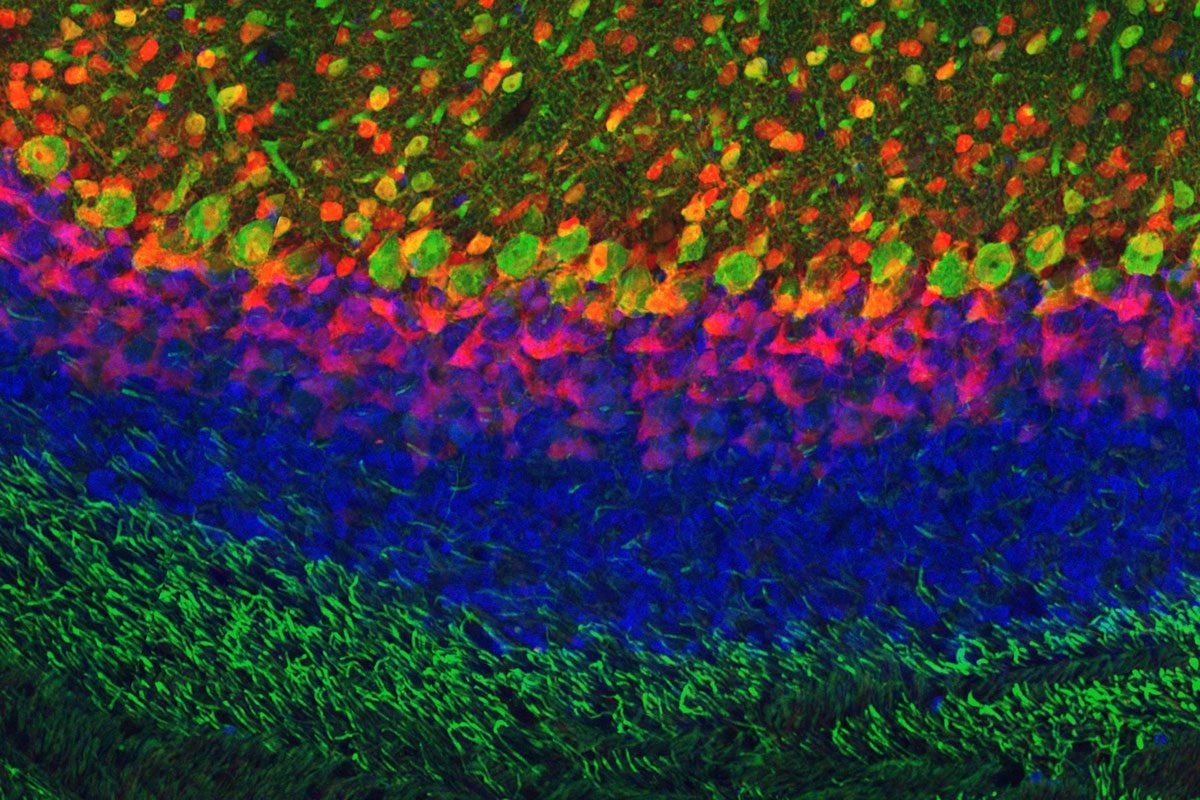
Grassland of Erlkönig
by LEE Seung Jun
Center for Synaptic Brain Dysfunctions
A sinister red flash of light sinks over the grasslands amidst stormy
weather.
This
photo is reminiscent of Schubert's " Erlkönig ", which is a piece of music inspired by
Goethe's
poem with the same name. This picture of the cerebellum of a mouse looks like the red
touch
of
Erlking reaching down into the blue night air that fills up the dynamically entangled
grassland.
The cerebellum is involved in muscle movement and maintenance of balance,
it
has a
fairly unique structure that matches with the dynamic description of this work. It has
several
clearly distinct layers, and the types of cells that make up each layer are also
different.
The
large green circles lining the upper middle part of the picture are Purkinje cells in
the
cerebellum. Parvalbumin protein, which is present in Purkinje cells, was stained
(Immunohistochemistry, green) before imaging. This staining method is a time-consuming
process
and is difficult to perform in living cells. In order to rapidly identify the cells
expressing
Parvalbumin without killing the organism, mice were genetically modified so that cells
expressing Parvalbumin co-express red fluorescent protein (tdTomato, red).
However,
the
two
signals do not appear to overlap completely (yellow color would result if they overlap),
which
is an interesting example of how artificially inserted genes may not actually function
as
intended. This unpredictable uncertainty is one of the most fascinating parts of living
processes.
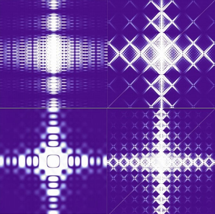
What a beautiful Diffraction pattern!
by KWON Min-chan, JUNG Chan-wook, EOM Seungjae, KIM Moon-gyeom, CHO
Hye-ri
Gyeongnam science high school
The picture above is obtained from the simulation of the Fraunhofer
diffraction
pattern of multiple concentric square holes. While numerous studies have been conducted
on
the
diffraction of various types of slits, the high school curriculum only teaches single
slit
or
double-slit diffraction. To overcome this, we simulated the diffraction pattern created
by
multiple concentric square-shaped slits by entering the irradiance function under the
given
conditions using a computer program called 'Mathematica'
Slits of various shapes can
be
produced. The picture on the bottom right is the pattern that can be produced using two
concentric 0.2 mm square holes with a gap of 0.2mm between the holes. “What a beautiful
Diffraction pattern!” was selected as the title of this work, because various beautiful
patterns
could be created using only concentric squares.
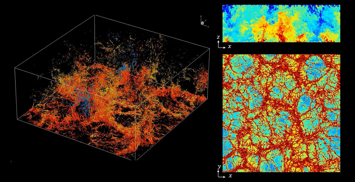
Turbulent cosmos
by CHOI Jung-il, KIM Ki-ha
Yonsei University
Natural convection is a flow that arises due to the difference in density
caused
by
temperature and the buoyancy caused by gravity. Rayleigh-Bénard convection is a standard
type of
natural convection occurring in a fluid that is heated at the lower layer and cooled at
the
upper layer. Rayleigh number (Ra) is used as an index to indicate the temperature
difference
between the two planes, with the larger number denoting a higher temperature difference
between
the two planes. The greater the temperature difference in turn results in stronger
buoyancy
and
more intense convection flow. When the Ra number is very high, turbulent flow can occur.
Turbulent flow is characterized by irregular fluctuation. One of the reasons why it is
difficult
to predict turbulent convection mathematically is because small and large movements of
fluid
affect each other. Therefore, to understand the turbulent flow accurately, it is
necessary
to
calculate even the smallest movements within the flow in detail.
This video is the
result
of
Rayleigh-Bénard convection implemented through computer simulation. Large-scale
calculations
using a supercomputer were required to capture all the minute movements within the
turbulence.
In the image, the red color represents higher temperature, while the blue denotes lower
temperature. Intricate small flow structures are visible among the large flow structures
within
the convection. The large flow structure generates convective flow and is responsible
for a
large portion of heat transfer. A cell structure is formed downwards flow of the cooler
fluid
meets the warmer layer below. The heat flow is generated when the warmer fluid becomes
displaced
by the cold flow and rises up due to buoyant forces.
The video was named 'Turbulent
Cosmos'
because the convection cells formed by the flow looks like a cosmos flower. It is
interesting to
see that some form of order can arise in the forms of these small complex flow
structures in
the
midst of the chaotic disorder of turbulent flow.
View video on
YouTube
2019 IBS Art in Science
* Scientists' Eyes : Observation and Imagination
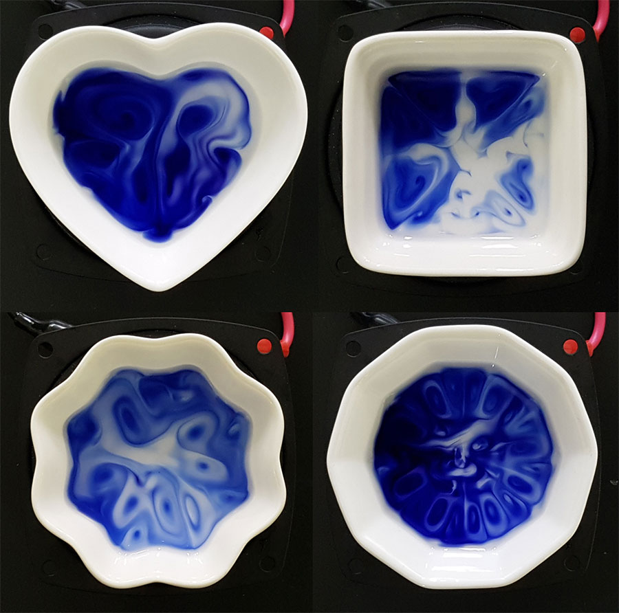
Painting with a Sound Brush
by Ilha Hwang
Center for Self-assembly and Complexity
Chemical reactions, in general, can be controlled by concentration, temperature, pH or external stimulus such as light. Is there any other way to control chemical reactions? While pondering this question, we got new inspiration from the sound of the music we heard over our speakers. We then tried to control complex chemical reactions using sound and during our attempts, we obtained interesting results and spectacular patterns. These photographs were taken during the experimental process of selectively controlling the oxidation reaction of a vividly blue organic dye molecule with sound. Now we paint using a sound frequency generator, a speaker, and different-shaped chambers. We believe that someday we can paint more complex yet aesthetic patterns with music or natural sound as a paint brush.
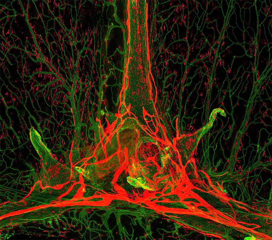
Bonfire in the Brain
by Jihoon Ahn
Center for Vascular Research
The brain is a tightly regulated and protected organ. Scientists have long been interested in the brain, but not much is known about the meninges—the protective membranes that cover it. The Center for Vascular Research looked closely at these thin layers of tissue covering the brain using a genetically engineered lymphatic reporter mouse model under a fluorescence microscope and has recently discovered a waste disposal system of the brain—meningeal lymphatic vessels and perivascular macrophages. The meningeal lymphatic vessels, shown in green, constantly sprouts and regresses, while perivascular macrophages, shown in orange red, keeps moving and surveilling the surface of the brain to prevent pathogens from entering the brain. These dynamic biological processes resemble a glowing bonfire with large flames and flying sparks.
Lung of Dionysus
by Seok Kang
Center for Vascular Research
Dionysus, also known as Bacchus in Roman mythology, is the ancient Greek god of wine, fertility, productivity, and grape-harvest. Grapes symbolize life and abundance due to the plant’s strong vitality and many clusters of grapes on a single vine. An interesting resemblance to grapes can be seen in the most important organ for respiration; lungs, which allow one of the most fundamental processes of life maintenance. Within the lungs are pulmonary alveoli which are small hallow air sacs where O2 and CO2 exchange occurs. An alveolus is located at the distal end of the airway and a cluster of alveolus, called alveoli, has an undeniable resemblance to a bunch of grapes. The image was captured using confocal microscopy after staining the vessel of a developing embryonic lung on embryonic day 12.5. The image was deformed by exaggerating the individual alveoli to more look like grapes. By coloring the image with eye catching neon colors, this image expresses the fest of Dionysus.
A Ray of Clarity
by Dongwoo Han
Center for Vascular Research
Lungs are essential respiratory organs for all living beings. They have a complex network of alveoli, vessels, and lymphatics. This work shows a photograph of a cleared mouse lung sample captured using an optical microscope. In order to observe the tissue completely intact—not sliced in any way, we used a technique called CLARITY that renders tissues transparent. We also stained CD31, a protein abundant in endothelial cells, to capture a complex network of blood vessels in lungs. However, because of the thickness of whole mounted lung tissue, the microscope brings only a certain spot into focus, much like a single ray of light. This ray of clarity resembles science as science is a process of investigation to find clear answers from amongst obscurity.
Differentiation of Affection
by Sejin Park
Center for RNA Research
New books are published every day, constantly changing the new release section in bookstores. A majority of them are books on human emotions, especially popular are publications on love and relationships for readers desperately seeking some helpful advice. Nonetheless, we will never fully know how affection works between people, how these feelings start, why they change, and where they will end up when everything is said and done. A heart shape cell block in the photograph was captured by chance in the course of a gene expression experiment for neural stem cell differentiation. The experiment itself was actually a failure as the result was not as expected but it left this interesting heart shape image of a cell block with what looks like twigs extending in all directions. It looks similar to the affection we have for someone—that feeling of emotion that keeps changing and spreading out naturally like water flowing. The cell-cell interactions observed through a microscope and the change of human emotions have something in common—both are unpredictable.
Glimpse of the Universe within a Plate
by Junyeop Roh
Center for Synaptic Brain Dysfunctions
When one looks up into the night sky, the glimpse into the vastness of the universe is often limited to the confines of the circular view allowed by the telescope’s lens. Remarkably, fluorescence-labeled cells grown in a circular plate resembled such view, with the cells serving as the stars. How the smallest units of life—cells—can look so much like the largest observable phenomena in the sky seems paradoxical. Green fluorescent protein-tagged membrane proteins overly expressing Chinese hamster ovary (CHO) cells were grown on plates and their images were captured by confocal microscope.
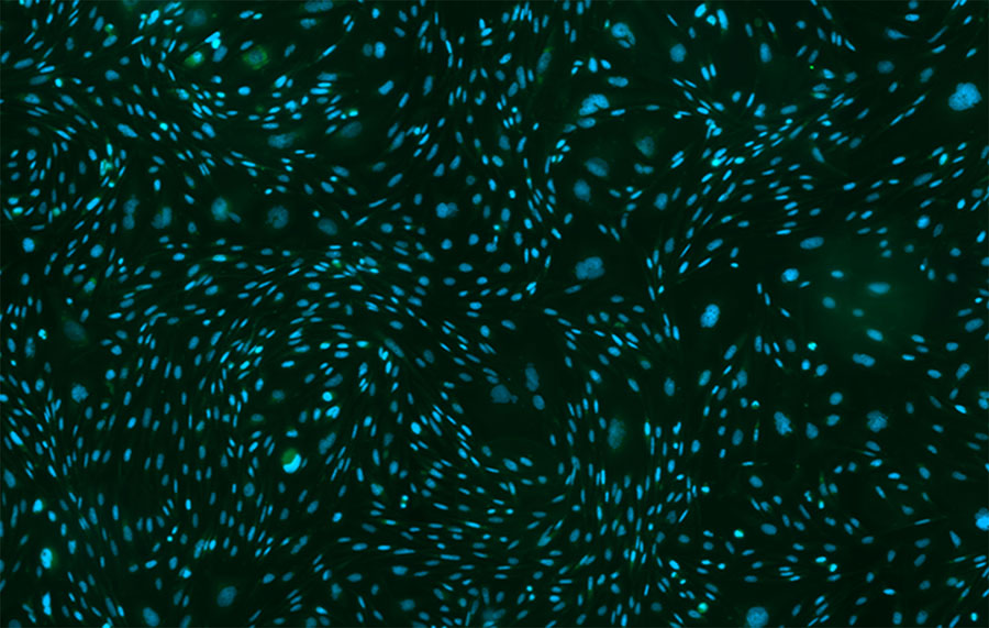
Starry Night
by Junyeop Roh
Center for Synaptic Brain Dysfunctions
Vincent van Gogh once stated, “I don’t know anything with certainty, but seeing the stars makes me dream.” One of the more iconic masterpieces of van Gogh is his The Starry Night, complete with swirling swaths of stars circling various elements of the night sky. When these DAPI, a blue-fluorescent DNS stain, and green fluorescent protein -labeled Chinese hamster ovary (CHO) cells were imaged by confocal microscopy, the resulting image showed groups of “stars” forming whirlpools against the contrastingly dark background—much like the stars of the night sky.

It‘s Okay.
by Joonhyuk Lee
Center for Cognition and Sociality
Occasionally we fail at something, but we learn from our failures and move forward. But sometimes, no matter how hard we try, we don't even see the signs of success, but instead fixate on the deeply rooted memories of past failures that continue to haunt us. The blue colors in the image are neurons called thalamic reticular nucleus. Some of these neurons, shown in red, stretch the thread-shaped axon, which serves to suppress fearful memory. When you are in a scary situation, these neurons increase their firing activity, allowing you to overcome the fearful situation. Whenever your past failure or fearful memories discourage and stop you from taking up new challenges, pay close attention to the message of support that is sent to you from the deep inside of your brain—it’s okay.
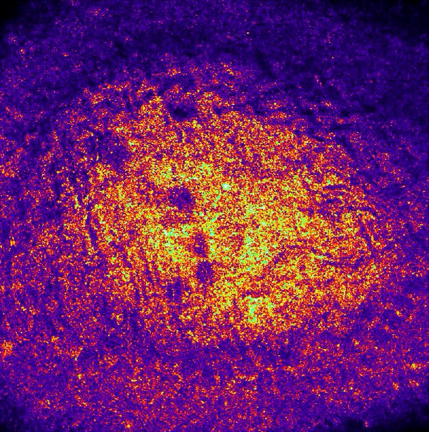
Zebrafish
by Jinhee Hong, Yonghyeon Jo, Jaecheol Jo
Center for Molecular
Spectroscopy
and Dynamics
They say eyes are the window of the mind. We can understand a person’s
emotions
and
thoughts by looking into each other’s eyes. This video clip is composed of confocal
reflectance
images of the eyes of zebrafish, which is a widely used animal model at our Center. The
pattern
of light reflected from the zebrafish’s cornea, lens, and vitreous chamber looks like
stars
and
a nebula in the Universe. Their eyes are less than 1 cm in size yet contain an immense,
light
year-scale of the Universe. Maybe the zebrafish are using their eyes to tell us they are
also
precious creatures, just like humans, and are part of the Earth and the
Universe.
View video on
YouTube
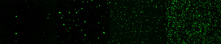
Green Galaxy
by Hyojin Kim
Center for Biomolecular and Cellular Structure
In modern times, the lights of cities fill the night sky, even at a late hour. Lights from office windows, street lamps along roads, and flashing neon signs exhaust and overwhelm our eyes. However, if you get a little off the beaten track, you can meet the real owners of the night sky. You can’t see well with eyes that are accustomed to bright city lights. But if you keep your eyes focused on the sky for a while, you will see a beautiful starry night. This photograph is of a virus-infected insect cell containing green fluorescent DNA. After a long period of time, the virus multiplies and more cells emit fluorescence, which resembles a green galaxy in the night sky. This is an essential step to make proteins, but researchers have nothing to do but wait during the process. The work can be arduous and the hours long, but if you endure you can make good proteins. These newly made proteins seem to say that if you close your eyes and take a deep breath, even for a moment in the midst of your busy and laborious time, one day it will be your time to shine like a glittering star.
Self-organized Octahedra
by Yaroslav Sobolev
Center for Soft and Living Matter
When jammed together, identical objects with symmetric shapes may self-assemble into formations that preserve order over large distances. The look of these packings is a fingerprint of the geometric shape of the underlying object, and simple objects can form intricate packings. Understanding the rules governing the outcomes of such self-assembly is important for designing structures that spontaneously self-build from basic blocks. In this photo, you can see how octahedral (eight-sided polyhedron with equal equilateral triangles as faces) that I have randomly thrown into a glass dish for storage, have—after a few accidental shakes—formed an ordered packing. This pattern of triangles is a known packing for octahedra on a flat surface, so it’s not anything new or unexpected, but I was still surprised how quickly this pattern formed with no guidance on my part whatsoever.
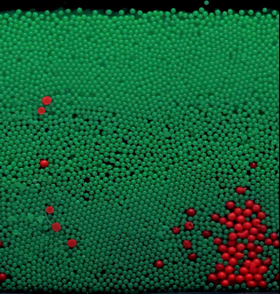
Granular Flame
by Yaroslav Sobolev
Center for Soft and Living Matter
It’s hard to believe, but there are no liquids involved in what you see
here.
These
are plastic balls only among air, with green balls smaller than red ones. Balls are
inside a
glass flask that is being vertically shaken, and one video frame is taken every few
shakes.
When
balls rub against each other, they become electrically charged, repel each other and
climb
up
the transparent glass wall. As a result, their motion starts to resemble convection or a
flame.
This is an interesting example of the intricate collective behaviors than are possible
in
simple
granular systems, including here in a mix of two sizes of balls in a glass
container.
View video on
YouTube
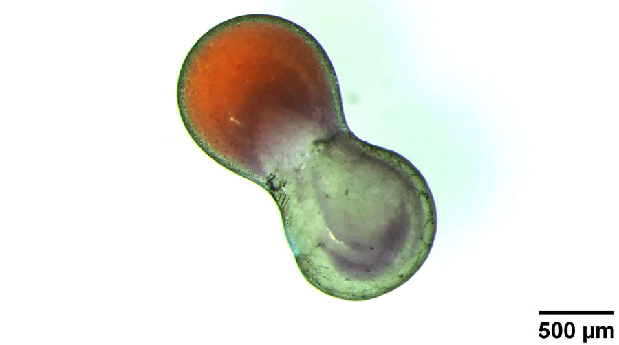
Convective Mixing
by Yaroslav Sobolev
Center for Soft and Living Matter
Mixing of liquids, essential for many chemical reactions, is notoriously
hard to
do
in tiny flasks when you only have a little bit of each liquid for your reaction. Mixing
even
plain water with dye in a sub-millimeter container is as difficult as mixing different
kinds
of
thick honey in a one-liter bottle. Here we show that heating by a laser can facilitate
mixing of
two droplets of water (one of them dyed orange, another containing acid) connected into
a
single
dumbbell-shaped droplet that is made possible by a special “nano-soap” we developed.
Nanoparticles of this “soap”, coating the droplet in a greenish ultrathin layer,
strongly
absorb
light from a laser (pointed at a spot in the lower left corner), heating the water
inside
and
making it swirl like in a teapot. As the contents inside mix, acid from the right part
makes
the
orange dye from the left part become violet and less vivid.
YouTube 영상 보기
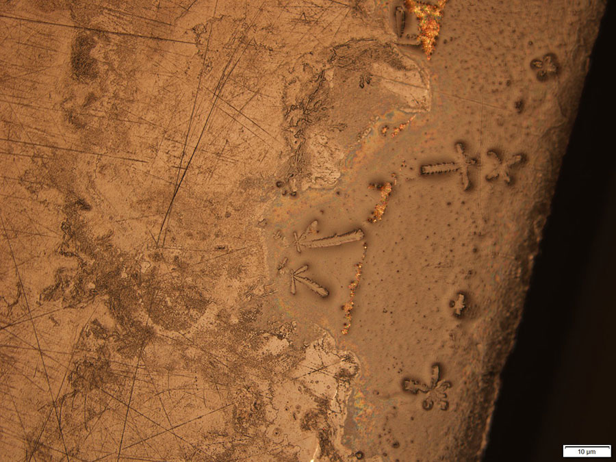
Footprints on Scintillating Crystal Surface
by Jungho So
Center for Underground Physics
A scintillating crystal can be used for a radiation detector by measuring its emission light as proportional to the energy of incident particles and radiation. The Center for Underground Physics has been developing a simultaneous measurement of heat and light from scintillating crystals at a temperature of 10 mK for an effective particle identification. The detector, once developed, will allow precise observation of neutrinoless double beta decay, bringing us one step closer to unraveling the mysteries of the universe. Making a clean, smooth, polished surface of the crystal is the first step towards the goal. As the image shows a surface that is not clean and smooth, this attempt was a failure. The image shows the surface of the crystal captured by a high resolution microscope. The patchy surface of the crystal was a total failure as we needed it to be smooth, but the result stirred up some artistic imagination. One might think that they are looking at the first human footprints on the Moon or Mars, or some fossils in a desert. As we are almost to the end to the experiment, we will soon be reminiscing when later looking at this picture as the footprints of our journey to unravel the mysteries of everything.
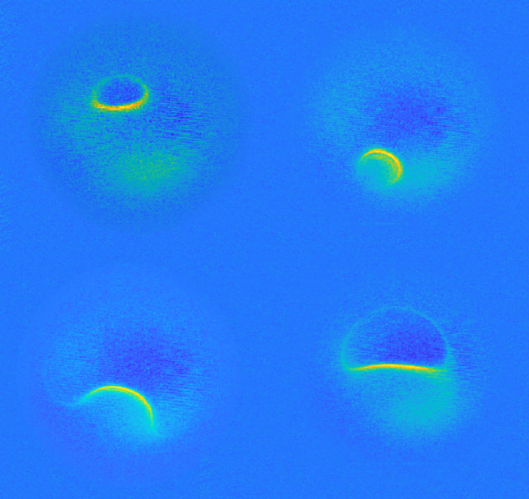
Patient Zero
by Philip Willke
Center for Quantum Nanoscience
The patient is long gone. It demised shortly after the experiment. What you see are four MRI scans, the last images ever taken of it. Was it sick? No. We just took these scans out of curiosity. Cruel? Not at all. No humans or animals were harmed in the making of these images. You are witnessing here the smallest MRI ever taken, displaying a single titanium atom. The image size is almost beyond human imagination, merely a billionth of a meter. The wave-like feature, gently sloshing from one side to the other, reflects the inside of the atom. It reveals the atoms’ magnetic field at different strengths. Thus, patient zero has not died in vain, but shared his most intimate secrets, guiding us to better understand the fundamental building blocks of nature.
Starry Quantum
by Shinjae Nam
Center for Quantum Nanoscience
Scott Lang, the title character of the movie Ant-Man, turns off the regulator on his suit to save his daughter. He then became smaller than the size of an atom and entered the extreme microscopic world—the quantum realm. Unlike Ant-Man, we cannot see atoms directly. However, by using a scanning tunneling microscope (STM), an atomic image can be obtained. In a microscopic world, things cannot be easily explained by the same laws of the world we live in. A flat-looking smooth gold surface can actually turn out to be not so flat if viewed by an STM. This picture is an STM image of a gold (111) surface which vanadyl phthalocyanine (VOPc) molecules are deposited. At the surface, the gold atoms change their arrangements and this results in a height difference. The Z-like patterns (herringbone patterns) are made by the height difference of rearranged gold atoms. The round shape particles are the deposited (vacuum evaporation) VOPc molecules. Most of them are located on the elbow of the herringbone pattern. These patterns show that there are rules in the atomic world as well. The microscopic world still has a lot to study, presenting scientists with many opportunities to push the limits of science and technology.
Atomic-scale Stained Glass
by Gyeongcheol Gye
Center for Artificial Low Dimensional Electronic
Systems
A charge density wave (CDW) is a phenomenon which atoms and electrons undergo a phase transition at low temperatures with new periodic structures different from those at high temperatures. This image shows two different CDW structures on the surface of niobium diselenide (NbSe2) taken by a scanning tunneling microscope (STM). The two charge density wave structures are distinguished into colored and uncolored areas with different colors indicating a difference in their location. The colorful areas of a charge density wave bring to mind stained glass in cathedrals.
The Nanoscale Vegetable Garden
(Subtitle : A Strange Co-existence of
Radishes
and Cabbages
by Eunseok Oh
Center for Artificial Low Dimensional Electronic
Systems
Periodic green peaks appearing on the curved red ground remind us of fresh growing vegetables at regular intervals in a field. Among the vegetables, when you look closely, it is easy to figure out that the shapes between the left and right areas are different. In the upper left area, single green peaks appear periodically, as if the radishes are with their leaves growing up. On the other hand, triple green peaks appear in the lower right area, as if the cabbages are with their leaves spreading out. These two kinds of vegetables coexist in the field and their area is divided by an interfacial line in the middle. This image was taken by a scanning tunneling microscope at cryogenic temperatures of -269 degrees Celsius (4 K), which shows the surface atomic structure of NbSe2 crystals in a charge density wave state. Peaks of various heights on the surface indicate Se atoms, and the green peaks with a specific period are due to the deformation of the atomic structure by a charge density wave. The coexistence of the two types of structures (the left radish area and the right cabbage area) of the charge density wave, which are energetically competing, provides an explanation for the cause of incommensuration of charge density wave of NbSe2 which has been unresolved for decades.
* CMS@CERN, The Art of Science
I. The Art of Science Series
Those visitors fortunate enough to visit the Compact Muon Solenoid (CMS) experiment at CERN, the
European
Laboratory for Particle Physics, when it is opened in all its glory have often commented it
could be
displayed as a work of art.
CMS is one of four large experiments embedded in the 27-km Large Hadron Collider (LHC), the
largest
and
most powerful particle accelerator ever built. It was designed to explore the secrets of our
universe,
and specifically to find evidence for the Brout-Englert-Higgs mass generation mechanism. The
discovery
of the associated Higgs boson in 2012 by the CMS and ATLAS experiments resulted in the awarding
of
the
2013 Nobel Prize to Francois Englert and Peter Higgs; unfortunately Robert Brout passed away in
2011.
CMS has the shape of a cylinder 20m in diameter and 25m long. It acts as a giant high-speed
camera,
taking 3D “photographs” of particle collisions in the centre of the experiment 40 million times
every
second with a spatial resolution on the micrometre scale.
As a scientist, I am fascinated by the massive scale of such high-tech instruments which allow
mankind
to decode nature with incredibly high precision.
As an artist, I am charmed by the intrinsic geometry, colours and scale of the architecture of
these
instruments which have the size of several storey high buildings.
The aesthetics of CMS are associated with its functionality. Its visual periodic multiplicities
emerge
from its physical measurement requirements, and the colour choice of the various components of
this
gigantic high precision camera make it appear like a piece of art on its own.
CMS - The Art of Science I
2013, Michael Hoch, Photo print on Alu Dibond behind Acrylic glass,, 80x80cm
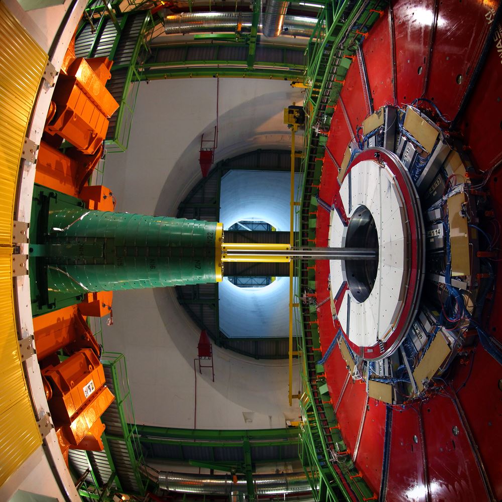
CMS - The Art of Science II
2013, Michael Hoch, Photo print on Alu Dibond behind Acrylic glass,, 80x80cm
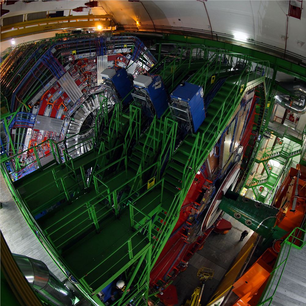
CMS - The Art of Science III
2013, Michael Hoch, Photo print on Alu Dibond behind Acrylic glass,, 80x80cm
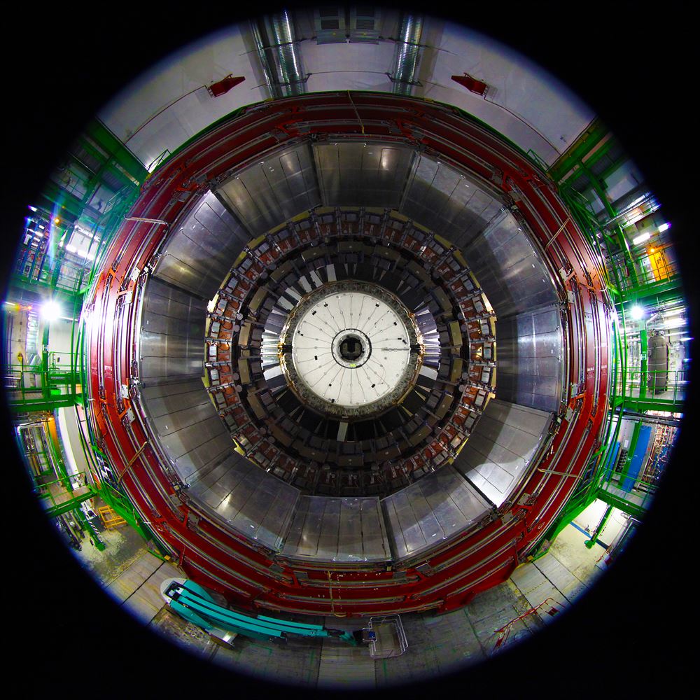
CMS - The Art of Science IV
2013, Michael Hoch, Photo print on Alu Dibond behind Acrylic glass,, 80x80cm
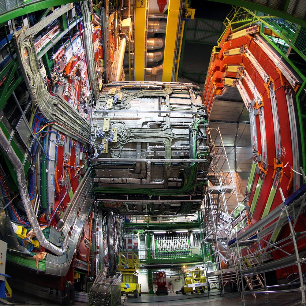
CMS - The Art of Science V
2013, Michael Hoch, Photo print on Alu Dibond behind Acrylic glass,, 80x80cm
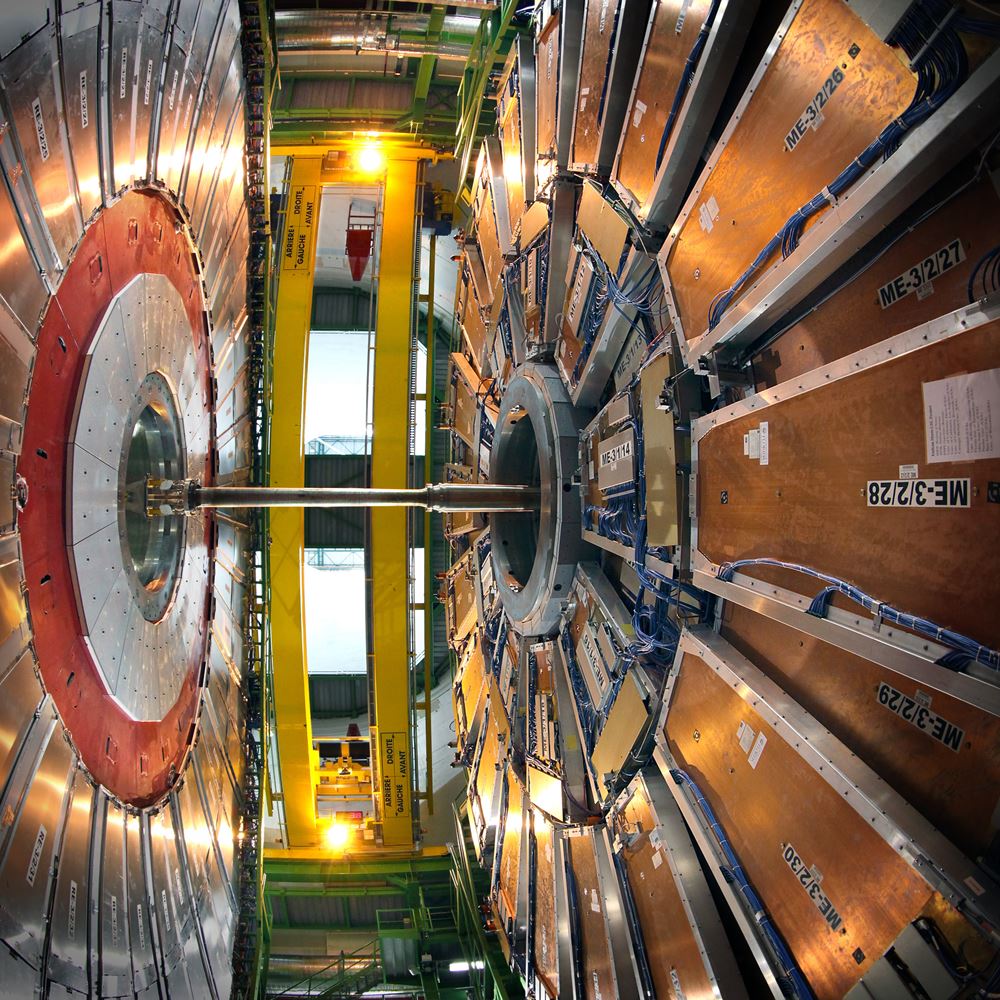
CMS - The Art of Science VI
2013, Michael Hoch, Photo print on Alu Dibond behind Acrylic glass,, 80x80cm
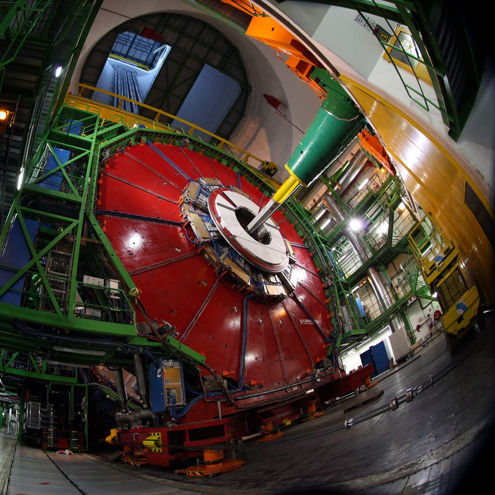
CMS - The Art of Science VII
2013, Michael Hoch, Photo print on Alu Dibond behind Acrylic glass,, 80x80cm
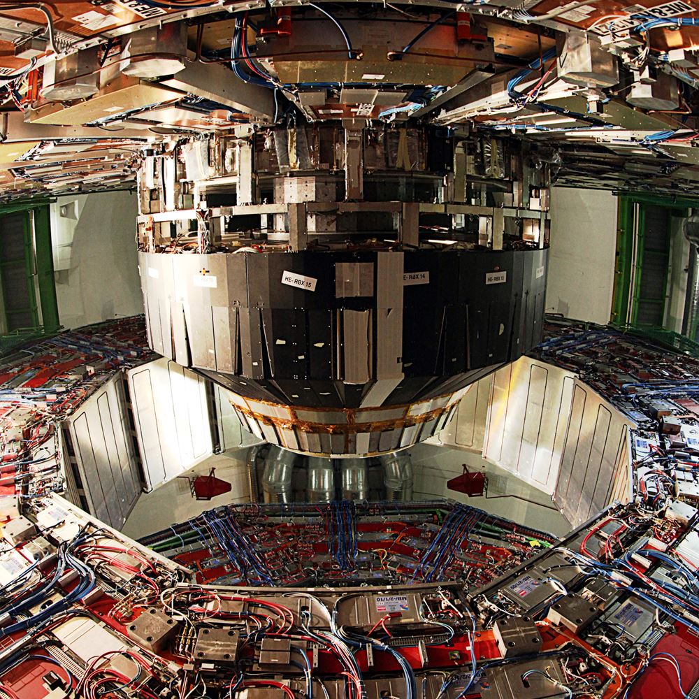
CMS - The Art of Science IIX
2013, Michael Hoch, Photo print on Alu Dibond behind Acrylic glass,, 80x80cm
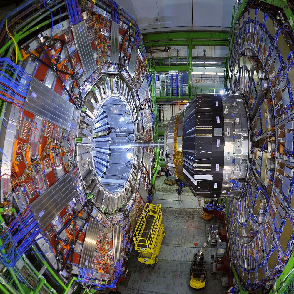
CMS - The Art of Science IX
2013, Michael Hoch, Photo print on Alu Dibond behind Acrylic glass,, 80x80cm
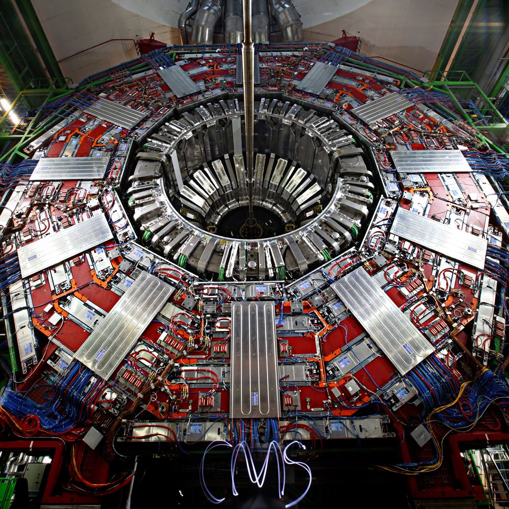
CMS – The Art of Science X
2013, Michael Hoch, Photo print on Alu Dibond behind Acrylic glass,, 80x80cm
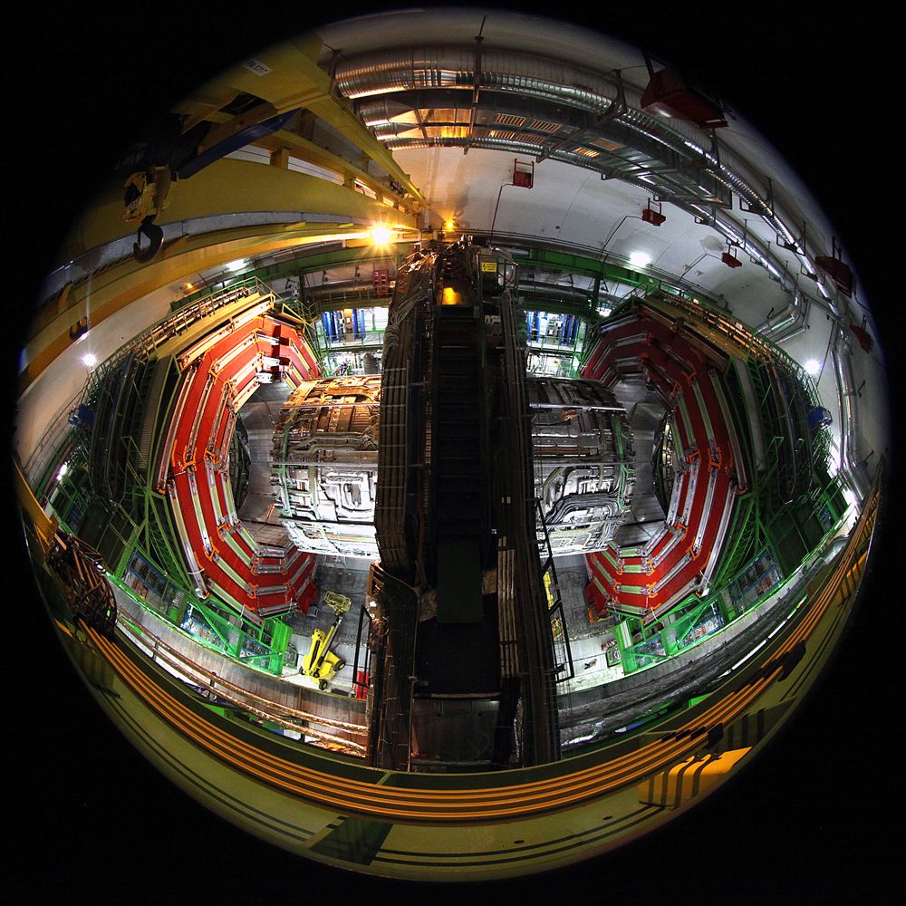
CMS - The Art of Science XI
2013, Michael Hoch, Photo print on Alu Dibond behind Acrylic glass,, 80x80cm
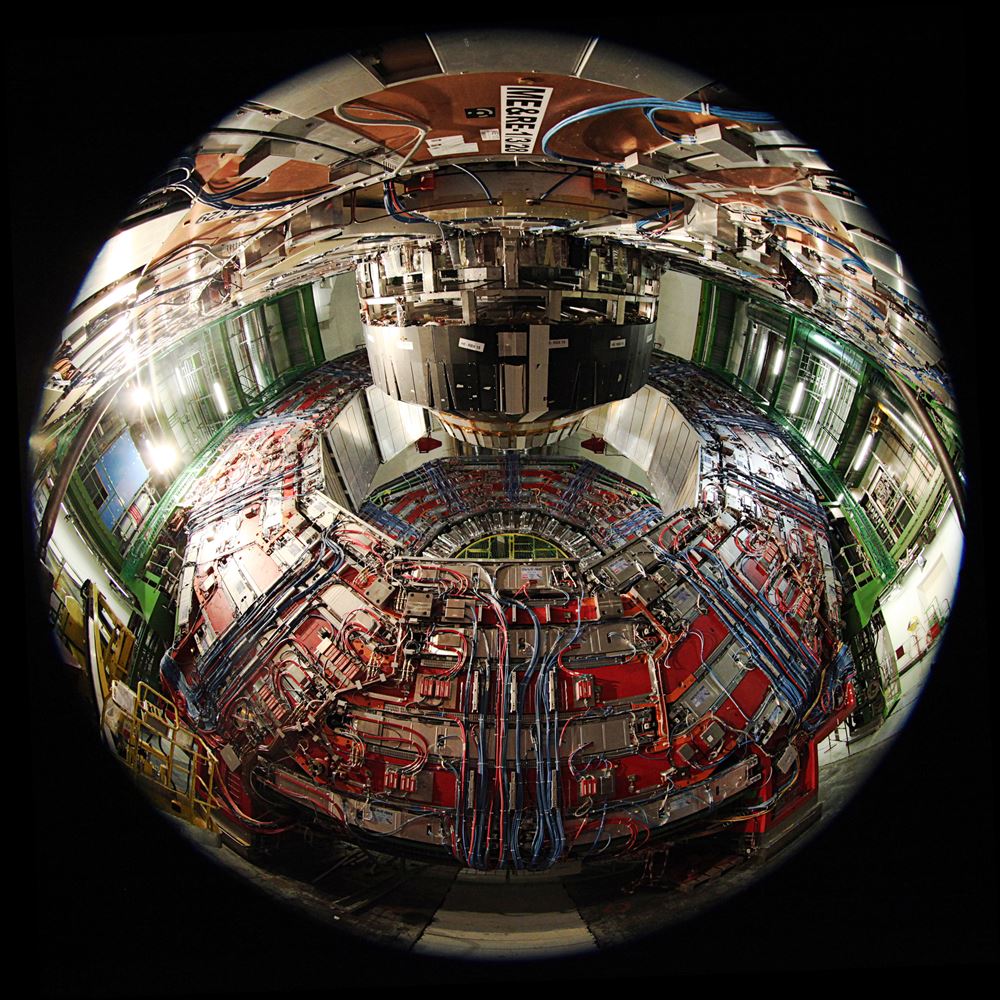
CMS – The Art of Science XII
2013, Michael Hoch, Photo print on Alu Dibond behind Acrylic glass,, 80x80cm
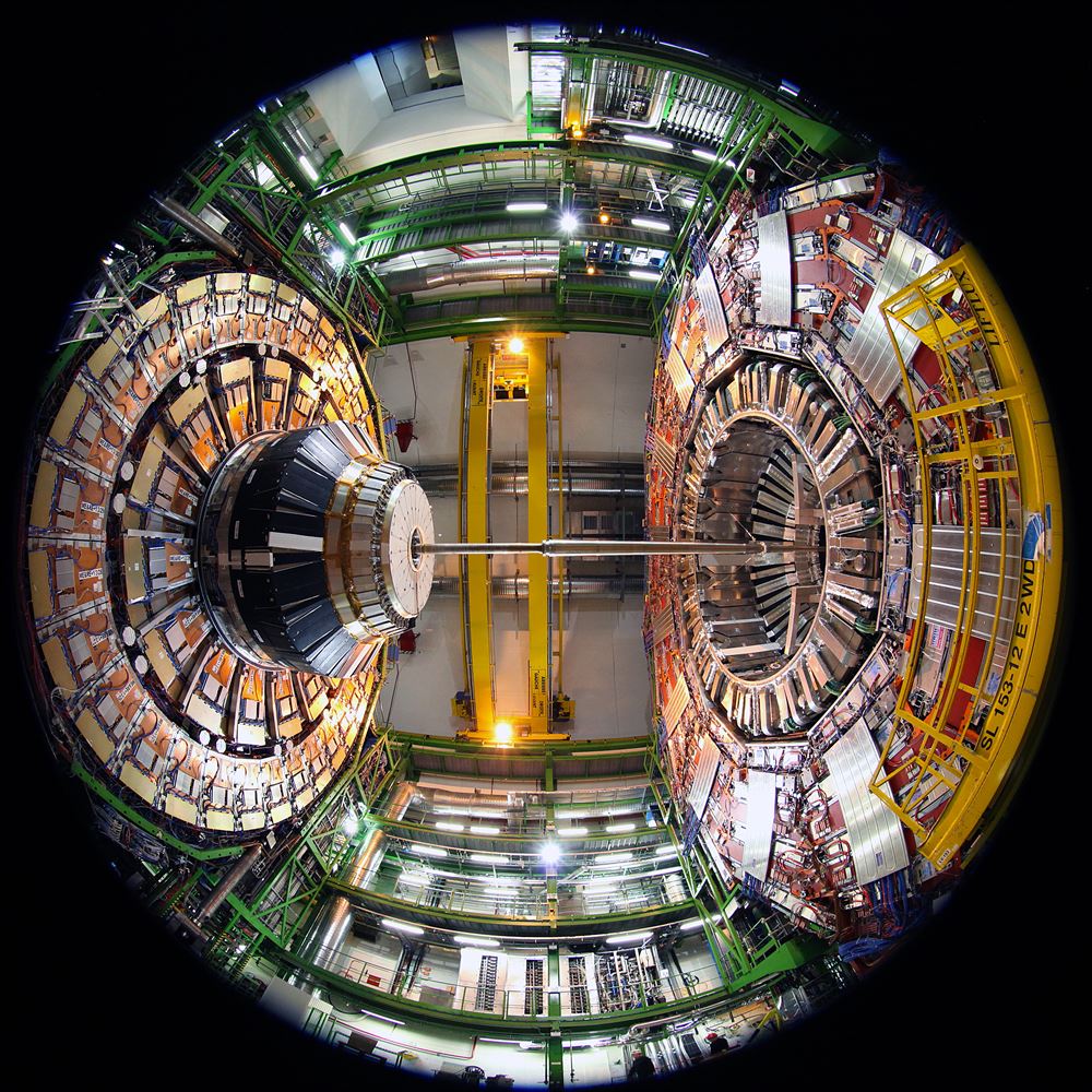
CMS – The Art of Science XIII
2013, Michael Hoch, Photo print on Alu Dibond behind Acrylic glass,, 80x80cm
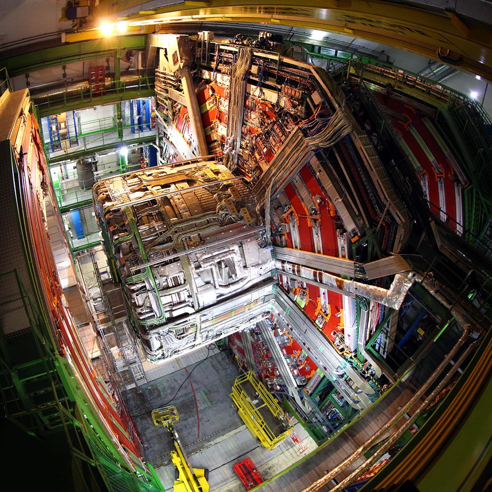
CMS – The Art of Science XIV
2013, Michael Hoch, Photo print on Alu Dibond behind Acrylic glass,, 80x80cm
II. The God Particle Hunting Machine Series
Both CMS and the LHC are not only engineering marvels operating at the forefront of science and
technology, but also have intrinsic geometry with exceptional aesthetics that can be viewed as
works
of
art. The aesthetics of CMS are associated with its functionality as a gigantic, high precision
camera.
Periodic multiplicities emerge out of the physical constraints and strict scientific precision,
which
allow us to unravel the secrets of nature.
In my artwork, the scientific apparatus designed
by
humans merges with the organic, apparently “quasi-chaotic“ appearance of nature. But nature in
itself is
by far not chaotic and follows strict rules. It is these rules that the apparatus and the
scientist
aim
to understand.
The God Particle Hunting Machine Daisy_YE1
Michael Hoch(2013) photo art print, Photo Paper print on Al Dipond, 200x66cm
The God Particle Hunting Machine Poppy_YE1
Michael Hoch(2013) photo art print, Photo Paper print on Al Dipond, 150x50cm
The God Particle Hunting Machine
Michael Hoch(2013) photo art print, Photo Paper print on Al Dipond, 150x50cm
The God Particle Hunting Machine Apple_YB2
Michael Hoch(2013) photo art print, Photo Paper print on Al Dipond, 150x50cm
III. Faces of CMS
CMS the modern tower of Babel, a marvel of forefront technology driven by the ultimate scientific
challenge to encode the most fundamental secrets of nature. It was built to find evidence for
the
Brouth-Englert-Higgs mass generation mechanism and was rewarded by the Nobel Prize for the
discovery
of
what is commonly known as the “God particle” or more scientifically the Higgs boson.
Its construction spanned more than 20 years. After 25 years when the first big scientific
achievements
have been harvested it counted more then 11,000 creative minds participating in this human
challenge
coming from 50 different nations and 200 university institutes. “The Faces of CMS” represents
the
human
aspect of this ultimate scientific and technological endeavour. It shows the large diversity of
characters and human brainpower needed. Women and men all ages worked together in teams,
inspired by
curiosity and passion by a common goal.
Faces of CMS
2010-2016, Michael Hoch, Photo Paper print on textile, 900x400cm
Korean CMS
2019, Michael Hoch, Photo art print, 60x45cm
IV. Matter – Anti-Matter Series
Looking back almost 14 billion years, to a time shortly after the Big Bang, it turns out that our mere existence is nothing but a near miss. At that time, not only matter was created, but also anti-matter. Not a tiny amount of it, but just 0.00000001% less than matter. Shortly after their creation, matter and anti-matter collided again, annihilated and disintegrated into pure radiation. If the universe were filled with radiation and nothing else, there would be no galaxies, no stars, no planets and no life. But nature decided to allow for a small violation of the perfect symmetry of matter and anti-matter. And this minute excess of matter is what our whole universe is made of. The series reflects the symmetric creation of matter and anti-matter during the Big Bang by asking the question, “If we are made of matter, where has all the anti-matter gone?”
Matter – Anti-Matter Symmetry 1
2012, Michael Hoch, photo art print, Photo Paper print on Alu Dibond, 100x100cm
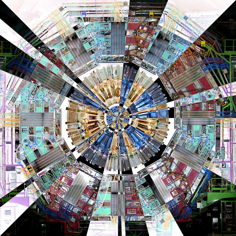
Matter – Anti-Matter Symmetry 2
2012, Michael Hoch, photo art print, Photo Paper print on Alu Dibond, 100x100cm
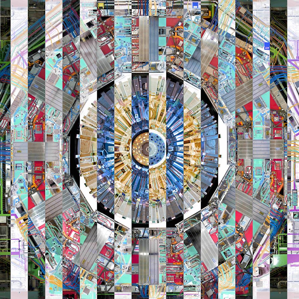
Matter – Anti-Matter Symmetry 3
2012, Michael Hoch, photo art print, Photo Paper print on Alu Dibond, 100x100cm
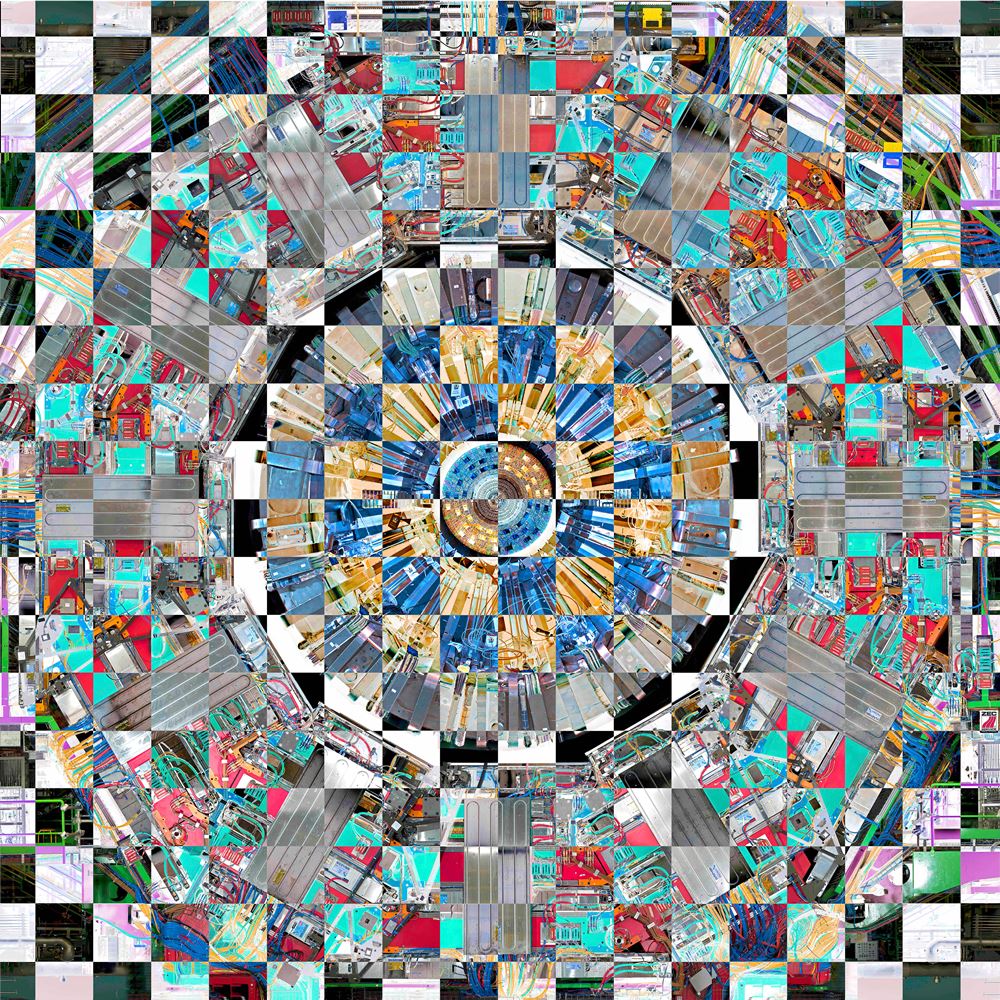
Matter – Anti-Matter Symmetry 4
2012, Michael Hoch, photo art print, Photo Paper print on Alu Dibond, 100x100cm
V. CMS be an Icon
Andy Warhol created in the 60th last century a series of iconic photos printed on with the same technic ‘silk screen printing’ and permutating the colors to reflecting the scientific aspect as an artistic expression.
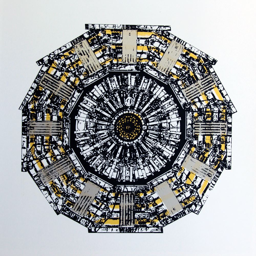
CMS graphic
Michael Hoch, Kathy Kissik(2016) Photo Paper print on silk screen, 80x80cm
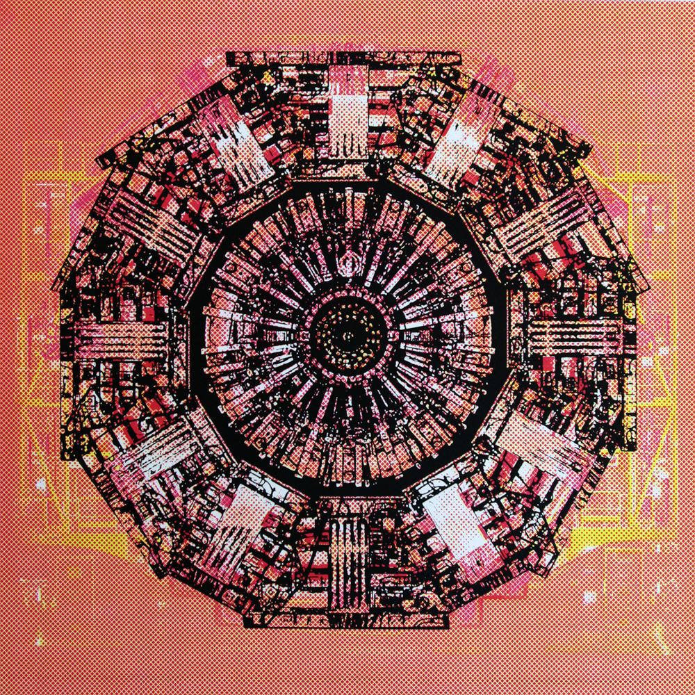
CMS colour Red
Michael Hoch, Kathy Kissik(2016) Photo Paper print on silk screen, 80x80cm
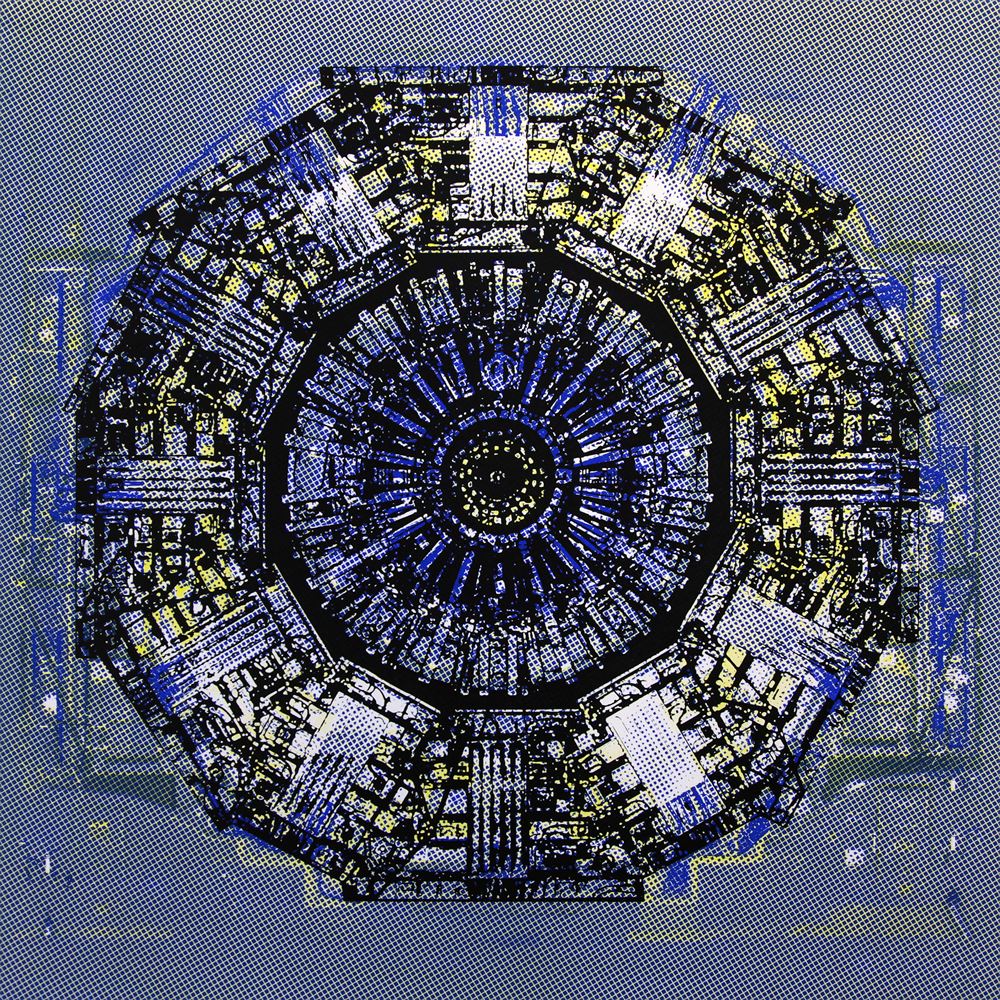
CMS colour Blue
Michael Hoch, Kathy Kissik(2016) Photo Paper print on silk screen, 80x80cm
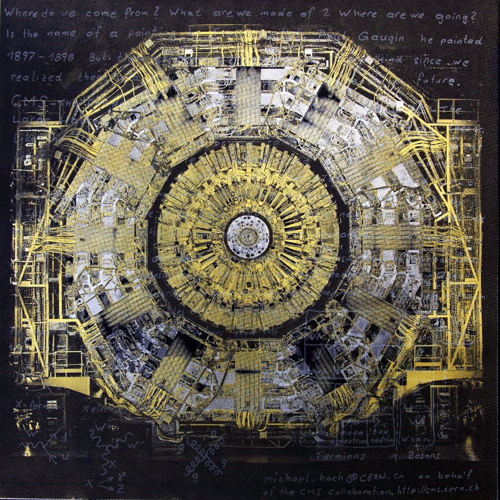
CMS metalic Physics
Michael Hoch, Kathy Kissik(2016) Photo Paper print on silk screen, 80x80cm
VI. CMS Videos
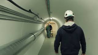
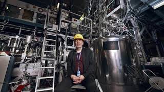
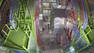
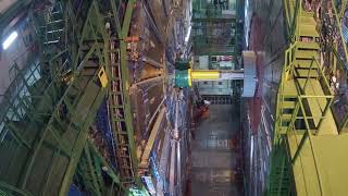
2018 IBS Art in Science
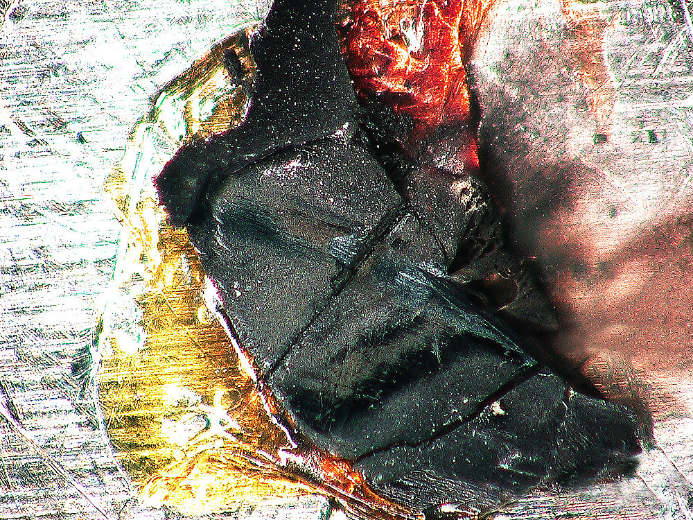
A Piece of Femto-Galaxy
by Min-Cheol Lee
Center for Correlated Electron Systems
A single crystalline Ca2RuO4, destroyed by an ultrafast laser beam, is modeled as a workpiece. The broken crystal shows a clear line along the path of the femtosecond laser pulse incident on the sample. Unlike conventional techniques, ultrafast spectroscopy can instantaneously introduce a large amount of energy by using femtosecond light. Particularly in the case of Ca2RuO4 with a strong correlation between electronic/lattice structures, huge structural changes can arise, such that the sample is destroyed not by physical contact but merely by the absorption of ultrafast light. The millimeter-sized black sample with red glue reminds me of a vast galaxy in the night sky.
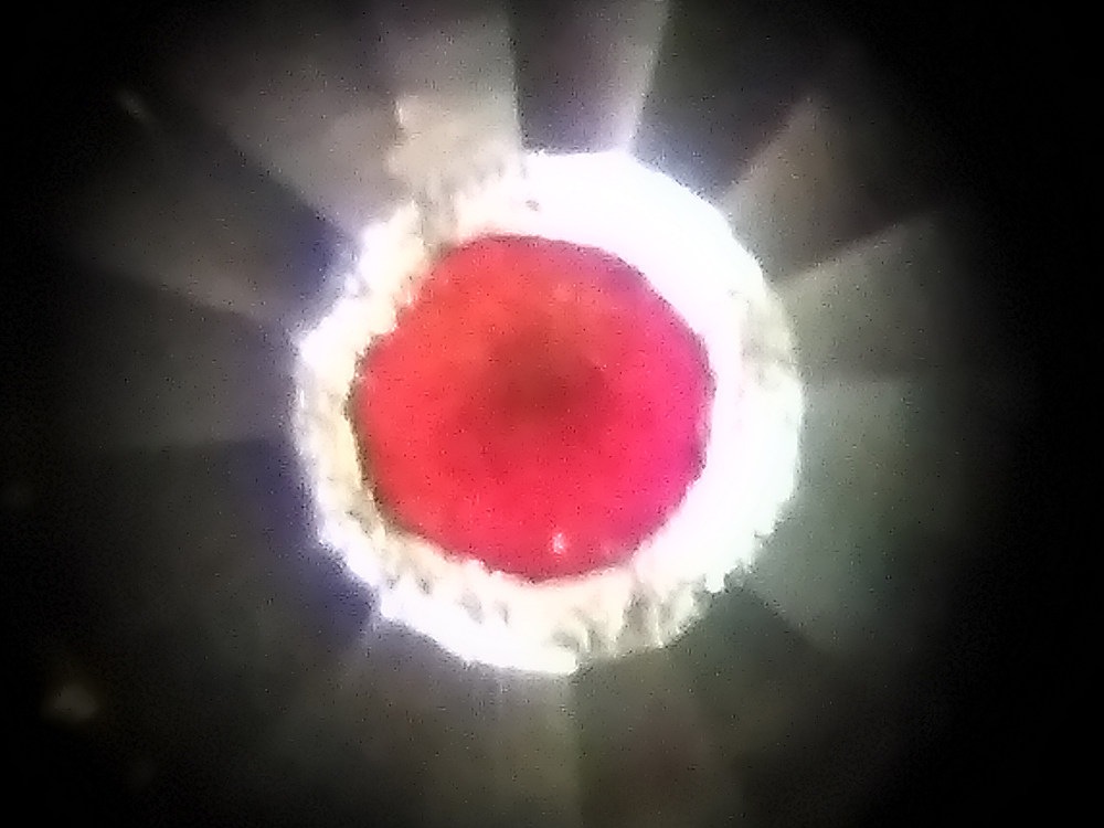
Diamonds and Wine
by Dr. Matthew Coak
Center for Correlated Electron Systems
Two-dimensional magnet MnPS3 is naturally a beautiful translucent lime green colour. As this photo, taken on the Diamond Light Source x-ray synchrotron in the UK, shows, squeezing this material between two diamonds changes it to a gorgeous deep red. With the light shining through it from behind, it looks like a droplet of the finest claret (Bordeaux). Surrounding the crystal in the centre, you can see the perfect facets of one of the two flawless natural diamonds. The crystal experienced 100,000 times atmospheric pressure - 100 times the pressure at the bottom of the Mariana Trench and enough to turn oxygen solid! The drop in energy changed the green to a red colour as MnPS3 was driven from an insulator to a metal, which was found to occur at 300,000 atmospheres.
Oil Painting Inside Donut
by Ki Hoon Lee
Center for Correlated Electron Systems
This figure demonstrates Berry curvature of the spin wave of a triangular lattice in nearly a 120 degree alignment. The spin wave forms a band with a dispersion in the crystal momentum space as the elementary excitation of magnetic materials. Berry’s curvature is considered an important quantity in modern condensed matter physics because it contains geometric and further topological properties of the various excitation states that form bands, including spin waves. In this figure, we placed a crystal momentum space on the surface of a torus or donut shape and tried to give a unique perspective by showing the Berry curvature from the inside. The color represents the size of curvature, and the point where the three black lines meet is the K point, which is the edge of the crystal momentum space.
Milkyway in CBST
by Jae-Joon Kim
Center for Correlated Electron Systems
Crx(Bi0.1Sb0.9)2Te3 (CBST) is the topological insulator that chromium is doped on (Bi0.1Sb0.9)2Te3. Due to local magnetic chromium dopants, a sample will change to global ferromagnetism. This topographic image was taken to find the magnetic domain boundary on a CBST sample with a scanning tunneling microscope (STM)*. It is only 384 nm x 216 nm in size but resembles the Milky Way.
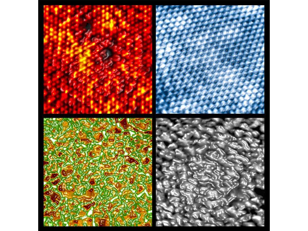
Is the catalyst alive?
by Jeongjin Kim
Center for Nanomaterials and Chemical
Reactions
The molecules that cause chemical reactions breathe new life on the atomic surface of catalyst materials. Much like how minuscule movement can trigger a huge typhoon at sea, an atomic-size wave packet, which is smallest measurement unit of material, can instigate tremendous effects where molecules meet atoms on a catalyst surface. Repeated collisions at the molecular level on a platinum-nickel catalyst surface with atomically-layered adsorptions of carbon monoxide and oxygen molecules result in the reconstruction of atomic layers and a new catalytic reaction.
Mankind in the Nanoworld
by Hyosun Lee
Center for Nanomaterials and Chemical
Reactions
The video shows the sequence of in situ transmission electron microscopy
(TEM)
images of platinum-cobalt (Pt3Co) bimetallic nanoparticles, where a cobalt
oxide
(CoO) layer is formed on the surface of the nanoparticles. The atoms in the
nanoparticles,
arranged in a regular array, seem to keep their position, but the atoms change slowly as
the
environment changes. Here, Pt3Co bimetallic nanoparticles are not observed in
a
high-vacuum environment, but in an oxygen gas (O2) environment measured in
real
time
while applying heat. In this research, we have found that the cobalt atoms move to the
surface
of the nanoparticles atom by atom and form a single CoO layer on the top surface during
the
catalytic reaction, resulting in the improvement of the catalytic activity. While
researching
these small nanoparticles, we are reminded that mankind gradually tries to find a way to
adapt
to surrounding changes and moves toward a better direction.
View video on
YouTube
The Night I Count the Stars
by Dongwook Kim
Center for Catalytic Hydrocarbon
Functionalizations
This diffraction pattern image came from a single crystal x-ray diffraction experiment. The harmony of aesthetic symmetry is an example of the beauty in nature. The diffraction with x-ray radiation to single crystal results in a well-arranged and symmetric pattern. The spreading spots are generated in a harmonious crystallographic condition. We get a sense of beauty looking at the symmetrical arrangement of dispersed diffraction spots give us a sense of beauty resembling stars in the universe.
Molecular Golf: Birdie or Bogey?
by Jiyong Park
Center for Catalytic Hydrocarbon
Functionalizations
Potential energy surface (PES) illustrates the energetics of a molecular system, on which the route and the outcome of a chemical reaction can be described. When color-coded, the surface resembles a golf-course with multiple hills. The lowest saddle point between the hills is the transition state (TS), which defines the minimum energy to access the product. Chemists can figuratively play golf to find the best route to reach the hole (product) from the teeing ground (starting material). Occasionally, the path passing the TS forks to lead two products, known as post-transition state bifurcation. When successful, much like a birdie, we obtain cyclopropane as a product; otherwise, we acquire cyclobutane, which is like getting a bogey.
CSLM in the Microworld
by Kibom Nam
Center for Soft and Living Matter
Optical tweezers are scientific instruments that can trap micro-size objects
with a
highly focused laser. Holographic optical tweezers can generate multiple optical traps
with
a
single laser source. It can generate any arbitrary position of optical traps. This work
shows
optically trapped 2-micrometer polystyrene particles in deionized water spelling out
"ibs.
CSLM"
an acronym for the Center for Soft and Living Matter.
View video on
YouTube
The Phases of DNA
by Anisha Shakya
Center for Soft and Living Matter
Due to its polyelectrolyte nature, DNA can form different phases upon interaction with multivalent cationic polymers. Despite their importance, the prediction and control over these phases remain elusive. This collage shows images of DNA condensed into liquid, multicompartment liquid-like, liquid-crystalline, and semi-solid phases. The variety of visually striking features seen under the microscope takes a scientist’s imagination on delightful artistic detours.
A Tree in the Forest
by Kyu-Hee Lee
Center for Cognition and Sociality
Many neurons come together to form the brain, just like how trees constitute a forest. By seeing only one tree you cannot know what a forest looks like, and even if you can observe the forest from the sky, you cannot know the types nor the individual roles of each tree. Investigating brain function is similar. This image is a cross-section of hippocampus from a mouse. Among the many neurons in a living mouse, one single CA1 pyramidal neuron was detected. I investigated its activity resulting from interactions with other neurons by using patch-clamp techniques. This technique is used to measure the potential of cell membrane via glass micropipette. To find the patched single neuron and to check its shape, I stained the brain slice with fluorescent dye. Finally, I found the single neuron that was standing alone but shining with green fluorescence. You can find the one ‘neuronal tree’ in the middle of the forest composed of many neurons.
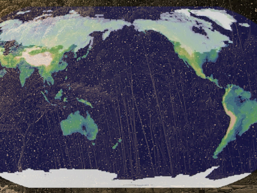
The Sound of Snow
by Axel Timmermann, Jung-Eun Chu, Karl Stein, Kyung-ja Ha
Center
for
Climate Physics
Snow is a crystalline form of water. It insolates soils and plants from
harsh
winter
conditions, it enhances earth’s reflectivity, and it can accumulate in cold areas to
form
ice
sheets, such as Greenland and Antarctica. In our warming world, the number of snow days
will
decrease considerably and climate computer model simulations show that towards the end
of
this
century snowfall events in the midlatitudes, such as the one in Busan on January 10th,
2018,
will become rare. When we think about snow, we often think about our childhood—the
experience of
building a snowman, the thrill of a snowball fight, or just the beauty of watching
snowflakes.
This video clip, which is accompanied by a piano improvisation, tries to remind us of
the
beauty
of snow as well as its fate in response to fossil fuel emissions.
View video
on
YouTube
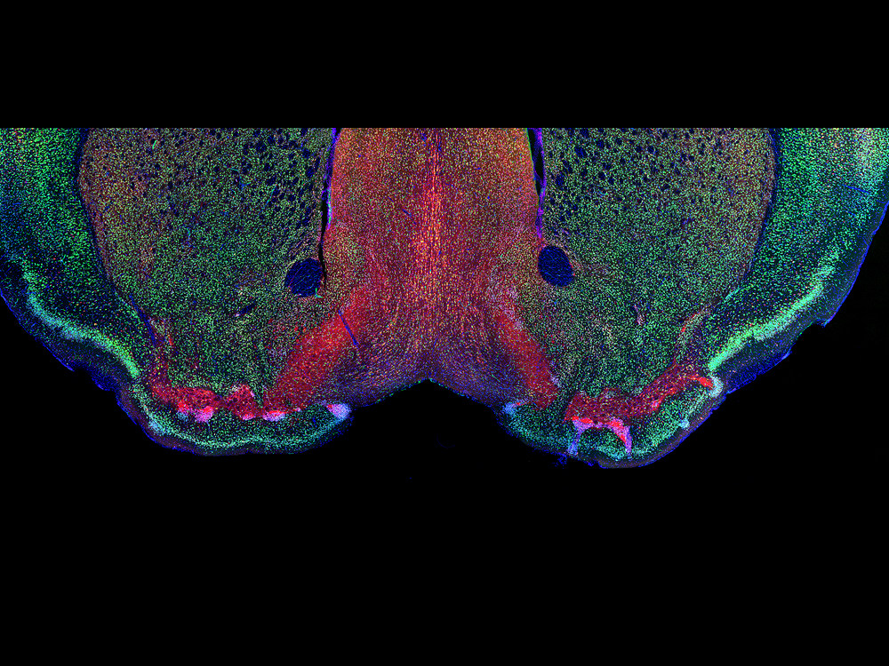
The Eyes of Morpheus
by Haram Park
Center for Synaptic Brain Dysfunctions
The Greeks of antiquity believed Morpheus was the god of dreams and would appear during sleep to shape and form our dreams. We spend one third of our lives asleep, and yet sleep is still a field relatively unknown to science. Why we sleep and how we dream are questions that are yet to be answered. What is known is that the lateral preoptic area (LPO), the region of the brain depicted in the image with red coloring, is critical for sleep to normally progress in the brain. Impairments of neurons in this area causes many disruptions in sleep, such as insomnia. Understanding the LPO will be the gateway to understanding the realm of Morpheus. (Green: NeuN, neuronal marker, Red: GAD, inhibitory neuron marker, Blue: DAPI, Nucleus).
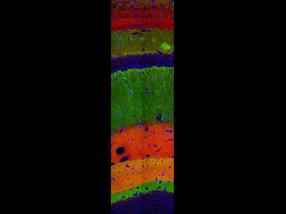
Layers Upon Layers
by Yeonsoo Choi
Center for Synaptic Brain Dysfunctions
A single neuron can receive thousands of signal inputs from other neurons and process them for downstream signalling. This image shows the layers of the hippocampus, the memory storage area of the brain. The colorful layers in the image do not depict different neurons but the different inputs a single neuron in the hippocampus can receive. Even now, as you read this, your brain is processing the layers upon layers of information from countless different regions and subregions to derive cognition from noise. (Green: excitatory neuron marker, Red: synaptic protein marker, Blue: nucleus).
Double Disaster
by Eunkyung Lie
Center for Synaptic Brain Dysfunctions
I wanted to express the unexpected happening that occurred during an
occurrence
of
the Morris water maze experiment, an animal behavior experiment. The experiment has a
mouse
which swims in water and visits a hidden escapable island, which is conducted to study
spatial
perception and memory. Through several trials, the mouse finds the island between 20
seconds
and
1 minute. The mouse in the video is too smart to go directly to the island and
eventually
escapes the 100 centimeter water tank. It is rare for ordinary mice to find the island,
and
even
if they do, they wait there as they do not always know where to go. I have expressed the
characteristics of the mice with the pop art of Andy Warhol’s Silver Car Crash
(Double
Disaster) and the video art of Nam June Paik’s Egg Grows. An unexpected
accident
that occurs during the experiment and widely varying results are a double disaster to
researchers.
View video on YouTube
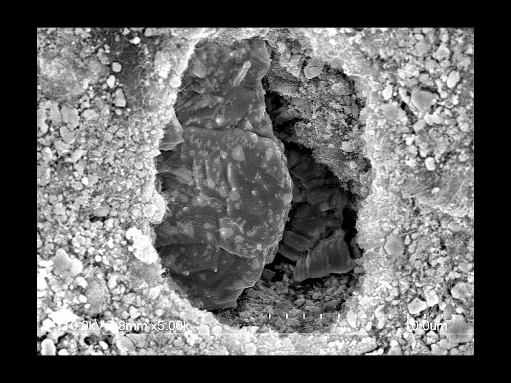
A Secret Door
by Mijoung Joung
RISP
The particles in the picture are lanthanum carbide (LaC2) and are used to generate rare isotopes. Isotope separation on-line (ISOL) is a method to generate rare isotopes that do not naturally occur. A variety of isotope targets are used in ISOL facilities around the world. The LaC2 target is a thin disk under development by the Rare Isotope Science Project (RISP). To achieve high yield of the rare isotope, the particles of LaC2 should be evenly spread over the whole area of the target disk to prevent local temperatures rise when the particles collide with the proton beam. Temperature rise in small area can cause thermal damage to the target disk. However, crystal structures made of agglomerate particles were observed in some parts of the target disk after fabrication. A hole was formed in the mass of particles, which brought to mind the secret cave that Ali Baba enters.
Self-portrait - the ALICE’s innermost particle detector looks at itself
by Magnus Mager
applied physicist at CERN
The pictured detector elements measure about 20cm by 40cm (diameter x
length)
and
will form the innermost part of a cylindrical particle detector that will soon (2020) be
installed in ALICE, an experiment at the Large Hadron Collider (LHC) located at the
European
Organization for Nuclear Research (CERN), in Geneva, Switzerland.
ALICE (A Large Ion
Collider
Experiment) studies the origin of the Universe, when matter was like a soup of quarks
and
gluons. ALICE’s innermost part will be tiled by about 25,000 CMOS particle sensors, each
of
1.5cm x 3cm in size. In everyday life, CMOS sensors are found in consumer products, like
webcams, but here they are used to take 12.5 Giga-Pixel images of particles coming from
high-energy collisions.
Beyond detecting charged particles, these sensors are also
sensitive
to sudden changes of light levels. This feature was utilised here in an old analogue SLR
camera,
where the 35mm film was replaced with the CMOS particle sensor, effectively turning it
into
a
digital camera.
The specific "aura" of the resulting photographs is caused by the
fully
binary operation of the sensor: pixels are either fully black or fully white. Moreover,
some
noise of white and black speckles is created by operating the camera at very low
thresholds.
Finally, a black square pattern is a result of opaque metal structures on top of the
sensor
that
is meant for the electrical interconnection of the sensor to the detector circuit
boards.
Cantor Bouquet
by CNRS (Centre National de la Recherche Scientifique)
Julia set of an exponential, an object of study in holomorphic dynamical systems. Its shape has been found to be the same as a model from continuum theory in topology: the Lelek fan--also nicknamed the Cantor bouquet--which is an infinite union of lines whose tips are densely distributed near every point on these lines.
Model of a critical orbit
by CNRS (Centre National de la Recherche Scientifique)
Pseudo-hedgehog. Model of the critical orbit of some holomorphic dynamical system. The black zone is made of myriads of strands radiating from the center, too densely packed to be resolved at this scale.
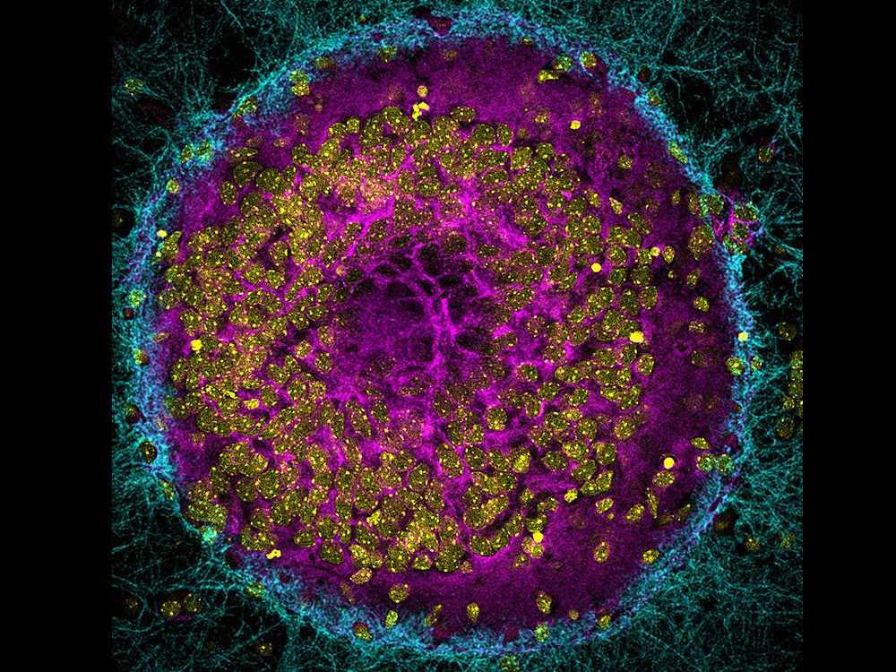
Image 1
by CNRS (Centre National de la Recherche Scientifique)
Hippocampal mouse neurons observed by confocal fluorescence microscopy and reconstructed in 2D by projecting the various optical sections onto the Z plane. The CAR protein was detected using a single antibody.
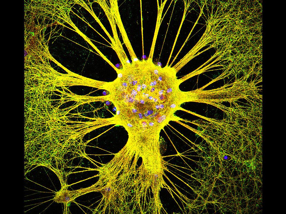
Image 2
by CNRS (Centre National de la Recherche Scientifique)
Hippocampal mouse neurons observed by confocal fluorescence microscopy and reconstructed in 2D by projecting 34 optical sections onto the Z plane. Using immunofluorescent staining, cell nuclei were stained blue with DAPI.
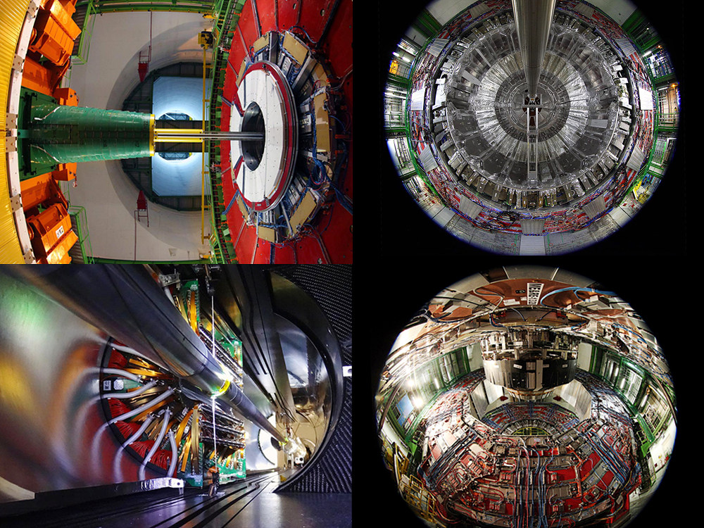
CMS –The Art of Science Architecture
by Michael Hoch
CERN
Those visitors fortunate enough to visit the CMS (Compact Muon Solenoid)
experiment
at CERN open in all its glory have often commented it could be displayed as a work of
art.
CMS
is inside the 27-km Large Hadron Collider (LHC) the largest and most powerful particle
accelerator ever built. It was designed to explore the secrets of our universe,
especially
to
find evidence for the Brout-Engler-Higgs mass generation mechanism and was rewarded by a
Nobel
Prize for the discovery. This detector has a shape of a cylinder is including services
20m
in
diameter and 25m long. It acts as a giant high-speed camera, taking 3D "photographs" of
particle
collisions from all directions up to 40 million times each second with a spatial
resolution
in
the micrometer scale.
As scientist I am fascinated by the massive scale and the
high-tech
instruments at CERN which allow mankind to decode nature in an incredibly high
precision. As
an
artist I am charmed by the intrinsic geometry as well colors and scale of the science
architecture of these experiments that have the size of mid-size houses. The aesthetics
of
CMS
are associated with its functionality as gigantic high precision camera. Periodic
multiplicities
emerge out of the physical measurements constrains and the strict scientific precision
which
allow us to unravel to secretes of nature.
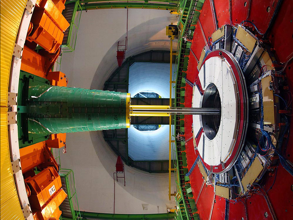
The colors of CMS
by Michael Hoch
CERN
The colors of CMS - view from the bottom of experimental cavern 100m below ground towards the surface.
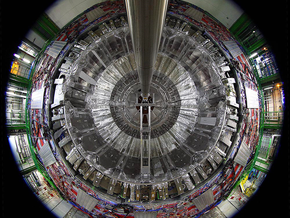
CMS Space Gate
by Michael Hoch
CERN
CMS Space Gate - 20m diameter of perfect circular geometry with LHC beam pipe.
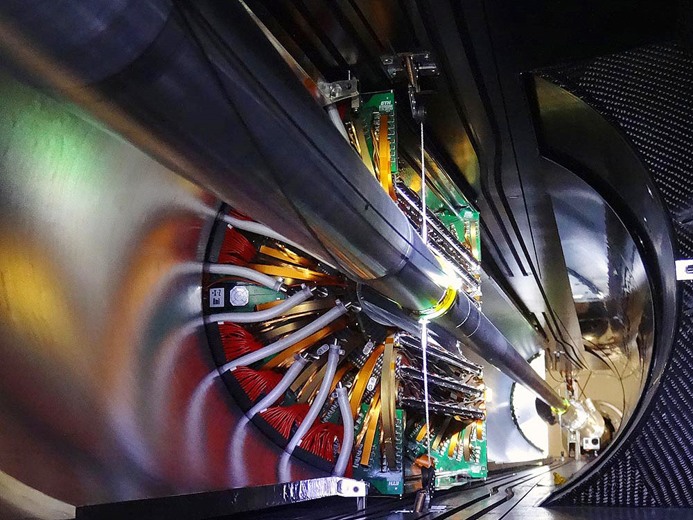
LHC P5 Interaction Point
by Michael Hoch
CERN
LHC P5 Interaction Point – ultra high resolution CMS pixel detector surrounding the LHC interaction vertex.
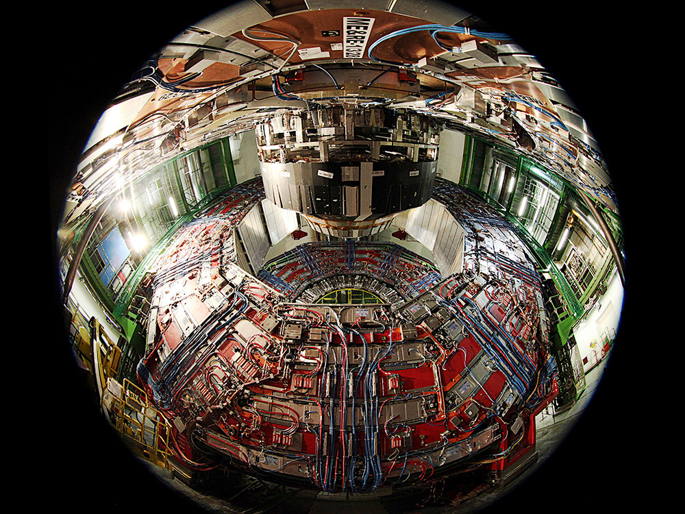
Cable Map
by Michael Hoch
CERN
Cable Map – to large CMS segments shifted apart to give access for services work and insight to the cable tracks, which look like the dense road map of modern city.
2017 IBS Art in Science
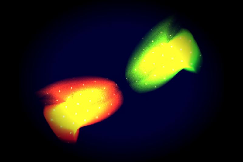
Nemo meets Marlin
by Soonmin Kang
Center for Correlated Electron Systems
Single crystal X-ray diffraction (SC-XRD) with 4 axes is an important experimental method to verify the structure of materials. It is possible to observe reciprocal lattice of material by performing SC-XRD and the limited reciprocal lattice occasionally shows interesting patterns in accordance with experimental conditions. The image contains the SC-XRD results of vanadium diselenides (VSe2) at 28 K and 300 K. The results seem to resemble a dramatic meeting of the clownfish character Nemo and his father Marlin who appear together in a Disney film. Blurry lines, dots, and splendid colors of the patterns contrast with the dark background, creating a dreamy atmosphere.
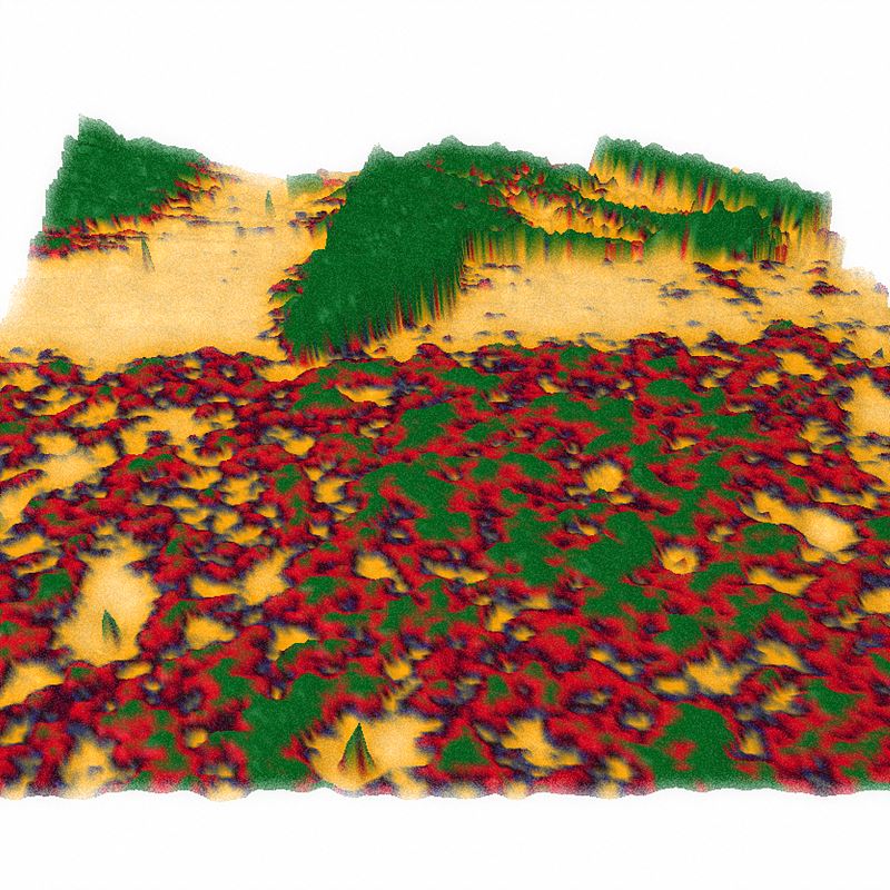
Autumn made of superconductor
by Suhan Son
Center for Correlated Electron Systems
Niobium diselenide (NbSe2), a material synthesized from niobium (Nb) and
selenium
(Se), is not only a quasi-two dimensional layered material, but also a superconductor
which
shows zero conductivity in low temperatures. In the figure, the atomic force microscope
(AFM)*
measurement data obtained after making NbSe2 into several tens of atomic layers by
mechanical
exfoliation is three-dimensionalized. The colors of the figure result from the thickness
differences between layers. It is reminiscent of a mountain where the autumn color comes
from a
combination of colorful colors.
* Atomic force microscope: It can
measure
atomic-scale forces between the surface and a probe tip and produce topographic maps
of
the
surface of the particle.
Spectrum of SI-STM 1 (Spectroscopic imaging scanning tunneling microscope)
by Kyoung Seok Lee
Center for Correlated Electron Systems
From design to completion of construction of a spectroscopic imaging - scanning tunneling microscope (SI-STM), we went through a colorful spectrum of memories; joy, excitement, frustration, hope, struggle, etc. The main image is an SI-STM system installed inside an acoustic and vibration isolation facility. The blue cylinder is a 14 T magnet liquid helium dewar, where a STM head and a cryostat are kept at an extremely low temperature. By this photomosaic, we attempt to visualize the fact that a scientific instrument can be built not only upon materialistic components, but also upon other intangible abstract elements as well. This mosaic is more meaningful since each and every pixel of it were crafted from real images of the researchers’ lives.
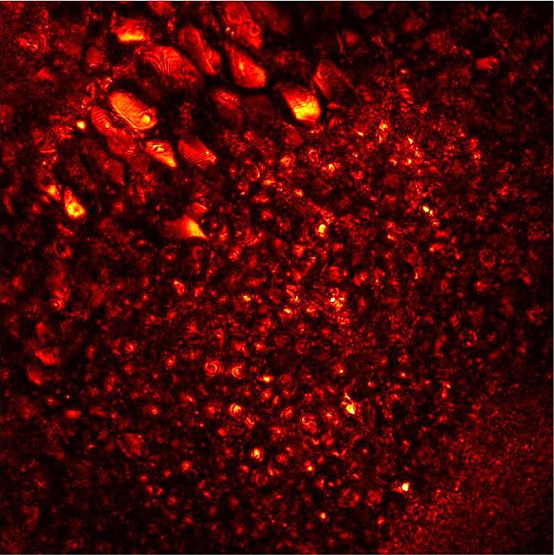
Leaves blowing in the wind : Inside the brain of a living zebrafish
by Moonseok Kim
Center for Molecular Spectroscopy and
Dynamics
This is a movie made of 3-dimensional images of the hindbrain of a living larval zebrafish two weeks after fertilization, which was acquired by label-free high-resolution computational adaptive optical microscopy. The complicated distribution of neurons within 100 μm2 of the field of view is shown from the top to a depth of 100 μm of the brain. Due to the refraction and multiple scattering of light inside the zebrafish, it was previously not possible to acquire high-resolution images with conventional label-free imaging techniques. In order to overcome such a limitation, we have developed a novel computational adaptive optical microscopy which can figure out light distortion and retrieve an original image by fundamental physics. The contrast of the diffraction-limited image is unprecedentedly enhanced, thereby even interference fringes at the membrane can be visualized. The magnificent view of the vanishing neurons reminds us of leaves blowing in the autumn wind.
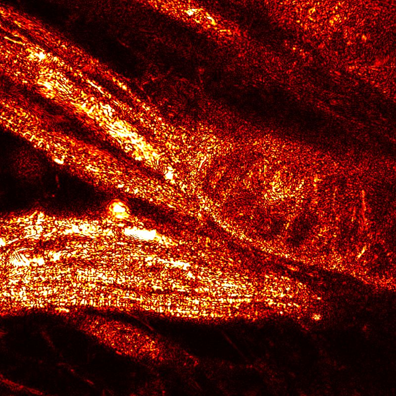
A muscular zebrafish
by Yonghyeon Jo
Center for Molecular Spectroscopy and
Dynamics
This is an image of the muscle tissue of the spinal cord area of the zebrafish 10 days after hatching obtained by a high-resolution aberration-compensated microscope technique with non-labeling. Healthy zebrafish swim fast even in small tanks. Thanks to sturdy muscles, zebrafish can swim vigorously. The separation of the erector spinae muscles and the definition of delicate muscle fibers are prominent. Whether he knew that he would soon be anesthetized and imprisoned in a hard agar, I even feel the will and spirit as he raised his muscles to resist his destiny. Asymmetry composition, flexible shape, and dynamics of the light create a sense of rhythm and space.
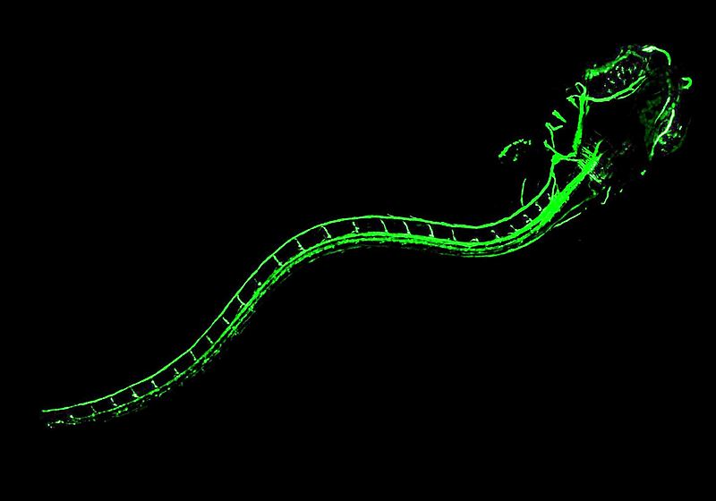
Z-dragon (Zebrafish-Dragon)
by Jin Hee Hong
Center for Molecular Spectroscopy and
Dynamics
The nervous system of vertebrates is surrounded by the myelin sheath that
forms
an
electrically insulating layer. This is essential for a rapid electrical signal
propagation.
This
image represents the myelination of zebrafish which is widely used as animal models.
Green
fluorescent protein (GFP) is expressed in cells that construct the myelin sheath of a
zebrafish
and the image is obtained by a confocal laser microscope*. The central nervous system,
consisting of the brain and spinal cord, and the peripheral nervous system of a
zebrafish 14
days after hatching are shining with green fluorescence. It looks like the shape of a
dragon
who
is eager to ascend. Strong contrast of colors and oblique composition give the sensation
of
speed and motion.
* Confocal microscopy: A type of microscopy that
uses a
laser along with particular optical components to create high-resolution images of
materials
stained with fluorescent probes.
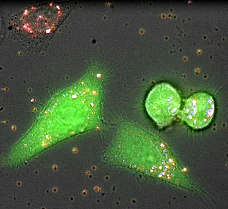
Now you see me : A trace of nanobiochemisty
by Seongchan Kim
Center for RNA Research
A drug delivery system based on RNA as a drug candidate is a novel strategy to treat various diseases at the molecular level, but still has an undesirable biological barrier of efficient delivery to the target site. We have developed a multifunctional RNA delivery vehicle enabling simultaneous bioimaging based on a highly biocompatible fluorescent nanoparticle (blue) and dye-labelled RNA (red) with simple preparation. Real-time study of fluorescence imaging in live cells (green) shows that the nanoparticle favorably localize in cytoplasm and successfully release RNA. This study suggests the excellent potential for simultaneous bioimaging and gene therapy system.
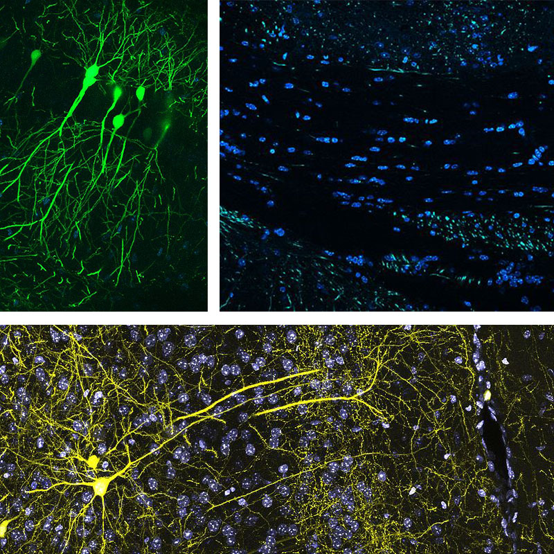
Der Teil und das Ganze trilogy : Contact, Sandbank, Take off at night
by Kyungdeok Kim
Center for Synaptic Brain Dysfunctions
These figures form a trilogy themed by a saying in Oriental philosophy, “There is small universe in each of our brains.” The first figure, “Take off at night”, provides similarity between the brain structure and a city landscape observed from the sky. The second figure, “Contact,” provides appreciation of synapses and the neurons which compose them. In the last figure, “Sandbank,” information is observed like a stream in which we can gaze. Neurons in the images of the trilogy shine by themselves expressing fluorescent proteins. The name of trilogy originated from an immortal classic, “Der Teil und das Ganze” (“the part and the whole” in English), of Werner Heisenberg (1901-1976), a quantum physicist.
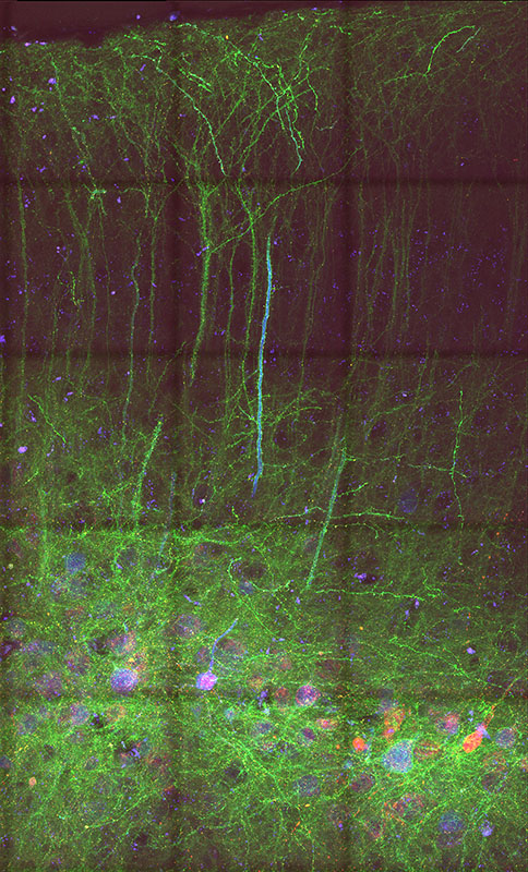
Garden in the brain : Connecting link
by Chaehyun Yook
Center for Synaptic Brain Dysfunctions
Reminiscent of a night forest with wild flowers, this figure shows intermingled structures in a mouse brain, connected to each other by synapses. These connect individual neuron and transmits neuronal activity, forming circuits in a complex neural network. Because of their sheer abundance and tiny size, visualizing synapses together with neuronal process structures necessitates specialized labeling strategies in light microscopy. This figure shows a confocal image of medial prefrontal cortex (mPFC) of Fmr1 knock out mouse, a mouse model for autism and intellectual disability, labeled by mGRASP, a labeling technique to visualize a synapse with its pre- and post-synaptic neurons. Synapses (red puncta) are located at the interface of pre- and post-synaptic neuronal processes (green), whose cell bodies (blue) are located in the deep layer. From this image, we can examine synapse distribution of local circuits of Fmr1-KO mPFC, and infer a connectivity correlation of neurological disorders.
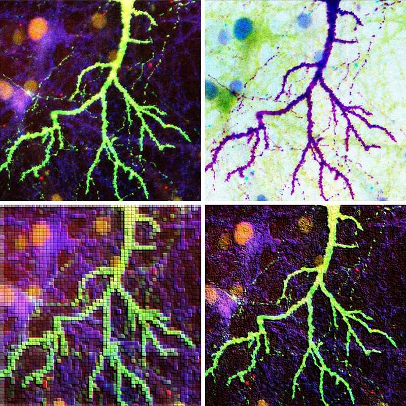
The flash
by Eunkyung Lie
Center for Synaptic Brain Dysfunctions
The four images are the dendrite of cultured pyramidal neuron, obtained from a hippocampus. The brain of human beings consists of about 100 billion nerve cells (neurons). Among them, only one billion pyramidal cells are related with long-term memory. By expressing the fluorescent EGFP proteins in cultured neurons, these images were captured while observing the shape or number of dendritic spines aiding study of the formation of synapses in which synaptic neurotransmission occur. The original (upper left) looks like lightning in the night sky and the other three images express glare, feeling, and afterimage by processing the original image. The four images with different colors and textures combine into one great artwork. The dendrite in the brain also looks like the roots of a plant.
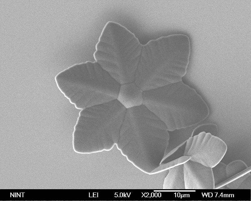
Fullerene flower
by Hee Cheul Choi
Center for Artificial Low Dimensional
Electronic
Systems
Fullerene is the smallest, stable carbon-based molecule. Recently, many
researchers
focus on solution-based self-crystallization of fullerene molecules due to their various
morphologies and crystal structures with different optical and electrical properties.
This
figure shows the first reported fullerene flower, which has unique flower-shaped
crystals
with
six symmetric petals followed by co-crystallization* of both C60 and C70 molecules. This
unique
crystal can be observed due to different solubility of C60 and C70
molecules in mesitylene, which promotes nucleation of C70 with sixfold
symmetry
in
the early stage, and it is followed by co-crystallization of both C60 and
C70 molecules.
* The process to grow crystal which
contains
more than two molecules.
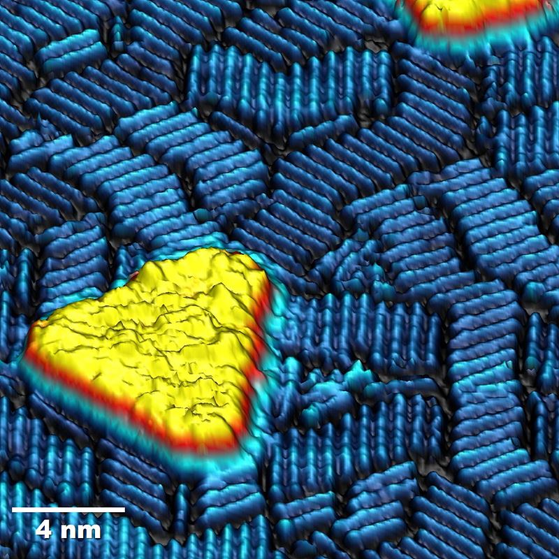
Nanoscale Escher’s stairs
by Jewook Park
Center for Artificial Low Dimensional Electronic
Systems
Scanning tunneling microscopy (STM) reveals self-assembled cobalt (Co) nano-islands and tantalum (Ta) nano-rods on a copper (Cu) (111) substrate. We prepared spin-polarized STM and observed ferromagnetic Co nano-islands (yellow). Ta nano-rods form staircase shapes. The atomic level surface image (3D rendering, false-colored) brings to mind the lithography print “Relativity” (1953) by the Dutch artist M. C. Escher. At the same time, the image also resembles a virtual space where a yellow island is floating in a blue ocean with waves rising and falling.
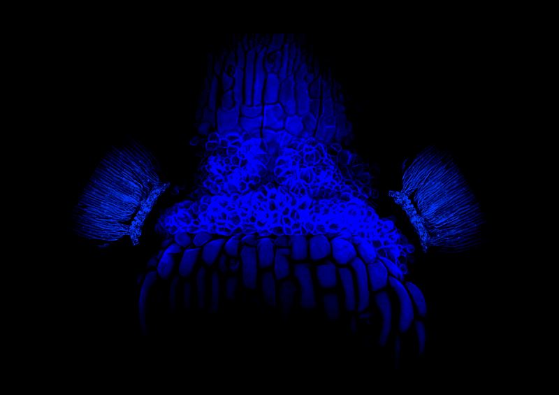
Blue Dokkaebi
by Yuree Lee
Center for Plant Aging Research
The picture shows cell wall of a receptacle after parts of floral organs are shed. An inflorescence of the plant Arabidopsis was stained with calcofluor white, a fluorescent dye that binds strongly to cell walls, and observed with a confocal microscope. Blue fluorescence generated by UV excitation wavelengths looks like the imaginary face of Dokkaebi; traditional Korean goblins. Mysterious blue light gives off a creepy look.
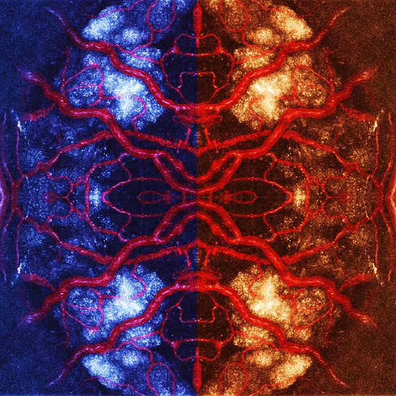
Imagining a brain aging : Color of aging
by Junsoo Park
Center for Plant Aging Research
Brains are developed inside heads, which make them difficult to observe. Due to this characteristic, observation of long-term scale biological events, such as a brain’s aging process, was believed to be nearly impossible. If we can relocate a brain in a location where we can directly observe it, wouldn’t it be possible to observe things that we previously couldn't? We implanted hypothalamic tissue on the iris of an eye. The red color indicates angiogenesis (development of new blood vessels, an indicator of successful implantation) of an implanted tissue . Structure and color were modified to our liking. What would be the shape and color of an aged brain? The image reminds us of an insect or the symmetry image of a mandala.
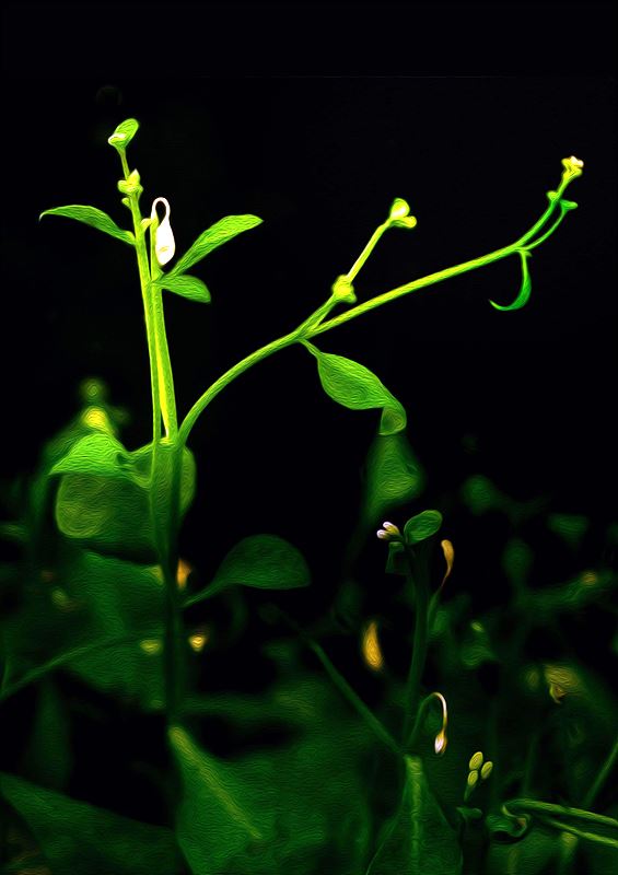
Sorrow of the weak
by Yuree Lee
Center for Plant Aging Research
Calcium is an essential nutrient for development of plants, and required for structural roles in cell walls and membranes, as well as an intracellular messenger in signal transaction. Insufficient calcium uptake degenerates plant development and growth, which is especially severe for flowers and young leaves. The picture shows the death of a young leaf due to the lack of calcium, which looks like a tear drop.
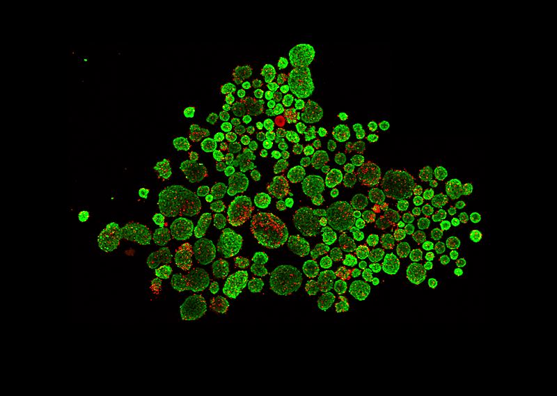
A Seoul map painted with pancreatic islets
by Jung Hoon Cho
Center for Plant Aging Research
Pancreatic islets situated in the pancreas produce several hormones (i.e. insulin, glucagon) playing a central role in retaining glucose homeostasis in our body. To determine the pancreatic islet cell viability after dimethyl sulfoxide (DMSO) treatment, we used live and dead cell markers, calcein AM (green) and EthD-1 (red). Coincidentally, this image looks like a city map of Seoul drawn with pancreatic islets.
The spinning dust
by Olgierd Cybulski
Center for Soft and Living Matter Bartosz
Grzybowski
Group
The four photographs show self-assembled patterns of micro-particles suspended in a heavy liquid at the top of a container which rotates about the vertical axis, with cyclically varying speed. The particles of three different sizes, densities and colors are lighter than the surrounding liquid, so they are pressed towards the transparent lid of the rotating container. The centrifugal force, resulted from rotation, pushes them toward the center, whereas the shear forces resulted from torsional shaking, correspond to the radial alignment of particles. Different patterns occur at different speed of rotation.
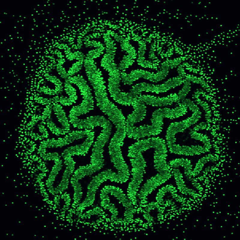
Colloidal brain
by Ruoyu Dong
Center for Soft and Living Matter
A colloidal cluster formed by acoustic trapping, explodes into this wrinkled structure when both an ultrasound and AC electric field are on simultaneously. This is the consequence of a tug of war between the ultrasound confining potential and the AC induced dipole-dipole interaction. It reminds us of the shape of the human cerebral cortex or a swelling fingerprint. They both share a similar formation mechanism; due to the limited space in a two dimensional plane, the structure tends to evolve in the third dimension, creating a convoluted pattern.
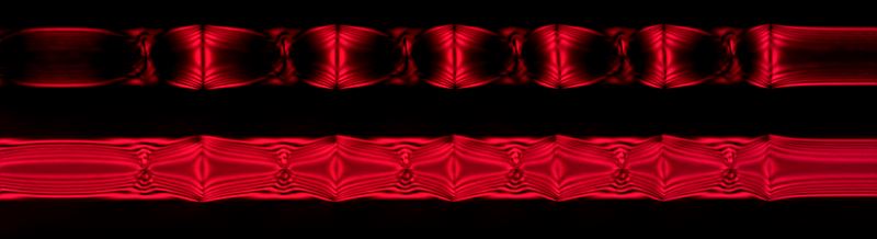
Red bamboo in a capillary : Topological defects in liquid crystals
by Sung-Jo Kim
Center for Soft and Living Matter
Confined liquid crystals struggle to find the most comfortable (lowest energy) structure. They want to minimize their elastic free energy while satisfying boundary conditions. These boundary conditions often present inevitable but beautiful imperfections called topological defects. The photo is a polarized optical microscopy* image of nematic Sunset Yellow (SSY) doped with a minuscule amount of polymer. In this chromonic liquid crystal of very small elastic modulus, chiral domains emerge spontaneously with topological defects between two domains. The shape resembles the structure of bamboo.* Polarized optical microscopy: uses filters to polarize light into waves that vibrate only in one direction
Platinum waterfall
by Yaroslav I. Sobolev
Center for Soft and Living Matter
A dull tip of a platinum wire was used as an electrode for applying high-voltage (3 kV) to a sessile water droplet. The purpose of these experiments was to study how the microscopic surface structure of water changes when it's highly charged. Here you see a magnified photo of the wire tip after one of the experiments. Wire is oriented vertically on the image (you can see the clean part of the wire on the top). Even though we used ultrapure water, some brown organic substance was found on the wire tip (bottom of the image) after 1 day under high voltage - probably due to impurities present in ambient air. This image seems like a piece of contemporary art. The contrast between sleek and rough surface and swelling silver waves maximize an abstract atmosphere.
Sunset over Hades
by Karel Goossens
Center for Multidimensional Carbon
Materials
‘Deep down in the Underworld, where restless souls are wandering in
eternity,
yet
another day comes to an end. The sky turns dark, but the flames never cease to give off
their
relentless heat, melting everything that comes too close.’ This is what came to my mind
when
this beautiful texture of a molten liquid crystal appeared in my polarized light optical
microscope*. The image provided some clues about the nanoscale organization of molecules
in
the
birefringent** sample, due to characteristic interference effects. It marked the
starting
point
for a detailed study by X-ray diffraction, which revealed that these molecules form an
intricate
hexagonal columnar mesophase with a triangular cylinder like morphology.
*
Polarized light microscopy: A useful method to generate contrast in birefringent
specimens
and to determine qualitative and quantitative aspects of crystallographic axes
present
in
various materials.
** Birefringent: Optical property of a material having a
refractive
index that depends on the polarization and propagation direction of light.
Psychedelic liquid crystals
by Karel Goossens
Center for Multidimensional Carbon
Materials
Watch out! Looking for too long through a microscope may cause a massive headache! Yet it is impossible to get bored while looking at the intriguing "fan-like defect" textures of liquid crystals when they are seen through crossed polarizers. The image is an artistic interpretation of the effects of introducing a so-called "full-wave retardation plate" in between the sample and the second polarizer (analyzer). By determining which color appears in which direction, it is possible to obtain information on the orientation of the rod-like aromatic cores of the molecules in a columnar liquid-crystalline mesophase. If the fan-like defects in the southwest-to-northeast direction appear yellow-orange while those in the southeast-to-northwest direction are colored blue-green, then the aromatic segments are most probably oriented parallel to the supramolecular columns. The reverse situation indicates that the aromatic parts are oriented perpendicularly with respect to the long axes of the columns.
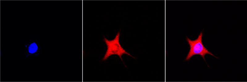
Red star
by Seon Gyeong Lee
Center for Genomic Integrity
Mammalians including human have 3 types of adipose tissue. The brown fat tissue generates heat and increases tissue size in response to a cold condition. Brown adipocyte has many mitochondria. The mitochondria cannot generate energy and die in mass when exposed to stress or cytotoxic conditions. We took a picture of a cell after knocking-down a gene which regulates genomic integrity. We stained mitochondria red and nucleus blue, using Mitotracker and DAPI, respectively. Merging image looks like a destroyed red star.
L'Elisir d'amore
by Jung Me Hwang
Center for Genomic Integrity
The title of this work is a love potion (L'elisir d'amore). The heart represents the dramatic encounter and survival of love on the battlefield. It is an image captured using CUPID technology (protein binding analysis technique) which analyzes the binding of proteins. Protein MSH2 (red) and protein MSH3 (green) in association with protein PKC migrate to the same place on the cell membrane after being exposed to the drug PMA. While observing their migration, this work captured the moment when both proteins reach the membrane, highlighting its heart shape. It is a love potion that comes from drugs. Both proteins play an important role by participating in various mechanisms by intracellular genetic factors, and with this technique, its process can be studied in detail.
Cell flower blooming on the nano-grass
by Chaejeong Heo, Chanho Jeong, Tae-il Kim
Center for
Neuroscience
Imaging Research
While we were developing geometry-controllable nanohairs that mimic cacti
spine-like
stooped hairs and studying cell migration behavior on these hairy structures, we decided
to
color a scanning electron microscope (SEM)* image that resembled a flower bud and grass.
The
decision to make the images into a 2x2 array with vivid colors was a nod to Andy
Warhol's
pop
art format. The work related to this image of neuroblastoma was published in Nanoscale
(Nanoscale, 2017, 13 Sep. DOI: 10.1039/C7NR04522K).
* Scanning
electron
microscope: An advanced microscope that uses a focused beam of electrons to produce
an
image
of a sample's surface.
The root of being, vessels
by Yoo Hyung Kim
Center for Vascular Research
Angiogenesis is an essential element for organ development and maturation. The blood vessels of the brain extend from the outermost surface into the parenchyma like a root of trees spreading out into the soil. At the same time, the blood vessels of the brain form a tighter barrier than the blood vessels of other organs. Overall, these are crucial processes that protect the brain while promoting brain maturation. The figure shows beautifully organized brain vessels of a postnatal 12-day-old mouse. Interestingly, by drawing a tree on the surface of the brain, the figure expresses the feeling that vessel development is essential for life, as root growth is essential for trees. The tree and complex of blood vessels in black and white are reminiscent of a water and ink painting.
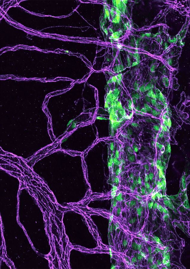
The stream of river flowing in our eyes
by Jaeryung Kim
Center for Vascular Research
This image, which looks like a green pillar surrounded by purple ivy, shows Schlemm's canal and blood vessels in an eye. Eyes maintains their shape and intraocular pressure by continuous production of aqueous humor. Schlemm's canal (green color) encircling peripheral cornea has a critical role for the drainage of aqueous humor into blood vessels (purple color). When normal drainage of aqueous humor is impaired, intraocular pressure is increased and the optic nerve is subsequently damaged, inducing glaucoma and vision loss. Researchers in IBS suggested a potential therapeutic target by demonstrating the new mechanism for development and progress of glaucoma. Although this seemingly river in the eye reminds us of a tear, we can feel hope in conjunction with these research results.
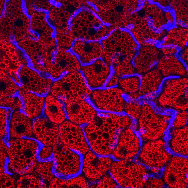
Raspberry fields of blood
by Hosung Bae
Center for Vascular Research
Adipose tissue is highly vascularized and most adipocytes (red) are supported by an extensive vascular network (blue). Brown adipose tissue has an especially large network of blood vessels. Mitochondria-packed brown adipocytes burn energy and produce heat. The image of adipocytes is reminiscent to raspberries, which have been known for burning fat. Blood vessels, sustaining brown fat, can be compared with stems that provide raspberries with necessary nutrients. The strong color contrast of red and blue makes the image look dynamic.
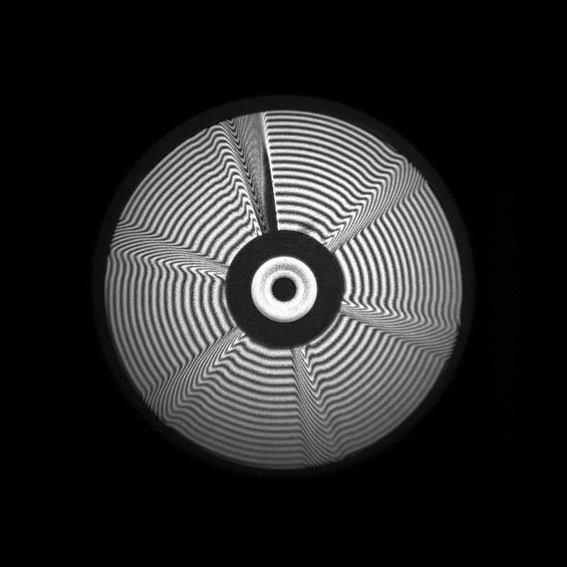
Light donut factory
by Cheonha Jeon
Center for Relativistic Laser Science
We have designed a spherical phase mirror that operates at an angle of 45 degrees to generate a donut shaped beam for light matter interaction. This is an interferometric image of the phase mirror surface. The surface is constructed of six different steps, and as the laser light reflects from the surface, the path difference is λ/3 for each step and 2λ for the combined steps. Due to this path difference, when focused, the beam will have a donut shaped profile. This high intensity donut profile beam will generate a pillar of electrons at the center as it interacts with matter. This will result in proton acceleration that focuses at the center and these accelerated protons can be used for proton therapy for cancer. The bending image with radiating circular waves and the interference pattern is associated with op art.
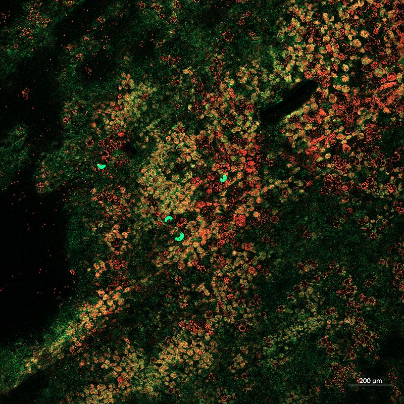
Is it real?
by Beum-Chang Kang
Center for Genome Engineering
When a gene of interest is introduced into a plant cell together with a
fluorescent
protein gene, the presence or absence of the gene can be determined through the
observation
of
fluorescence. The most difficult factor to overcome while observing the fluorescence of
transformed plant tissue with confocal microscopy is autofluorescence from plant cells.
Autofluorescence may look green like a fluorescent protein, but the real color is
different.
The
photograph shows the expression of a fluorescent protein introduced in stomata in plant
tissues
using confocal microscopy*. The image looks like vivid yellow-green stains on an aerial
photo of
mountains covered with autumn leaves, creating a disparate and peculiar
atmosphere.
* Confocal laser microscopy: A type of microscopy that
uses a laser along
with
particular optical components to create high-resolution images of materials stained
with
fluorescent probes.
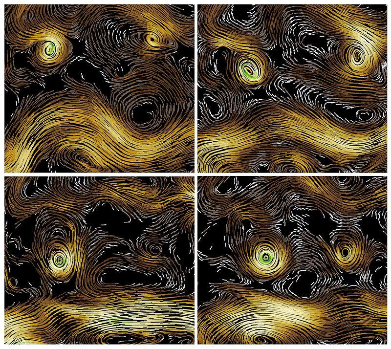
Stormy night
by Seung-Bu Park, Sun-Seon Lee, Jung-Eun Chu
Center for Climate
Physics
Hurricanes (typhoons) are feared for their destructive power, but when simulated on computers they exhibit unprecedented natural beauty. This image shows the time-evolution of a pair of computer-generated typhoons over the Indian Ocean and their interaction with a meandering westerly wind jet. The numerical model used for this animation captures the dynamics of the global coupled atmosphere/ocean/land and ice system with a resolution of about 25 km. ICCP scientists conducted the numerical experiment on a supercomputer in the United States. The title "Stormy Night" was inspired by Vincent van Gogh's masterpiece, The Starry Night.
A message from the past
by Axel Timmermann, Karl Stein, Elke Zeller
Center for Climate
Physics
"Li circuli delli rami degli alberi segati mostrano il numero delli suoi
anni, e
quali furono più umidi o più secchi la maggiore o minore loro
grossezza."
(The
circles of the branches of the sawn trees show the number of their years, and which ones
were
damp or dry the greater or lesser their thickness.)
Leonardo da Vinci, Trattato
della
Pittura
Tree growth only occurs in the outermost section of the tree, in a
layer
under the bark that is only a few cells thick. Outside tropical regions, the rate of
tree
growth
changes throughout the year, creating distinct annual rings. Leonardo da Vinci first
related
the
thickness of the annual rings to the amount of rainfall received during the year. ICCP
scientists use tree ring data to gain information about past climates and past changes
in
ecosystems. The image shows 4 sections of a tree core sampled by Axel Timmermann from a
high
elevation site in Hawaii. In addition to using the visual images, scientists also
analyze
stable
carbon isotopes within the tree rings, which give them an insight into subseasonal
climate
variations and changes in photosynthesis.
2016 IBS Art in Science
The Beauty of Evolution:the Structure of the Drosha Protein
by Sung Chul Kwon
Center for RNA Research)
A type of regulatory RNA called microRNA involved in almost all biological processes such as development, aging, cancer, differentiation and immune response, is generated by two consecutive processing steps involving Drosha and Dicer proteins. This figure shows the bottom part of a Drosha protein whose structure has been solved 12 years after its first identification. Drosha has the same backbone structure as Dicer (red and green), plus some unique regions (yellow and white). Thanks to this information, we can infer how Drosha diverged from the ancestral Dicer and obtained its new function.
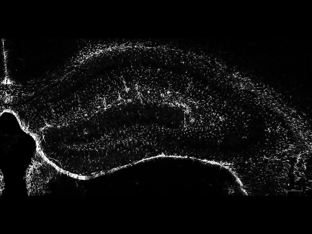
Milky Way in the Brain
by Haram Park
Center for Synaptic Brain Dysfunctions
The hippocampus is the "sea horse" shaped structure located bilaterally within mammalian brain. It is part of the limbic system, and is thought to contribute to information consolidation, spatial memory, and both short and long term memory. In the image, we stained star-shaped astrocytes, known as astroglia in the hippocampal region of the brain with GFAP, glial fibrillary acidic protein. The stained cells are represented in white and they look like white stars set within a contrastingly dark background, akin to the milky way observed in the night sky.
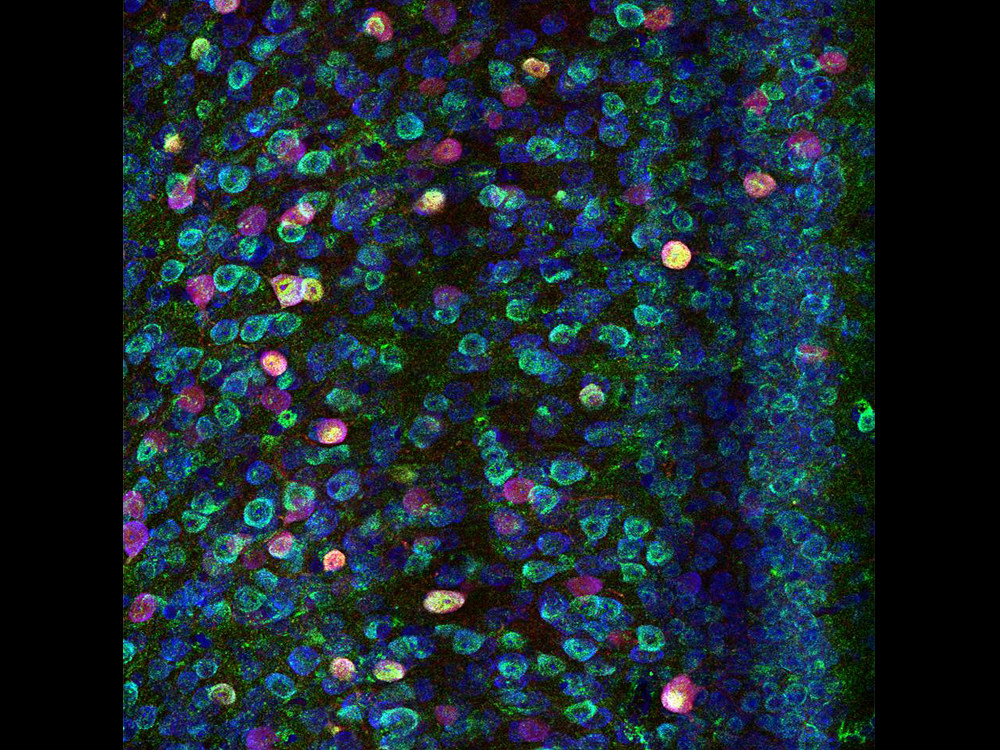
The Pond
by Junyeop Roh
Center for Synaptic Brain Dysfunctions
Claude Monet has been immortalized by his renowned painting of the pond dotted by colorful water lilies. When calcium-binding proteins (Parvalbumin, in red), synaptic proteins (PTP delta, in green) and DNA (in blue) are stained in the layer 2/3 of mouse medial prefrontal cortex region, the resultant image is remarkably reminiscent of the masterpiece. The medial prefrontal cortex has been extensively studied as the region mediating social behavior, decision making and, in humans, personality expression.
Chamber for the Mystery Matter
by Chang Hyon Ha
Center for Underground Physics
The photo shows the innermost view of the dark matter experiment, called COSINE, situated 700 meters below the surface at Yangyang underground laboratory. The cylindrical cells in the middle center encase ultra-pure NaI(Tl) crystals which are used to collect a faint light induced by the interaction of dark matter and the crystal nuclei. In order to distinguish the signal from external noise entering outside of the crystals, we position the crystals in a large chamber where all surfaces are laminated by reflective films that work like a mirror. Then, photons from the noise or any other light in the chamber can easily bounce back and forth and be contained, creating the exotic image.
World of Diversity
by Young-Im Kim,Jihoon Choi
Center for Axion and Precision
Physics
This work of art shows that various resonant modes are generated in a variety of sizes resonator visually. A resonator is a device that records a specific frequency and vibration using a resonance phenomenon that is amplified when a specific frequency is found. Various studies using such resonance phenomenon is being done. Resonance is a phenomenon that can be found everywhere in our world. In one of the resonator to various resonance modes exist similar to our world people have a variety of looks and emotions.
Drowning Lung
by Seung-Jun Lee, Gou Young Koh
Center for Vascular
Research
The image is a visualization of vascular leakage in the lungs of a mouse model of sepsis. An infection or injury can lead to sepsis where severe inflammatory response and vascular leakage occur throughout the body. These septic challenges lead to multiple organ failure, including accumulation of fluid in the lungs. We have demonstrated the beneficiary role of the endothelial tyrosine kinase receptor Tie2 in ameliorating vascular leakage and sepsis.
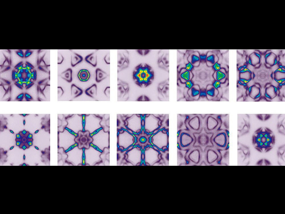
Material World through Kaleidoscope
by Beom Seo Kim
Center for Correlated Electron Systems
Molybdenum di-telluride (MoTe2) has drawn growing attention due to its application possibility. It has six-fold symmetry in the electronic structure due to its crystal structure. The electronic structure of MoTe2 was measured by angle resolved photoemission and images were constructed by folding constant energy contour of the experimental data. Each image represents constant energy contour at a specific binding energy. While the image drastically changes over different binding energies, the six-fold symmetry remains intact. The six-fold symmetry is reminiscent of the colors of a peacock or a view through a kaleidoscope.
The Scream of Electrons
by Shoresh Soltani
Center for Correlated Electron Systems
Portraits of electron pockets living in a peculiar quantum world on the surface of strontium titanate. These ghostly beings which resemble The Scream by Edvard Munch, reveal themselves when we shed synchrotron light on a flat, shiny and clean surface of an single crystal in very low temperature and ultrahigh vacuum. They are responsible for electrical conductance on the surface of an insulating strontium titanate crystal which is the basis of oxide electronics. Different colors in each image, speak for different electron densities.
The Nose of Plant
by Taek Han Yoon
Center for Plant Aging Research
Plant's respiratory organs are called 'stomata'. They are responsible for photosynthesis and therefore are essential to the life of most of the species on Earth. Via Field Emission Scanning Electron Microscopy(FE SEM)* imaging technique, we were able to reveal that stomata are shaped like a heart....maybe a hint to Mother Earth’s love?
2015 IBS Art in Science
Autumn Graphene Leaves on Copper Snow
by Youngwoo Kwon, Rodney S. Ruoff
Center for Multidimensional
Carbon
Materials
Copper foil is one the most common substrates for graphene growth. Because of the unique crystalline structure of graphene ('2D honeycomb' structure), growth of graphene happens in certain patterns such as hexagons or, as in this case, hexagrams (also referred to as "sexagrams"). This Image by Scanning Electron Microscope (SEM)* shows the pattern of graphene growth at the micron scale - quite on time to greet the fall season with beautiful autumn/winter leaves on a copper landscape on a campus such as UNIST!
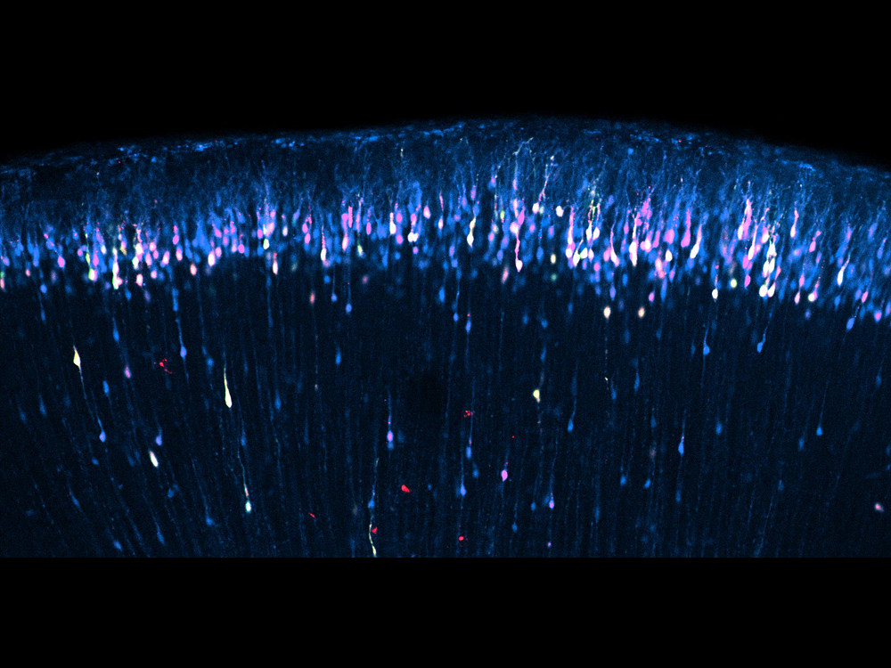
Star Shower in the Brain
by Sangkyu Lee, Hyeonjin Jung
Center for Cognition and
Sociality
Brain's outer layer near the skull consists of cerebral cortex neurons. Fluorescent proteins of three different colors (green, red, far-red) were expressed in the neurons under developmental stage by injecting expression plasmids into ventricle regions of uterus (embryonic day 15) followed by electroporation with stick electrodes. This fluorescence image was taken after cross-section of brain sample.
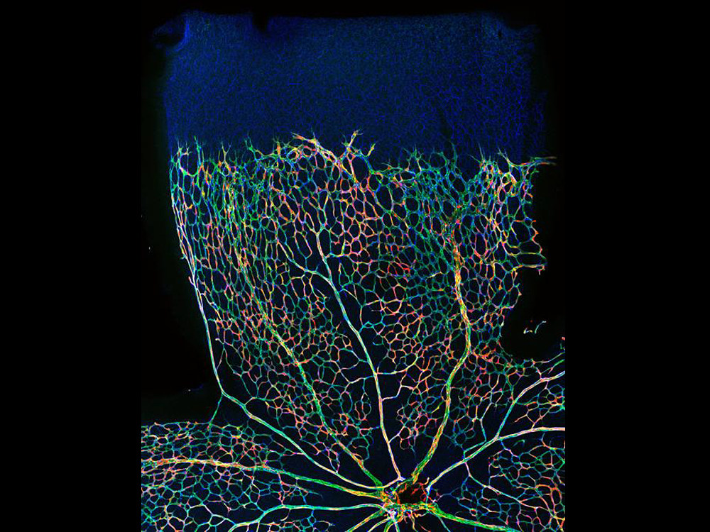
Rainbow in the Eye
by Do Young Park
Center for Vascular Research
Retinal vessels grow from center to periphery after birth. Retinal vessels are composed of vascular endothelial cells and pericytes. Pericytes cover endothelial cells and are important for angiogenesis, survival of endothelial cells, and barrier function. In this image, retinal blood vessels (in green) of mouse retina at postnatal day 5 show sprouting peripherally. Pericytes (red) are shown in mosaic pattern from Cre-induced loxP recombination to make tdtomato signals in pericytes.
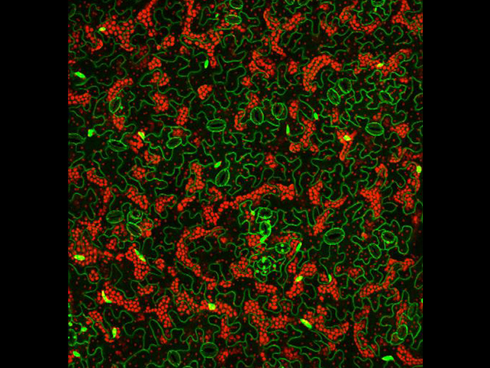
Nature Puzzle
by Sunghyun Hong, Sowook Seo, Soohyun Jung
Center for Plant Aging
Research
Fluorescences in Cotyledon epidermis of 2 weeks grown Arabidopsis expressing H2B-GFP (Nucleus localized) and SYP122-GFP (Plasma membrane) were detected by LSM780 fluorescent confocal microscopy. Bright green dots are nuclei of epidermal cells and green lines are membranes. Small paired cells containing smaller green spots are guard cells which control gas exchange by opening and closing the stomata. Red fluorescent spots are chloroplasts which absorb sunlight to produce food and contain various fluorescent compounds such as anthocyanin causing autofluorescence. Shapes of epidermal cells are irregular like pieces of jigsaw puzzle.