2018 IBS Art in Science
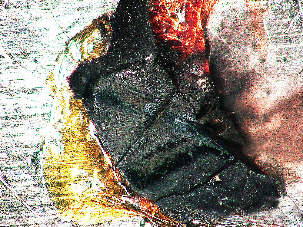
A Piece of Femto-Galaxy
by Min-Cheol Lee
Center for Correlated Electron Systems
A single crystalline Ca2RuO4, destroyed by an ultrafast laser beam, is modeled as a workpiece. The broken crystal shows a clear line along the path of the femtosecond laser pulse incident on the sample. Unlike conventional techniques, ultrafast spectroscopy can instantaneously introduce a large amount of energy by using femtosecond light. Particularly in the case of Ca2RuO4 with a strong correlation between electronic/lattice structures, huge structural changes can arise, such that the sample is destroyed not by physical contact but merely by the absorption of ultrafast light. The millimeter-sized black sample with red glue reminds me of a vast galaxy in the night sky.
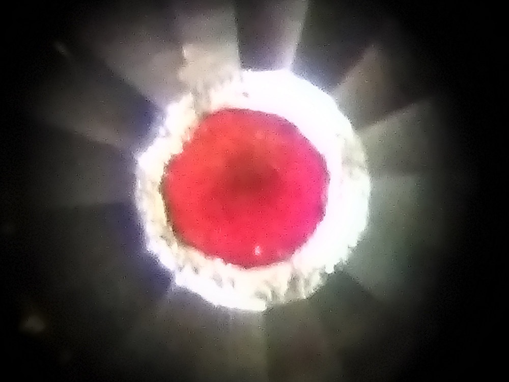
Diamonds and Wine
by Dr. Matthew Coak
Center for Correlated Electron Systems
Two-dimensional magnet MnPS3 is naturally a beautiful translucent lime green colour. As this photo, taken on the Diamond Light Source x-ray synchrotron in the UK, shows, squeezing this material between two diamonds changes it to a gorgeous deep red. With the light shining through it from behind, it looks like a droplet of the finest claret (Bordeaux). Surrounding the crystal in the centre, you can see the perfect facets of one of the two flawless natural diamonds. The crystal experienced 100,000 times atmospheric pressure - 100 times the pressure at the bottom of the Mariana Trench and enough to turn oxygen solid! The drop in energy changed the green to a red colour as MnPS3 was driven from an insulator to a metal, which was found to occur at 300,000 atmospheres.
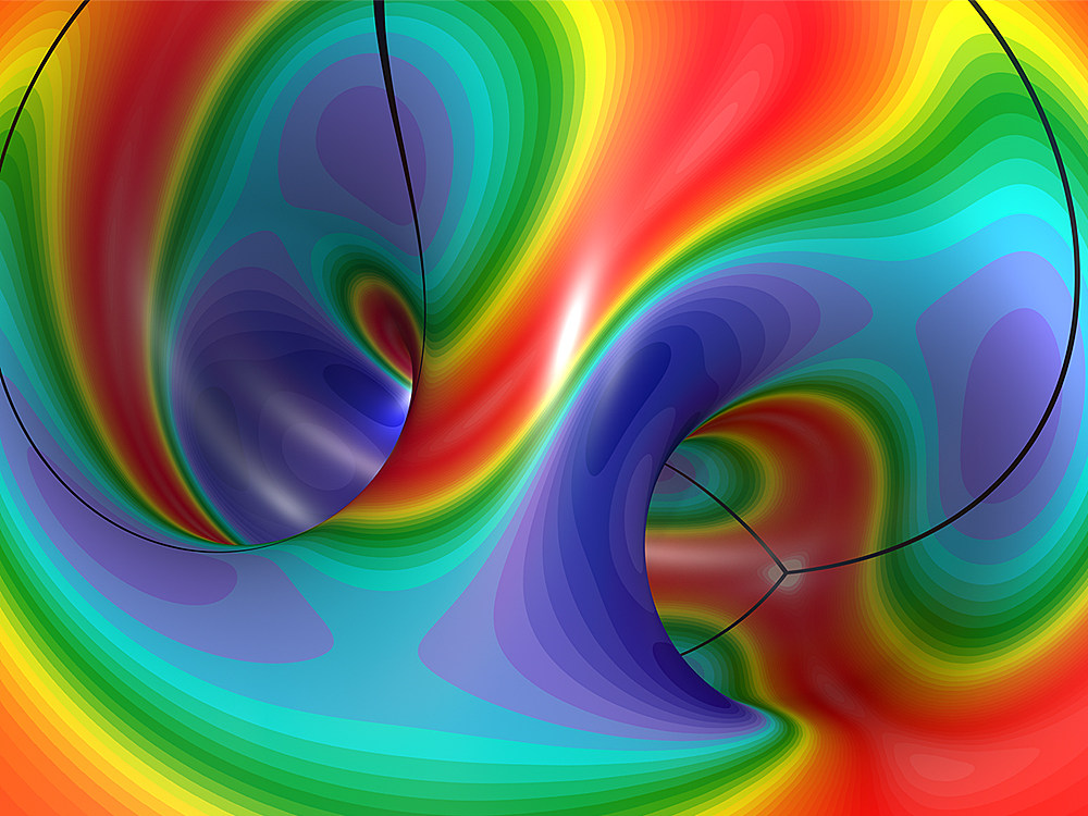
Oil Painting Inside Donut
by Ki Hoon Lee
Center for Correlated Electron Systems
This figure demonstrates Berry curvature of the spin wave of a triangular lattice in nearly a 120 degree alignment. The spin wave forms a band with a dispersion in the crystal momentum space as the elementary excitation of magnetic materials. Berry’s curvature is considered an important quantity in modern condensed matter physics because it contains geometric and further topological properties of the various excitation states that form bands, including spin waves. In this figure, we placed a crystal momentum space on the surface of a torus or donut shape and tried to give a unique perspective by showing the Berry curvature from the inside. The color represents the size of curvature, and the point where the three black lines meet is the K point, which is the edge of the crystal momentum space.
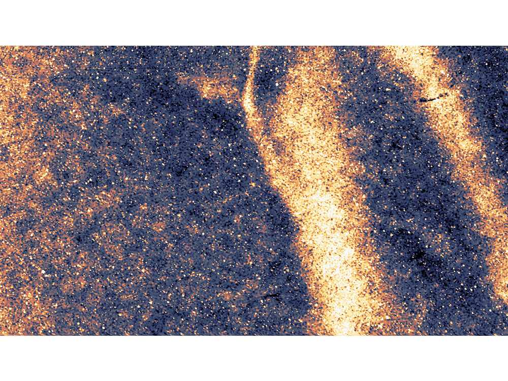
Milkyway in CBST
by Jae-Joon Kim
Center for Correlated Electron Systems
Crx(Bi0.1Sb0.9)2Te3 (CBST) is the topological insulator that chromium is doped on (Bi0.1Sb0.9)2Te3. Due to local magnetic chromium dopants, a sample will change to global ferromagnetism. This topographic image was taken to find the magnetic domain boundary on a CBST sample with a scanning tunneling microscope (STM)*. It is only 384 nm x 216 nm in size but resembles the Milky Way.
Is the catalyst alive?
by Jeongjin Kim
Center for Nanomaterials and Chemical Reactions
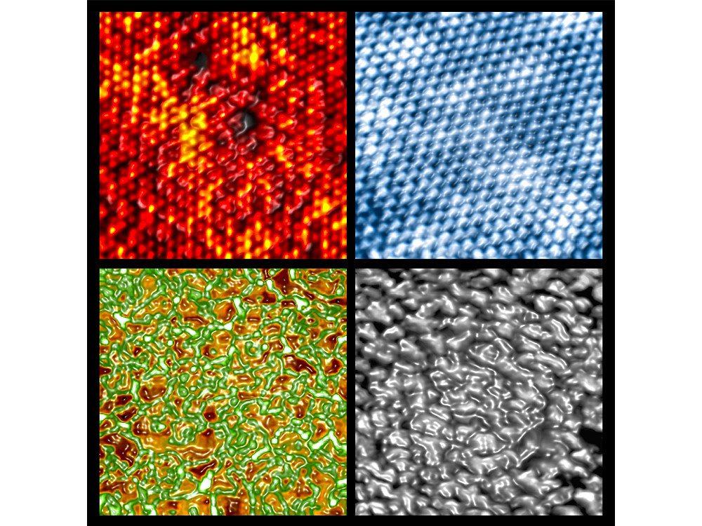
The molecules that cause chemical reactions breathe new life on the atomic surface of catalyst materials. Much like how minuscule movement can trigger a huge typhoon at sea, an atomic-size wave packet, which is smallest measurement unit of material, can instigate tremendous effects where molecules meet atoms on a catalyst surface. Repeated collisions at the molecular level on a platinum-nickel catalyst surface with atomically-layered adsorptions of carbon monoxide and oxygen molecules result in the reconstruction of atomic layers and a new catalytic reaction.
Mankind in the Nanoworld
by Hyosun Lee
Center for Nanomaterials and Chemical Reactions
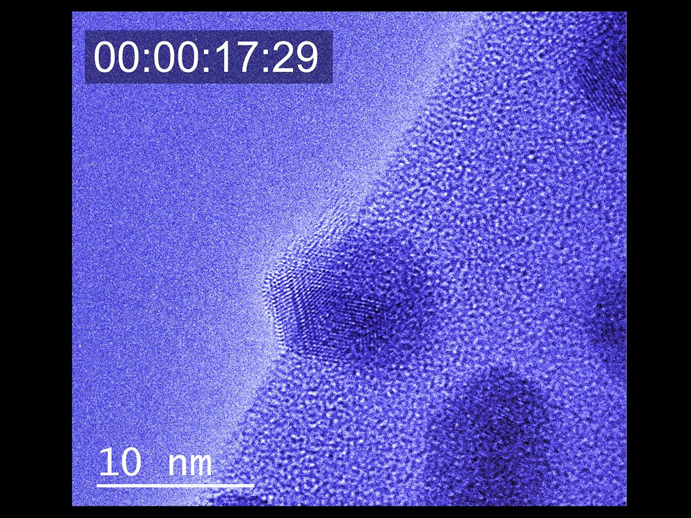
The video shows the sequence of in situ transmission electron microscopy (TEM) images of platinum-cobalt (Pt3Co) bimetallic nanoparticles, where a cobalt oxide (CoO) layer is formed on the surface of the nanoparticles. The atoms in the nanoparticles, arranged in a regular array, seem to keep their position, but the atoms change slowly as the environment changes. Here, Pt3Co bimetallic nanoparticles are not observed in a high-vacuum environment, but in an oxygen gas (O2) environment measured in real time while applying heat. In this research, we have found that the cobalt atoms move to the surface of the nanoparticles atom by atom and form a single CoO layer on the top surface during the catalytic reaction, resulting in the improvement of the catalytic activity. While researching these small nanoparticles, we are reminded that mankind gradually tries to find a way to adapt to surrounding changes and moves toward a better direction.
View video on YouTube
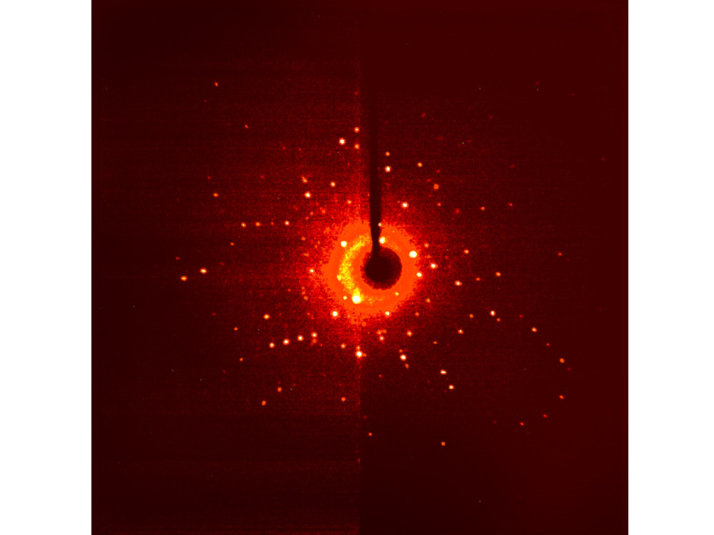
The Night I Count the Stars
by Dongwook Kim
Center for Catalytic Hydrocarbon Functionalizations
This diffraction pattern image came from a single crystal x-ray diffraction experiment. The harmony of aesthetic symmetry is an example of the beauty in nature. The diffraction with x-ray radiation to single crystal results in a well-arranged and symmetric pattern. The spreading spots are generated in a harmonious crystallographic condition. We get a sense of beauty looking at the symmetrical arrangement of dispersed diffraction spots give us a sense of beauty resembling stars in the universe.
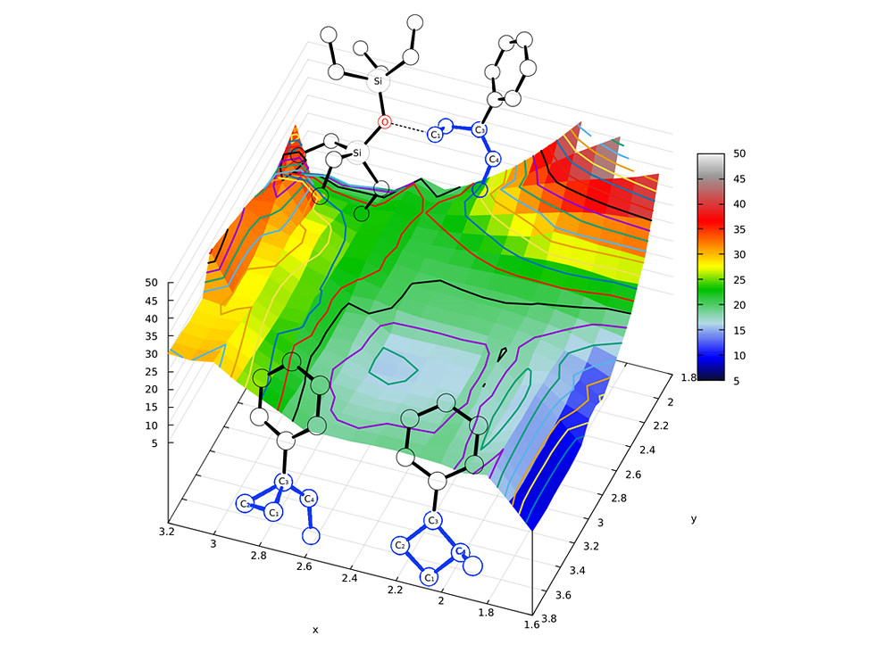
Molecular Golf: Birdie or Bogey?
by Jiyong Park
Center for Catalytic Hydrocarbon Functionalizations
Potential energy surface (PES) illustrates the energetics of a molecular system, on which the route and the outcome of a chemical reaction can be described. When color-coded, the surface resembles a golf-course with multiple hills. The lowest saddle point between the hills is the transition state (TS), which defines the minimum energy to access the product. Chemists can figuratively play golf to find the best route to reach the hole (product) from the teeing ground (starting material). Occasionally, the path passing the TS forks to lead two products, known as post-transition state bifurcation. When successful, much like a birdie, we obtain cyclopropane as a product; otherwise, we acquire cyclobutane, which is like getting a bogey.
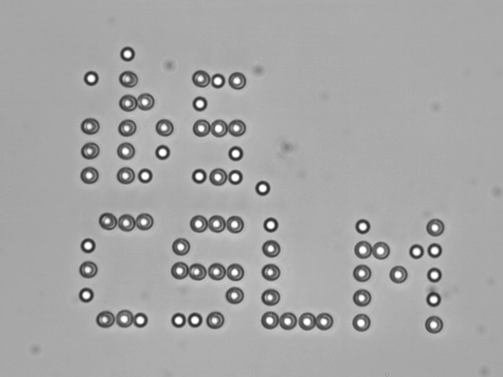
CSLM in the Microworld
by Kibom Nam
Center for Soft and Living Matter
Optical tweezers are scientific instruments that can trap micro-size objects with a highly focused laser. Holographic optical tweezers can generate multiple optical traps with a single laser source. It can generate any arbitrary position of optical traps. This work shows optically trapped 2-micrometer polystyrene particles in deionized water spelling out "ibs. CSLM" an acronym for the Center for Soft and Living Matter.
View video on YouTube
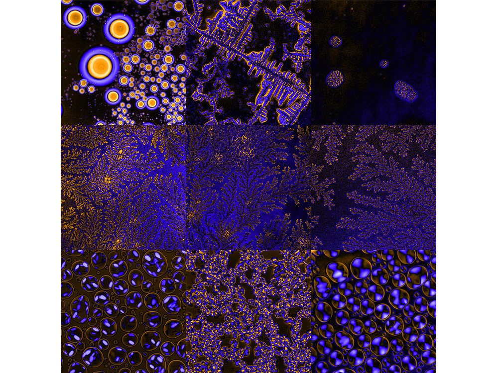
The Phases of DNA
by Anisha Shakya
Center for Soft and Living Matter
Due to its polyelectrolyte nature, DNA can form different phases upon interaction with multivalent cationic polymers. Despite their importance, the prediction and control over these phases remain elusive. This collage shows images of DNA condensed into liquid, multicompartment liquid-like, liquid-crystalline, and semi-solid phases. The variety of visually striking features seen under the microscope takes a scientist’s imagination on delightful artistic detours.
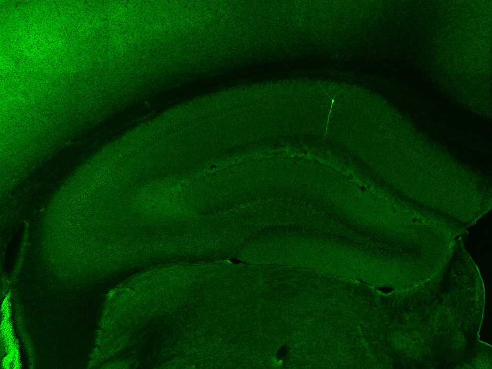
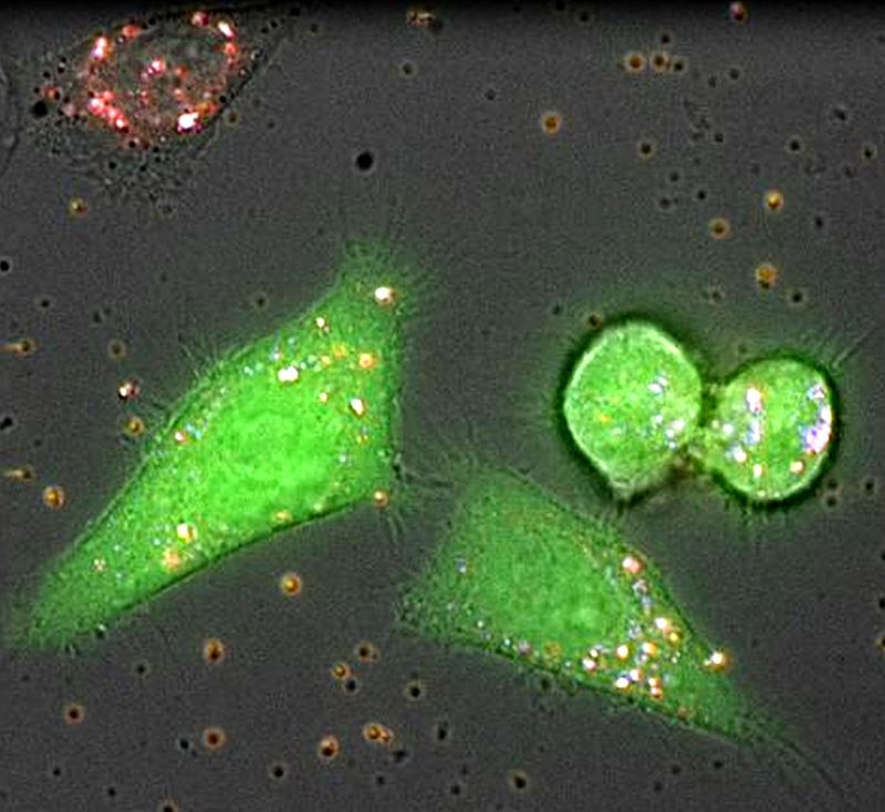
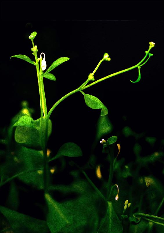
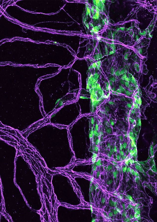
A Tree in the Forest
by Kyu-Hee Lee
Center for Cognition and Sociality
Many neurons come together to form the brain, just like how trees constitute a forest. By seeing only one tree you cannot know what a forest looks like, and even if you can observe the forest from the sky, you cannot know the types nor the individual roles of each tree. Investigating brain function is similar. This image is a cross-section of hippocampus from a mouse. Among the many neurons in a living mouse, one single CA1 pyramidal neuron was detected. I investigated its activity resulting from interactions with other neurons by using patch-clamp techniques. This technique is used to measure the potential of cell membrane via glass micropipette. To find the patched single neuron and to check its shape, I stained the brain slice with fluorescent dye. Finally, I found the single neuron that was standing alone but shining with green fluorescence. You can find the one ‘neuronal tree’ in the middle of the forest composed of many neurons.
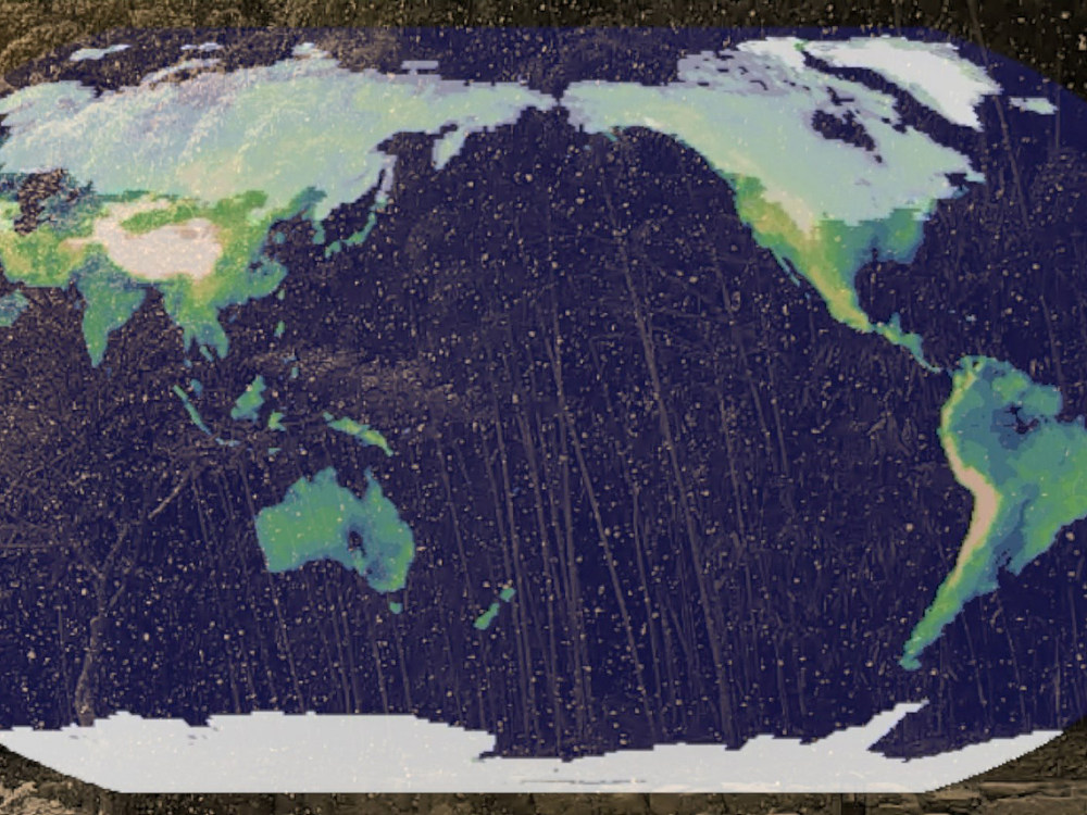
The Sound of Snow
by Axel Timmermann, Jung-Eun Chu, Karl Stein, Kyung-ja Ha
Center for Climate Physics
Snow is a crystalline form of water. It insolates soils and plants from harsh winter conditions, it enhances earth’s reflectivity, and it can accumulate in cold areas to form ice sheets, such as Greenland and Antarctica. In our warming world, the number of snow days will decrease considerably and climate computer model simulations show that towards the end of this century snowfall events in the midlatitudes, such as the one in Busan on January 10th, 2018, will become rare. When we think about snow, we often think about our childhood—the experience of building a snowman, the thrill of a snowball fight, or just the beauty of watching snowflakes. This video clip, which is accompanied by a piano improvisation, tries to remind us of the beauty of snow as well as its fate in response to fossil fuel emissions.
View video on YouTube
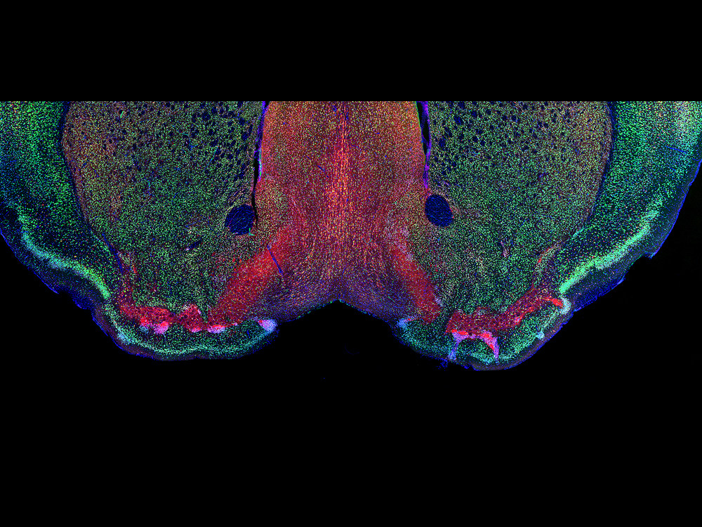
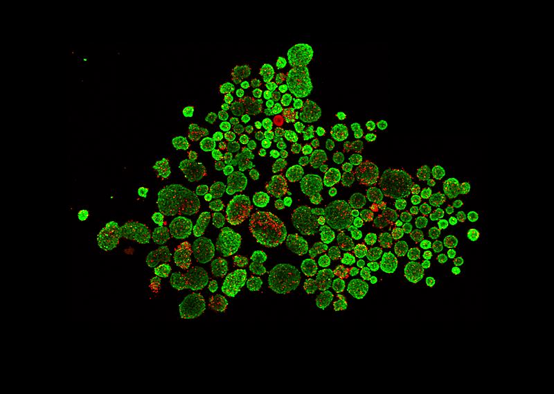
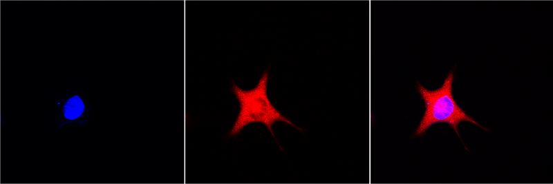
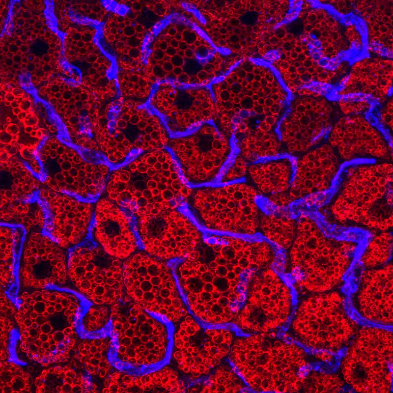
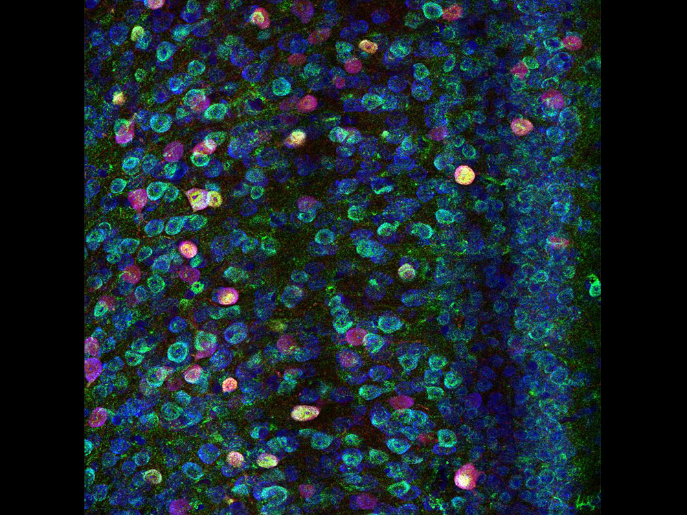
The Eyes of Morpheus
by Haram Park
Center for Synaptic Brain Dysfunctions
The Greeks of antiquity believed Morpheus was the god of dreams and would appear during sleep to shape and form our dreams. We spend one third of our lives asleep, and yet sleep is still a field relatively unknown to science. Why we sleep and how we dream are questions that are yet to be answered. What is known is that the lateral preoptic area (LPO), the region of the brain depicted in the image with red coloring, is critical for sleep to normally progress in the brain. Impairments of neurons in this area causes many disruptions in sleep, such as insomnia. Understanding the LPO will be the gateway to understanding the realm of Morpheus. (Green: NeuN, neuronal marker, Red: GAD, inhibitory neuron marker, Blue: DAPI, Nucleus).
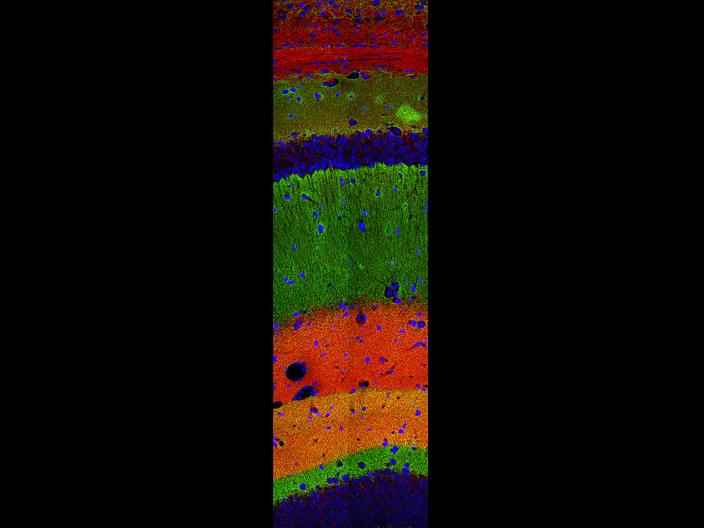
Layers Upon Layers
by Yeonsoo Choi
Center for Synaptic Brain Dysfunctions
A single neuron can receive thousands of signal inputs from other neurons and process them for downstream signalling. This image shows the layers of the hippocampus, the memory storage area of the brain. The colorful layers in the image do not depict different neurons but the different inputs a single neuron in the hippocampus can receive. Even now, as you read this, your brain is processing the layers upon layers of information from countless different regions and subregions to derive cognition from noise. (Green: excitatory neuron marker, Red: synaptic protein marker, Blue: nucleus).
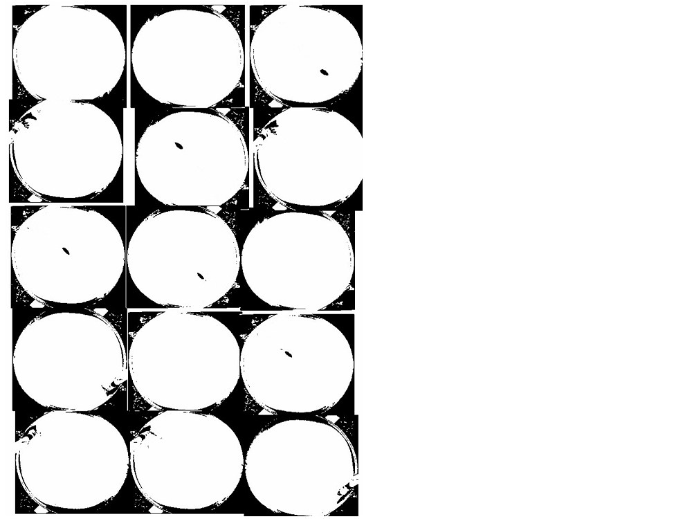
Double Disaster
by Eunkyung Lie
Center for Synaptic Brain Dysfunctions
I wanted to express the unexpected happening that occurred during an occurrence of the Morris water maze experiment, an animal behavior experiment. The experiment has a mouse which swims in water and visits a hidden escapable island, which is conducted to study spatial perception and memory. Through several trials, the mouse finds the island between 20 seconds and 1 minute. The mouse in the video is too smart to go directly to the island and eventually escapes the 100 centimeter water tank. It is rare for ordinary mice to find the island, and even if they do, they wait there as they do not always know where to go. I have expressed the characteristics of the mice with the pop art of Andy Warhol’s Silver Car Crash (Double Disaster) and the video art of Nam June Paik’s Egg Grows. An unexpected accident that occurs during the experiment and widely varying results are a double disaster to researchers.
View video on YouTube
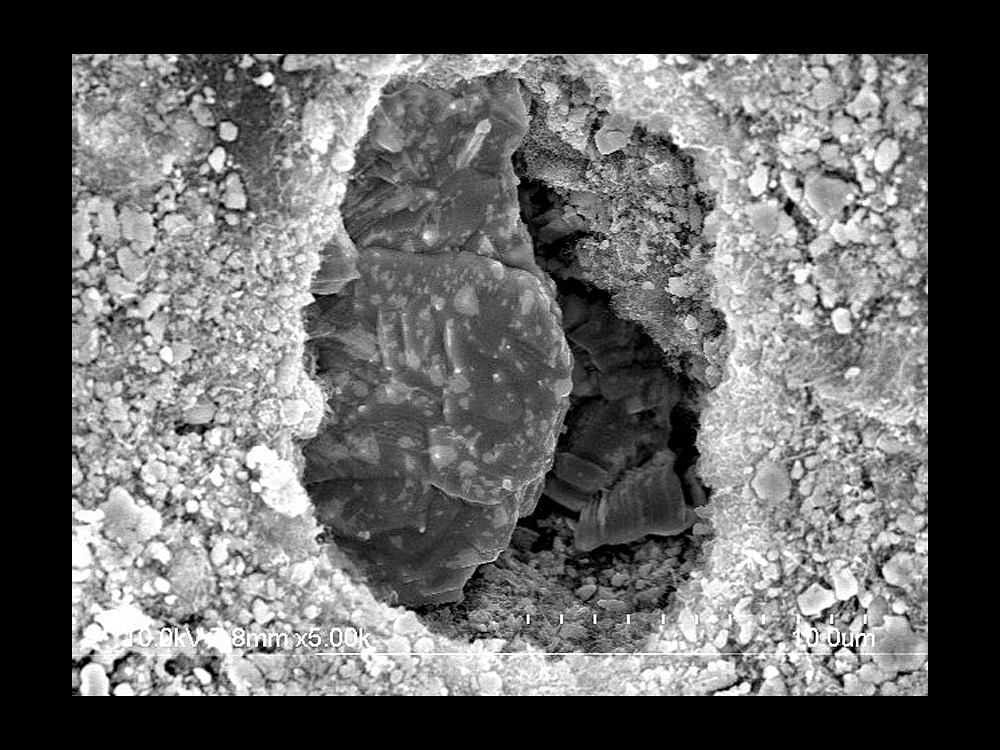
A Secret Door
by Mijoung Joung
RISP
The particles in the picture are lanthanum carbide (LaC2) and are used to generate rare isotopes. Isotope separation on-line (ISOL) is a method to generate rare isotopes that do not naturally occur. A variety of isotope targets are used in ISOL facilities around the world. The LaC2 target is a thin disk under development by the Rare Isotope Science Project (RISP). To achieve high yield of the rare isotope, the particles of LaC2 should be evenly spread over the whole area of the target disk to prevent local temperatures rise when the particles collide with the proton beam. Temperature rise in small area can cause thermal damage to the target disk. However, crystal structures made of agglomerate particles were observed in some parts of the target disk after fabrication. A hole was formed in the mass of particles, which brought to mind the secret cave that Ali Baba enters.
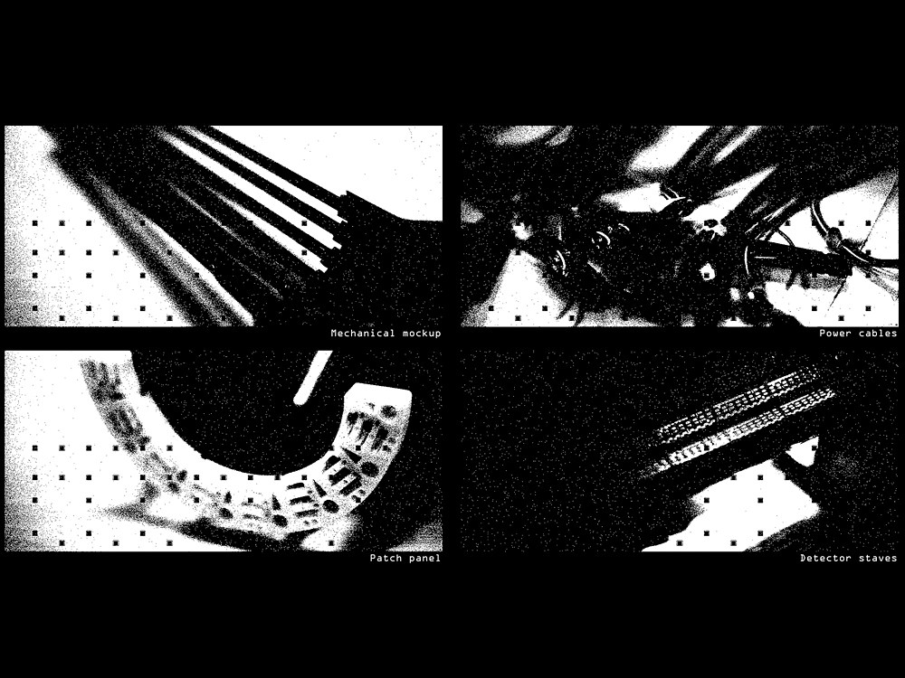
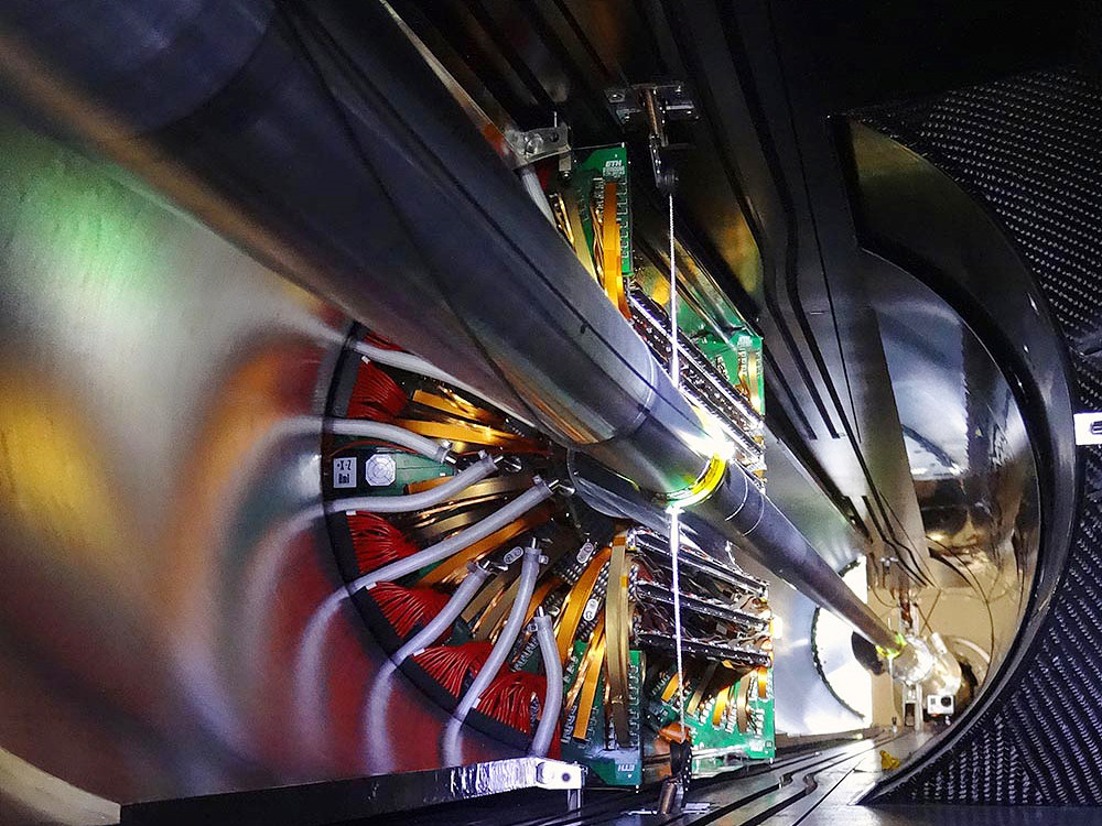
Self-portrait - the ALICE’s innermost particle detector looks at itself
by Magnus Mager
applied physicist at CERN
The pictured detector elements measure about 20cm by 40cm (diameter x length) and will form the innermost part of a cylindrical particle detector that will soon (2020) be installed in ALICE, an experiment at the Large Hadron Collider (LHC) located at the European Organization for Nuclear Research (CERN), in Geneva, Switzerland.
ALICE (A Large Ion Collider Experiment) studies the origin of the Universe, when matter was like a soup of quarks and gluons. ALICE’s innermost part will be tiled by about 25,000 CMOS particle sensors, each of 1.5cm x 3cm in size. In everyday life, CMOS sensors are found in consumer products, like webcams, but here they are used to take 12.5 Giga-Pixel images of particles coming from high-energy collisions.
Beyond detecting charged particles, these sensors are also sensitive to sudden changes of light levels. This feature was utilised here in an old analogue SLR camera, where the 35mm film was replaced with the CMOS particle sensor, effectively turning it into a digital camera.
The specific "aura" of the resulting photographs is caused by the fully binary operation of the sensor: pixels are either fully black or fully white. Moreover, some noise of white and black speckles is created by operating the camera at very low thresholds. Finally, a black square pattern is a result of opaque metal structures on top of the sensor that is meant for the electrical interconnection of the sensor to the detector circuit boards.
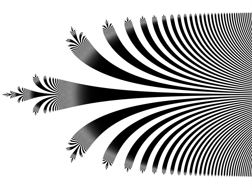
Cantor Bouquet
by CNRS (Centre National de la Recherche Scientifique)
Julia set of an exponential, an object of study in holomorphic dynamical systems. Its shape has been found to be the same as a model from continuum theory in topology: the Lelek fan--also nicknamed the Cantor bouquet--which is an infinite union of lines whose tips are densely distributed near every point on these lines.
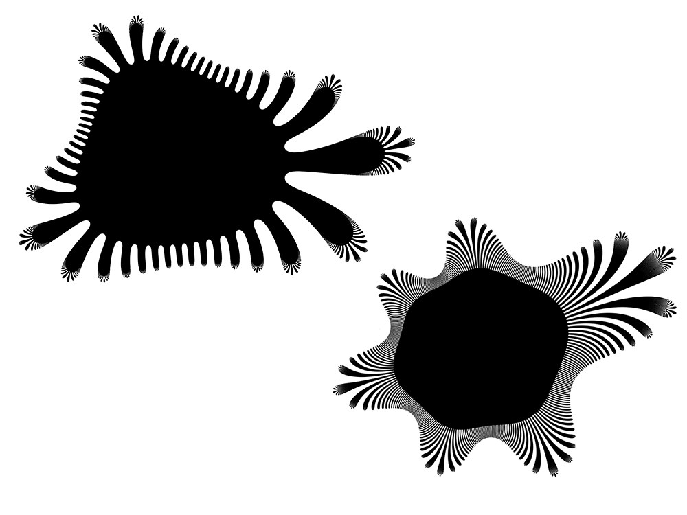
Model of a critical orbit
by CNRS (Centre National de la Recherche Scientifique)
Pseudo-hedgehog. Model of the critical orbit of some holomorphic dynamical system. The black zone is made of myriads of strands radiating from the center, too densely packed to be resolved at this scale.
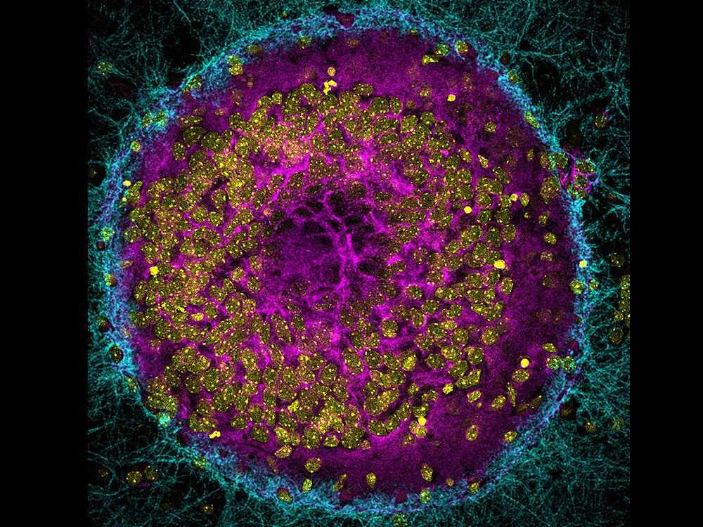
Image 1
by CNRS (Centre National de la Recherche Scientifique)
Hippocampal mouse neurons observed by confocal fluorescence microscopy and reconstructed in 2D by projecting the various optical sections onto the Z plane. The CAR protein was detected using a single antibody.
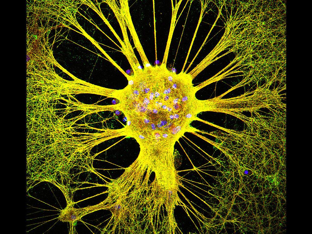
Image 2
by CNRS (Centre National de la Recherche Scientifique)
Hippocampal mouse neurons observed by confocal fluorescence microscopy and reconstructed in 2D by projecting 34 optical sections onto the Z plane. Using immunofluorescent staining, cell nuclei were stained blue with DAPI.
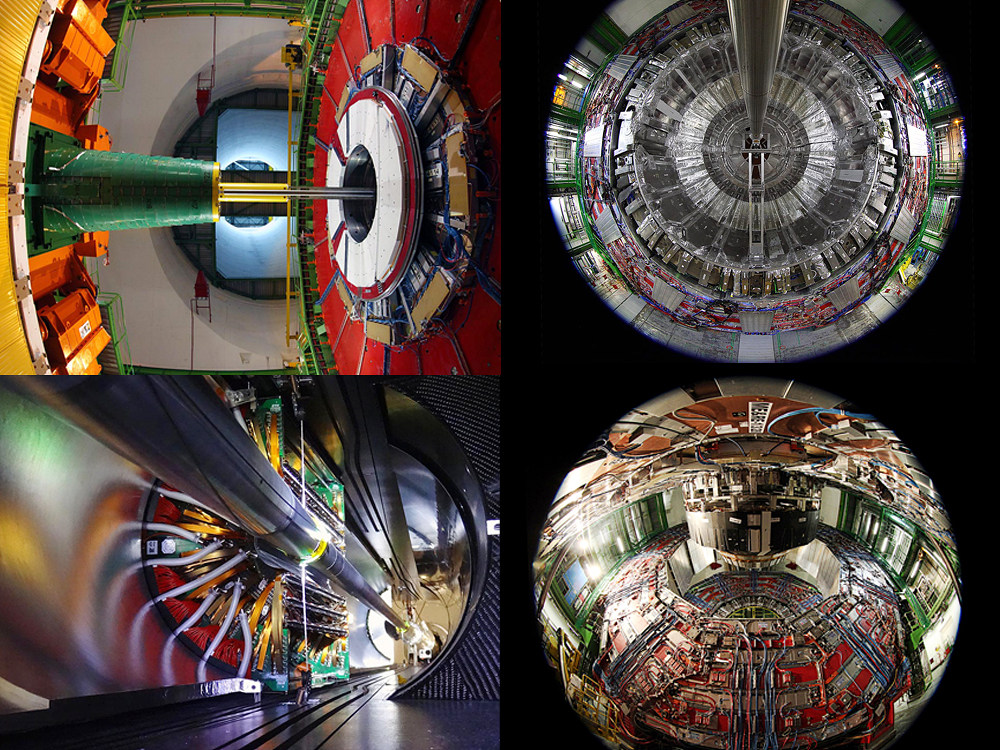
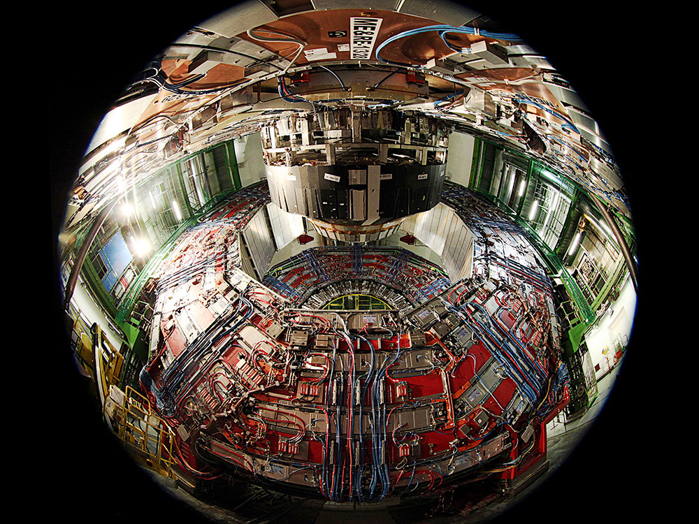
CMS –The Art of Science Architecture
by Michael Hoch
CERN
Those visitors fortunate enough to visit the CMS (Compact Muon Solenoid) experiment at CERN open in all its glory have often commented it could be displayed as a work of art. CMS is inside the 27-km Large Hadron Collider (LHC) the largest and most powerful particle accelerator ever built. It was designed to explore the secrets of our universe, especially to find evidence for the Brout-Engler-Higgs mass generation mechanism and was rewarded by a Nobel Prize for the discovery. This detector has a shape of a cylinder is including services 20m in diameter and 25m long. It acts as a giant high-speed camera, taking 3D "photographs" of particle collisions from all directions up to 40 million times each second with a spatial resolution in the micrometer scale.
As scientist I am fascinated by the massive scale and the high-tech instruments at CERN which allow mankind to decode nature in an incredibly high precision. As an artist I am charmed by the intrinsic geometry as well colors and scale of the science architecture of these experiments that have the size of mid-size houses. The aesthetics of CMS are associated with its functionality as gigantic high precision camera. Periodic multiplicities emerge out of the physical measurements constrains and the strict scientific precision which allow us to unravel to secretes of nature.
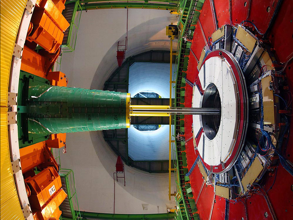
The colors of CMS
by Michael Hoch
CERN
The colors of CMS - view from the bottom of experimental cavern 100m below ground towards the surface.
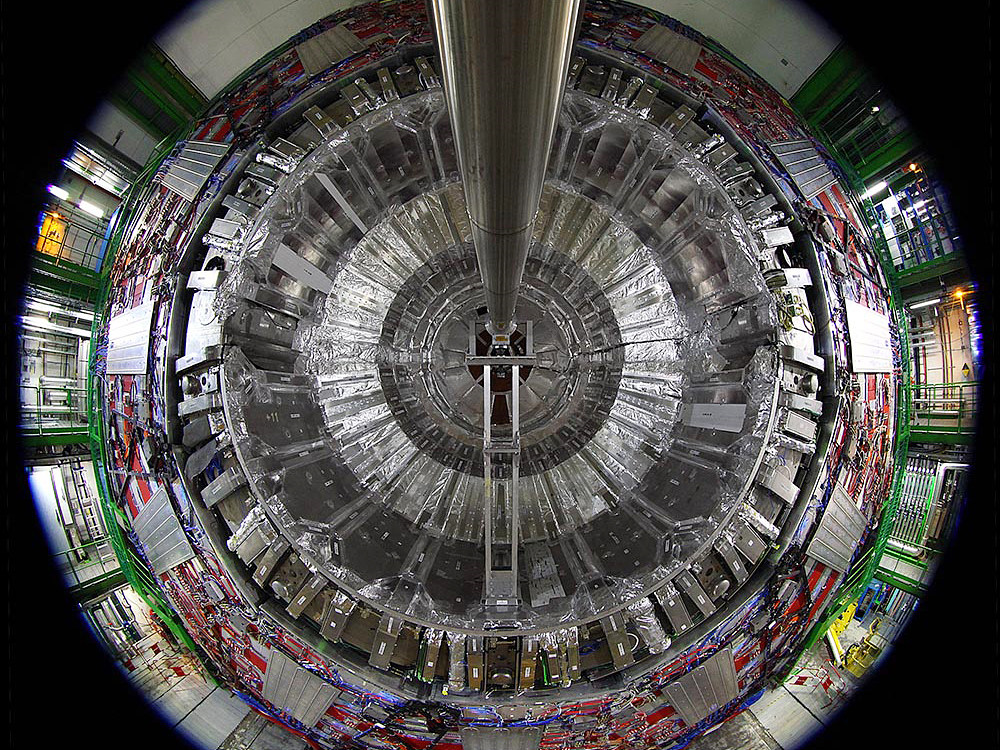
CMS Space Gate
by Michael Hoch
CERN
CMS Space Gate - 20m diameter of perfect circular geometry with LHC beam pipe.
LHC P5 Interaction Point
by Michael Hoch
CERN
LHC P5 Interaction Point – ultra high resolution CMS pixel detector surrounding the LHC interaction vertex.
Cable Map
by Michael Hoch
CERN
Cable Map – to large CMS segments shifted apart to give access for services work and insight to the cable tracks, which look like the dense road map of modern city.
2017 IBS Art in Science
Nemo meets Marlin
by Soonmin Kang
Center for Correlated Electron Systems
Single crystal X-ray diffraction (SC-XRD) with 4 axes is an important experimental method to verify the structure of materials. It is possible to observe reciprocal lattice of material by performing SC-XRD and the limited reciprocal lattice occasionally shows interesting patterns in accordance with experimental conditions. The image contains the SC-XRD results of vanadium diselenides (VSe2) at 28 K and 300 K. The results seem to resemble a dramatic meeting of the clownfish character Nemo and his father Marlin who appear together in a Disney film. Blurry lines, dots, and splendid colors of the patterns contrast with the dark background, creating a dreamy atmosphere.
Autumn made of superconductor
by Suhan Son
Center for Correlated Electron Systems
Niobium diselenide (NbSe2), a material synthesized from niobium (Nb) and selenium (Se), is not only a quasi-two dimensional layered material, but also a superconductor which shows zero conductivity in low temperatures. In the figure, the atomic force microscope (AFM)* measurement data obtained after making NbSe2 into several tens of atomic layers by mechanical exfoliation is three-dimensionalized. The colors of the figure result from the thickness differences between layers. It is reminiscent of a mountain where the autumn color comes from a combination of colorful colors.
* Atomic force microscope: It can measure atomic-scale forces between the surface and a probe tip and produce topographic maps of the surface of the particle.
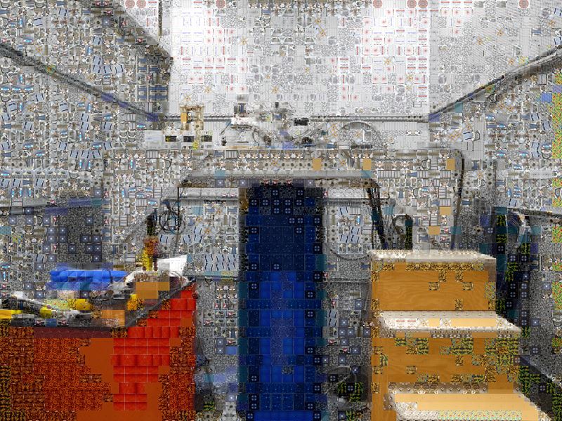
Spectrum of SI-STM 1 (Spectroscopic imaging scanning tunneling microscope)
by Kyoung Seok Lee
Center for Correlated Electron Systems
From design to completion of construction of a spectroscopic imaging - scanning tunneling microscope (SI-STM), we went through a colorful spectrum of memories; joy, excitement, frustration, hope, struggle, etc. The main image is an SI-STM system installed inside an acoustic and vibration isolation facility. The blue cylinder is a 14 T magnet liquid helium dewar, where a STM head and a cryostat are kept at an extremely low temperature. By this photomosaic, we attempt to visualize the fact that a scientific instrument can be built not only upon materialistic components, but also upon other intangible abstract elements as well. This mosaic is more meaningful since each and every pixel of it were crafted from real images of the researchers’ lives.
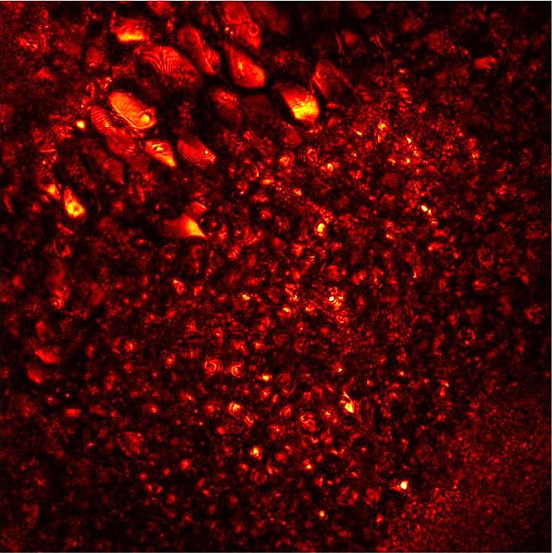
Leaves blowing in the wind : Inside the brain of a living zebrafish
by Moonseok Kim
Center for Molecular Spectroscopy and Dynamics
This is a movie made of 3-dimensional images of the hindbrain of a living larval zebrafish two weeks after fertilization, which was acquired by label-free high-resolution computational adaptive optical microscopy. The complicated distribution of neurons within 100 μm2 of the field of view is shown from the top to a depth of 100 μm of the brain. Due to the refraction and multiple scattering of light inside the zebrafish, it was previously not possible to acquire high-resolution images with conventional label-free imaging techniques. In order to overcome such a limitation, we have developed a novel computational adaptive optical microscopy which can figure out light distortion and retrieve an original image by fundamental physics. The contrast of the diffraction-limited image is unprecedentedly enhanced, thereby even interference fringes at the membrane can be visualized. The magnificent view of the vanishing neurons reminds us of leaves blowing in the autumn wind.
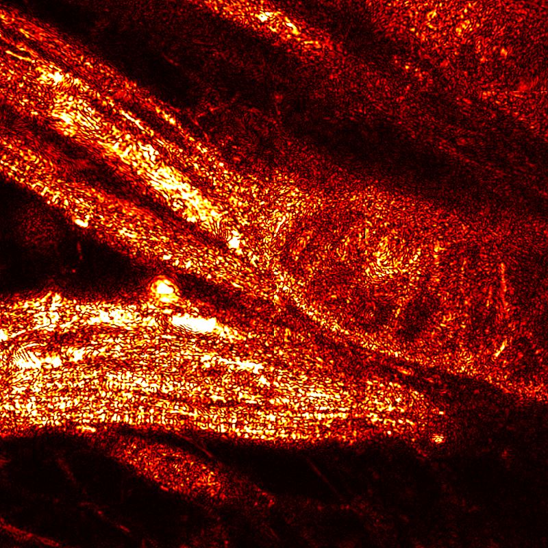
A muscular zebrafish
by Yonghyeon Jo
Center for Molecular Spectroscopy and Dynamics
This is an image of the muscle tissue of the spinal cord area of the zebrafish 10 days after hatching obtained by a high-resolution aberration-compensated microscope technique with non-labeling. Healthy zebrafish swim fast even in small tanks. Thanks to sturdy muscles, zebrafish can swim vigorously. The separation of the erector spinae muscles and the definition of delicate muscle fibers are prominent. Whether he knew that he would soon be anesthetized and imprisoned in a hard agar, I even feel the will and spirit as he raised his muscles to resist his destiny. Asymmetry composition, flexible shape, and dynamics of the light create a sense of rhythm and space.
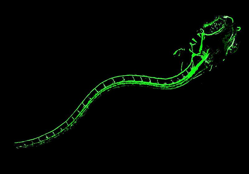
Z-dragon (Zebrafish-Dragon)
by Jin Hee Hong
Center for Molecular Spectroscopy and Dynamics
The nervous system of vertebrates is surrounded by the myelin sheath that forms an electrically insulating layer. This is essential for a rapid electrical signal propagation. This image represents the myelination of zebrafish which is widely used as animal models. Green fluorescent protein (GFP) is expressed in cells that construct the myelin sheath of a zebrafish and the image is obtained by a confocal laser microscope*. The central nervous system, consisting of the brain and spinal cord, and the peripheral nervous system of a zebrafish 14 days after hatching are shining with green fluorescence. It looks like the shape of a dragon who is eager to ascend. Strong contrast of colors and oblique composition give the sensation of speed and motion.
* Confocal microscopy: A type of microscopy that uses a laser along with particular optical components to create high-resolution images of materials stained with fluorescent probes.
Now you see me : A trace of nanobiochemisty
by Seongchan Kim
Center for RNA Research
A drug delivery system based on RNA as a drug candidate is a novel strategy to treat various diseases at the molecular level, but still has an undesirable biological barrier of efficient delivery to the target site. We have developed a multifunctional RNA delivery vehicle enabling simultaneous bioimaging based on a highly biocompatible fluorescent nanoparticle (blue) and dye-labelled RNA (red) with simple preparation. Real-time study of fluorescence imaging in live cells (green) shows that the nanoparticle favorably localize in cytoplasm and successfully release RNA. This study suggests the excellent potential for simultaneous bioimaging and gene therapy system.
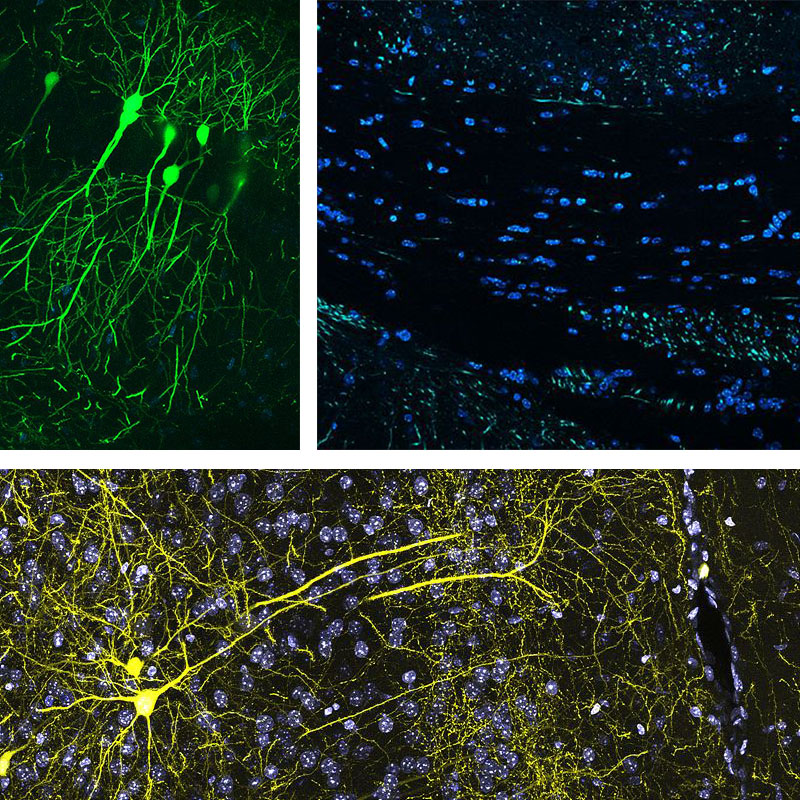
Der Teil und das Ganze trilogy : Contact, Sandbank, Take off at night
by Kyungdeok Kim
Center for Synaptic Brain Dysfunctions
These figures form a trilogy themed by a saying in Oriental philosophy, “There is small universe in each of our brains.” The first figure, “Take off at night”, provides similarity between the brain structure and a city landscape observed from the sky. The second figure, “Contact,” provides appreciation of synapses and the neurons which compose them. In the last figure, “Sandbank,” information is observed like a stream in which we can gaze. Neurons in the images of the trilogy shine by themselves expressing fluorescent proteins. The name of trilogy originated from an immortal classic, “Der Teil und das Ganze” (“the part and the whole” in English), of Werner Heisenberg (1901-1976), a quantum physicist.
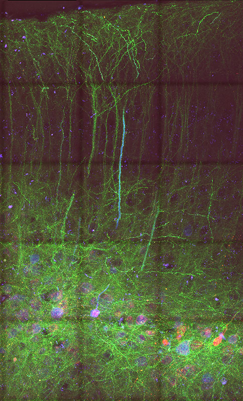
Garden in the brain : Connecting link
by Chaehyun Yook
Center for Synaptic Brain Dysfunctions
Reminiscent of a night forest with wild flowers, this figure shows intermingled structures in a mouse brain, connected to each other by synapses. These connect individual neuron and transmits neuronal activity, forming circuits in a complex neural network. Because of their sheer abundance and tiny size, visualizing synapses together with neuronal process structures necessitates specialized labeling strategies in light microscopy. This figure shows a confocal image of medial prefrontal cortex (mPFC) of Fmr1 knock out mouse, a mouse model for autism and intellectual disability, labeled by mGRASP, a labeling technique to visualize a synapse with its pre- and post-synaptic neurons. Synapses (red puncta) are located at the interface of pre- and post-synaptic neuronal processes (green), whose cell bodies (blue) are located in the deep layer. From this image, we can examine synapse distribution of local circuits of Fmr1-KO mPFC, and infer a connectivity correlation of neurological disorders.
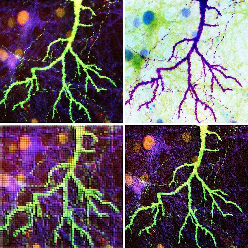
The flash
by Eunkyung Lie
Center for Synaptic Brain Dysfunctions
The four images are the dendrite of cultured pyramidal neuron, obtained from a hippocampus. The brain of human beings consists of about 100 billion nerve cells (neurons). Among them, only one billion pyramidal cells are related with long-term memory. By expressing the fluorescent EGFP proteins in cultured neurons, these images were captured while observing the shape or number of dendritic spines aiding study of the formation of synapses in which synaptic neurotransmission occur. The original (upper left) looks like lightning in the night sky and the other three images express glare, feeling, and afterimage by processing the original image. The four images with different colors and textures combine into one great artwork. The dendrite in the brain also looks like the roots of a plant.
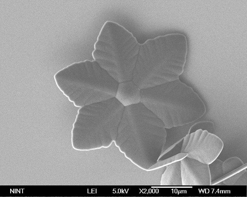
Fullerene flower
by Hee Cheul Choi
Center for Artificial Low Dimensional Electronic Systems
Fullerene is the smallest, stable carbon-based molecule. Recently, many researchers focus on solution-based self-crystallization of fullerene molecules due to their various morphologies and crystal structures with different optical and electrical properties. This figure shows the first reported fullerene flower, which has unique flower-shaped crystals with six symmetric petals followed by co-crystallization* of both C60 and C70 molecules. This unique crystal can be observed due to different solubility of C60 and C70 molecules in mesitylene, which promotes nucleation of C70 with sixfold symmetry in the early stage, and it is followed by co-crystallization of both C60 and C70 molecules.
* The process to grow crystal which contains more than two molecules.
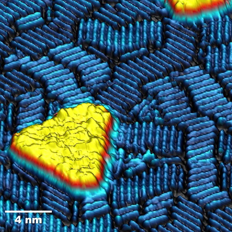
Nanoscale Escher’s stairs
by Jewook Park
Center for Artificial Low Dimensional Electronic Systems
Scanning tunneling microscopy (STM) reveals self-assembled cobalt (Co) nano-islands and tantalum (Ta) nano-rods on a copper (Cu) (111) substrate. We prepared spin-polarized STM and observed ferromagnetic Co nano-islands (yellow). Ta nano-rods form staircase shapes. The atomic level surface image (3D rendering, false-colored) brings to mind the lithography print “Relativity” (1953) by the Dutch artist M. C. Escher. At the same time, the image also resembles a virtual space where a yellow island is floating in a blue ocean with waves rising and falling.
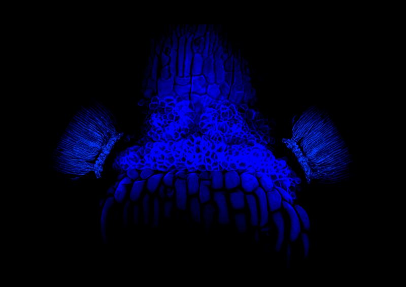
Blue Dokkaebi
by Yuree Lee
Center for Plant Aging Research
The picture shows cell wall of a receptacle after parts of floral organs are shed. An inflorescence of the plant Arabidopsis was stained with calcofluor white, a fluorescent dye that binds strongly to cell walls, and observed with a confocal microscope. Blue fluorescence generated by UV excitation wavelengths looks like the imaginary face of Dokkaebi; traditional Korean goblins. Mysterious blue light gives off a creepy look.
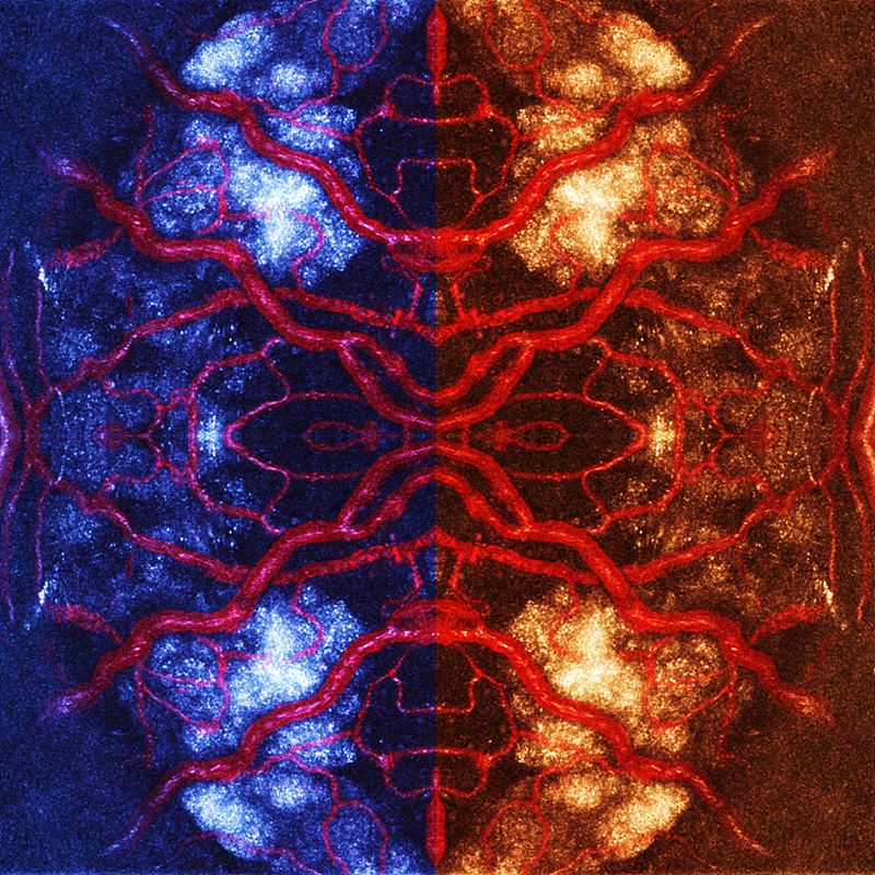
Imagining a brain aging : Color of aging
by Junsoo Park
Center for Plant Aging Research
Brains are developed inside heads, which make them difficult to observe. Due to this characteristic, observation of long-term scale biological events, such as a brain’s aging process, was believed to be nearly impossible. If we can relocate a brain in a location where we can directly observe it, wouldn’t it be possible to observe things that we previously couldn't? We implanted hypothalamic tissue on the iris of an eye. The red color indicates angiogenesis (development of new blood vessels, an indicator of successful implantation) of an implanted tissue . Structure and color were modified to our liking. What would be the shape and color of an aged brain? The image reminds us of an insect or the symmetry image of a mandala.
Sorrow of the weak
by Yuree Lee
Center for Plant Aging Research
Calcium is an essential nutrient for development of plants, and required for structural roles in cell walls and membranes, as well as an intracellular messenger in signal transaction. Insufficient calcium uptake degenerates plant development and growth, which is especially severe for flowers and young leaves. The picture shows the death of a young leaf due to the lack of calcium, which looks like a tear drop.
A Seoul map painted with pancreatic islets
by Jung Hoon Cho
Center for Plant Aging Research
Pancreatic islets situated in the pancreas produce several hormones (i.e. insulin, glucagon) playing a central role in retaining glucose homeostasis in our body. To determine the pancreatic islet cell viability after dimethyl sulfoxide (DMSO) treatment, we used live and dead cell markers, calcein AM (green) and EthD-1 (red). Coincidentally, this image looks like a city map of Seoul drawn with pancreatic islets.
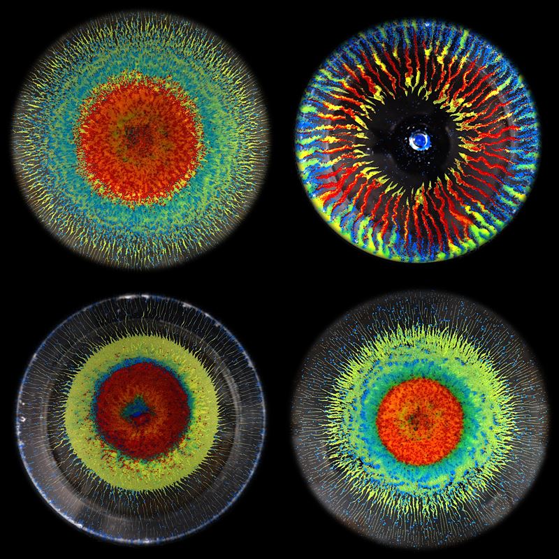
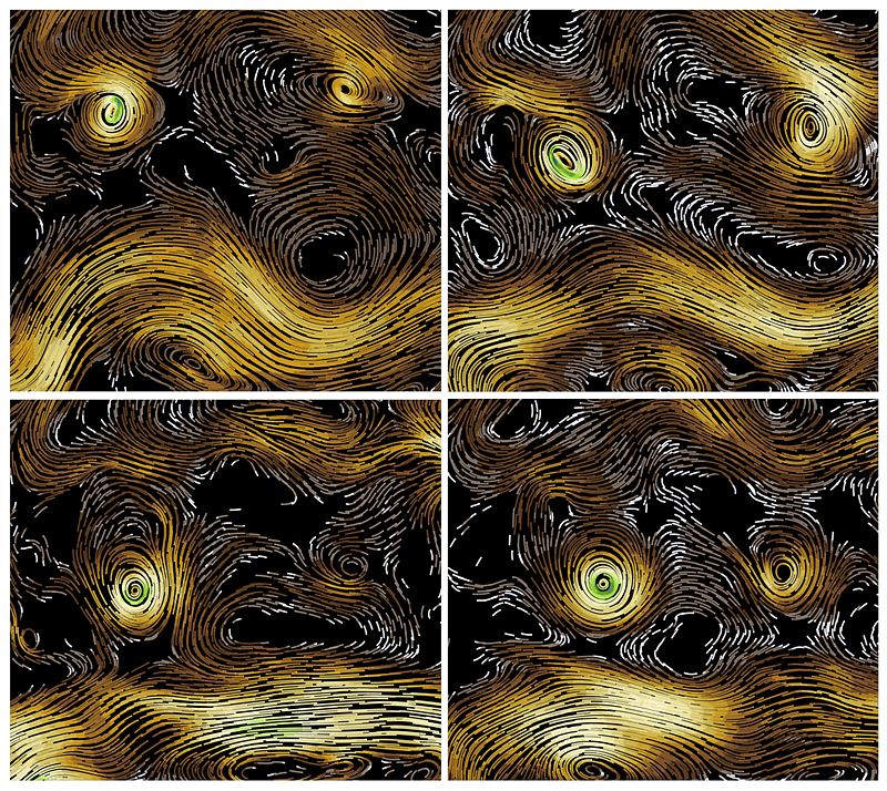
The spinning dust
by Olgierd Cybulski
Center for Soft and Living Matter Bartosz Grzybowski Group
The four photographs show self-assembled patterns of micro-particles suspended in a heavy liquid at the top of a container which rotates about the vertical axis, with cyclically varying speed. The particles of three different sizes, densities and colors are lighter than the surrounding liquid, so they are pressed towards the transparent lid of the rotating container. The centrifugal force, resulted from rotation, pushes them toward the center, whereas the shear forces resulted from torsional shaking, correspond to the radial alignment of particles. Different patterns occur at different speed of rotation.
Colloidal brain
by Ruoyu Dong
Center for Soft and Living Matter
A colloidal cluster formed by acoustic trapping, explodes into this wrinkled structure when both an ultrasound and AC electric field are on simultaneously. This is the consequence of a tug of war between the ultrasound confining potential and the AC induced dipole-dipole interaction. It reminds us of the shape of the human cerebral cortex or a swelling fingerprint. They both share a similar formation mechanism; due to the limited space in a two dimensional plane, the structure tends to evolve in the third dimension, creating a convoluted pattern.
Red bamboo in a capillary : Topological defects in liquid crystals
by Sung-Jo Kim
Center for Soft and Living Matter
Confined liquid crystals struggle to find the most comfortable (lowest energy) structure. They want to minimize their elastic free energy while satisfying boundary conditions. These boundary conditions often present inevitable but beautiful imperfections called topological defects. The photo is a polarized optical microscopy* image of nematic Sunset Yellow (SSY) doped with a minuscule amount of polymer. In this chromonic liquid crystal of very small elastic modulus, chiral domains emerge spontaneously with topological defects between two domains. The shape resembles the structure of bamboo.* Polarized optical microscopy: uses filters to polarize light into waves that vibrate only in one direction
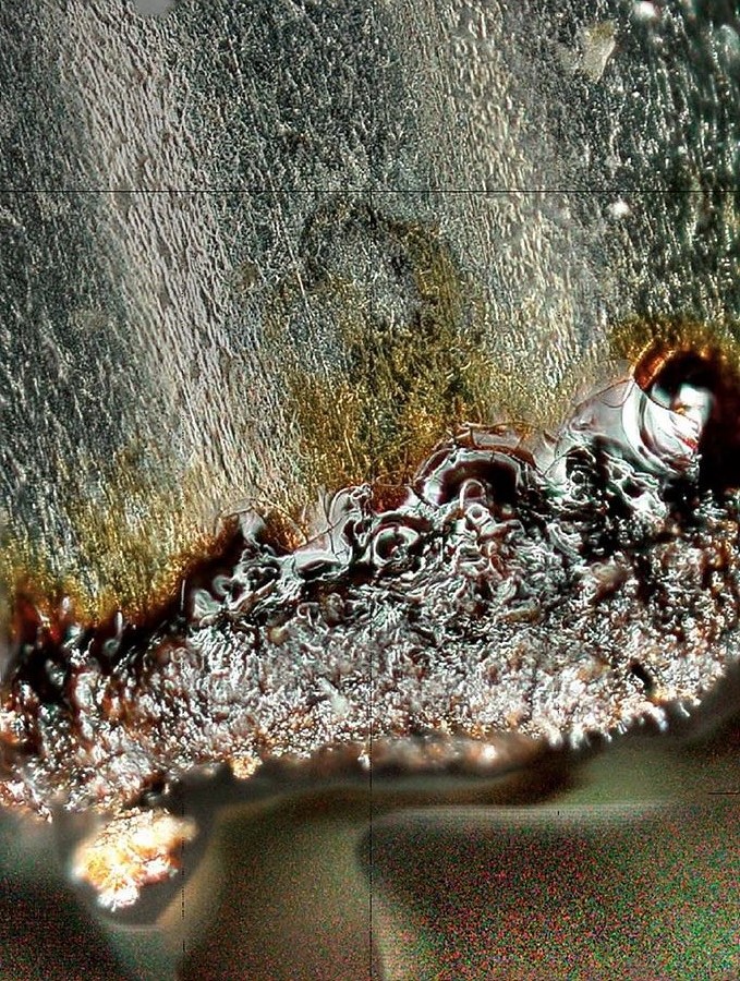
Platinum waterfall
by Yaroslav I. Sobolev
Center for Soft and Living Matter
A dull tip of a platinum wire was used as an electrode for applying high-voltage (3 kV) to a sessile water droplet. The purpose of these experiments was to study how the microscopic surface structure of water changes when it's highly charged. Here you see a magnified photo of the wire tip after one of the experiments. Wire is oriented vertically on the image (you can see the clean part of the wire on the top). Even though we used ultrapure water, some brown organic substance was found on the wire tip (bottom of the image) after 1 day under high voltage - probably due to impurities present in ambient air. This image seems like a piece of contemporary art. The contrast between sleek and rough surface and swelling silver waves maximize an abstract atmosphere.
Sunset over Hades
by Karel Goossens
Center for Multidimensional Carbon Materials
‘Deep down in the Underworld, where restless souls are wandering in eternity, yet another day comes to an end. The sky turns dark, but the flames never cease to give off their relentless heat, melting everything that comes too close.’ This is what came to my mind when this beautiful texture of a molten liquid crystal appeared in my polarized light optical microscope*. The image provided some clues about the nanoscale organization of molecules in the birefringent** sample, due to characteristic interference effects. It marked the starting point for a detailed study by X-ray diffraction, which revealed that these molecules form an intricate hexagonal columnar mesophase with a triangular cylinder like morphology.
* Polarized light microscopy: A useful method to generate contrast in birefringent specimens and to determine qualitative and quantitative aspects of crystallographic axes present in various materials.
** Birefringent: Optical property of a material having a refractive index that depends on the polarization and propagation direction of light.
Psychedelic liquid crystals
by Karel Goossens
Center for Multidimensional Carbon Materials
Watch out! Looking for too long through a microscope may cause a massive headache! Yet it is impossible to get bored while looking at the intriguing "fan-like defect" textures of liquid crystals when they are seen through crossed polarizers. The image is an artistic interpretation of the effects of introducing a so-called "full-wave retardation plate" in between the sample and the second polarizer (analyzer). By determining which color appears in which direction, it is possible to obtain information on the orientation of the rod-like aromatic cores of the molecules in a columnar liquid-crystalline mesophase. If the fan-like defects in the southwest-to-northeast direction appear yellow-orange while those in the southeast-to-northwest direction are colored blue-green, then the aromatic segments are most probably oriented parallel to the supramolecular columns. The reverse situation indicates that the aromatic parts are oriented perpendicularly with respect to the long axes of the columns.
Red star
by Seon Gyeong Lee
Center for Genomic Integrity
Mammalians including human have 3 types of adipose tissue. The brown fat tissue generates heat and increases tissue size in response to a cold condition. Brown adipocyte has many mitochondria. The mitochondria cannot generate energy and die in mass when exposed to stress or cytotoxic conditions. We took a picture of a cell after knocking-down a gene which regulates genomic integrity. We stained mitochondria red and nucleus blue, using Mitotracker and DAPI, respectively. Merging image looks like a destroyed red star.
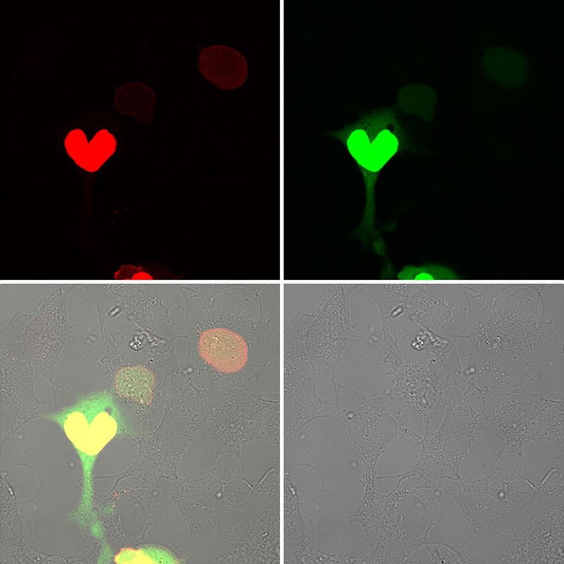
L'Elisir d'amore
by Jung Me Hwang
Center for Genomic Integrity
The title of this work is a love potion (L'elisir d'amore). The heart represents the dramatic encounter and survival of love on the battlefield. It is an image captured using CUPID technology (protein binding analysis technique) which analyzes the binding of proteins. Protein MSH2 (red) and protein MSH3 (green) in association with protein PKC migrate to the same place on the cell membrane after being exposed to the drug PMA. While observing their migration, this work captured the moment when both proteins reach the membrane, highlighting its heart shape. It is a love potion that comes from drugs. Both proteins play an important role by participating in various mechanisms by intracellular genetic factors, and with this technique, its process can be studied in detail.
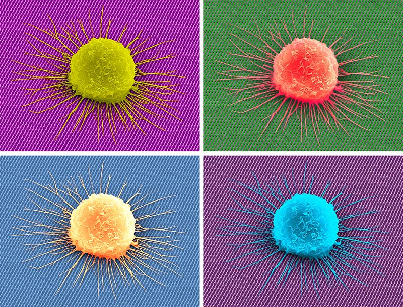
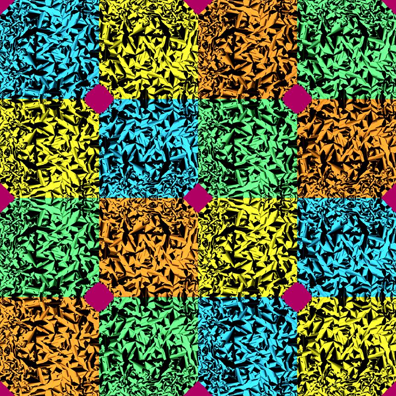
Cell flower blooming on the nano-grass
by Chaejeong Heo, Chanho Jeong, Tae-il Kim
Center for Neuroscience Imaging Research
While we were developing geometry-controllable nanohairs that mimic cacti spine-like stooped hairs and studying cell migration behavior on these hairy structures, we decided to color a scanning electron microscope (SEM)* image that resembled a flower bud and grass. The decision to make the images into a 2x2 array with vivid colors was a nod to Andy Warhol's pop art format. The work related to this image of neuroblastoma was published in Nanoscale (Nanoscale, 2017, 13 Sep. DOI: 10.1039/C7NR04522K).
* Scanning electron microscope: An advanced microscope that uses a focused beam of electrons to produce an image of a sample's surface.
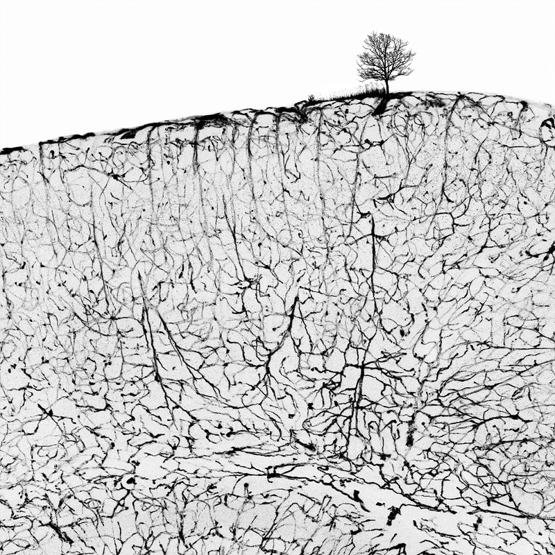
The root of being, vessels
by Yoo Hyung Kim
Center for Vascular Research
Angiogenesis is an essential element for organ development and maturation. The blood vessels of the brain extend from the outermost surface into the parenchyma like a root of trees spreading out into the soil. At the same time, the blood vessels of the brain form a tighter barrier than the blood vessels of other organs. Overall, these are crucial processes that protect the brain while promoting brain maturation. The figure shows beautifully organized brain vessels of a postnatal 12-day-old mouse. Interestingly, by drawing a tree on the surface of the brain, the figure expresses the feeling that vessel development is essential for life, as root growth is essential for trees. The tree and complex of blood vessels in black and white are reminiscent of a water and ink painting.
The stream of river flowing in our eyes
by Jaeryung Kim
Center for Vascular Research
This image, which looks like a green pillar surrounded by purple ivy, shows Schlemm's canal and blood vessels in an eye. Eyes maintains their shape and intraocular pressure by continuous production of aqueous humor. Schlemm's canal (green color) encircling peripheral cornea has a critical role for the drainage of aqueous humor into blood vessels (purple color). When normal drainage of aqueous humor is impaired, intraocular pressure is increased and the optic nerve is subsequently damaged, inducing glaucoma and vision loss. Researchers in IBS suggested a potential therapeutic target by demonstrating the new mechanism for development and progress of glaucoma. Although this seemingly river in the eye reminds us of a tear, we can feel hope in conjunction with these research results.
Raspberry fields of blood
by Hosung Bae
Center for Vascular Research
Adipose tissue is highly vascularized and most adipocytes (red) are supported by an extensive vascular network (blue). Brown adipose tissue has an especially large network of blood vessels. Mitochondria-packed brown adipocytes burn energy and produce heat. The image of adipocytes is reminiscent to raspberries, which have been known for burning fat. Blood vessels, sustaining brown fat, can be compared with stems that provide raspberries with necessary nutrients. The strong color contrast of red and blue makes the image look dynamic.
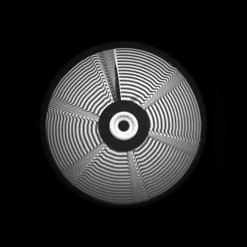
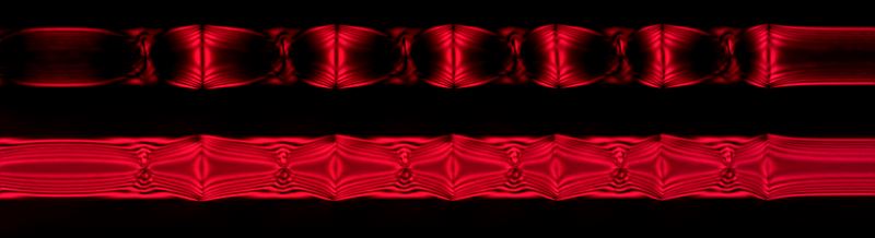
Light donut factory
by Cheonha Jeon
Center for Relativistic Laser Science
We have designed a spherical phase mirror that operates at an angle of 45 degrees to generate a donut shaped beam for light matter interaction. This is an interferometric image of the phase mirror surface. The surface is constructed of six different steps, and as the laser light reflects from the surface, the path difference is λ/3 for each step and 2λ for the combined steps. Due to this path difference, when focused, the beam will have a donut shaped profile. This high intensity donut profile beam will generate a pillar of electrons at the center as it interacts with matter. This will result in proton acceleration that focuses at the center and these accelerated protons can be used for proton therapy for cancer. The bending image with radiating circular waves and the interference pattern is associated with op art.
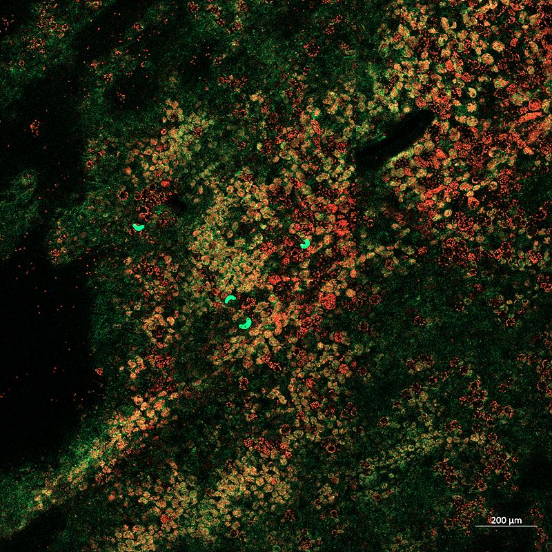
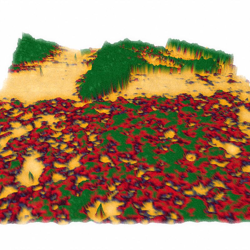
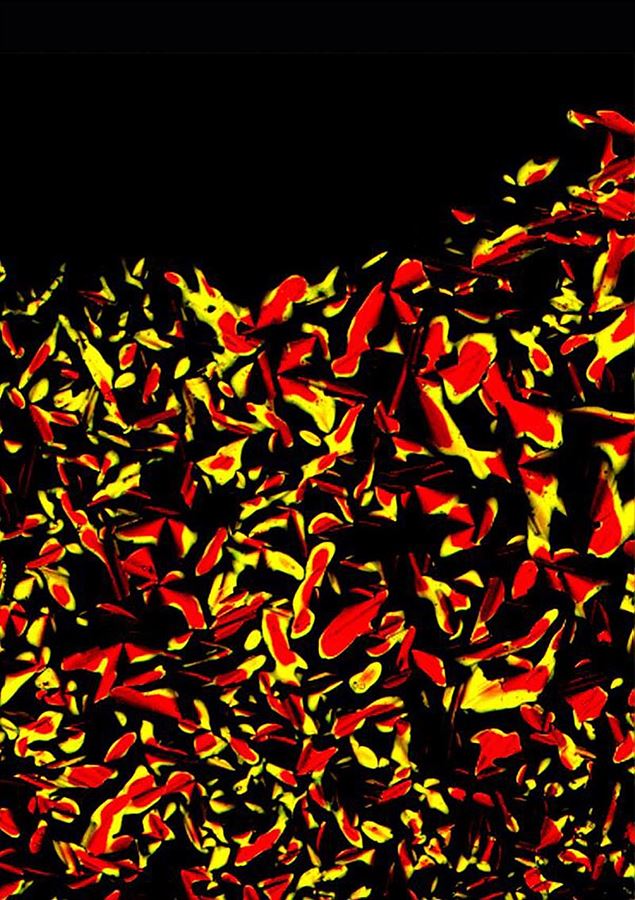
Is it real?
by Beum-Chang Kang
Center for Genome Engineering
When a gene of interest is introduced into a plant cell together with a fluorescent protein gene, the presence or absence of the gene can be determined through the observation of fluorescence. The most difficult factor to overcome while observing the fluorescence of transformed plant tissue with confocal microscopy is autofluorescence from plant cells. Autofluorescence may look green like a fluorescent protein, but the real color is different. The photograph shows the expression of a fluorescent protein introduced in stomata in plant tissues using confocal microscopy*. The image looks like vivid yellow-green stains on an aerial photo of mountains covered with autumn leaves, creating a disparate and peculiar atmosphere.
* Confocal laser microscopy: A type of microscopy that uses a laser along with particular optical components to create high-resolution images of materials stained with fluorescent probes.
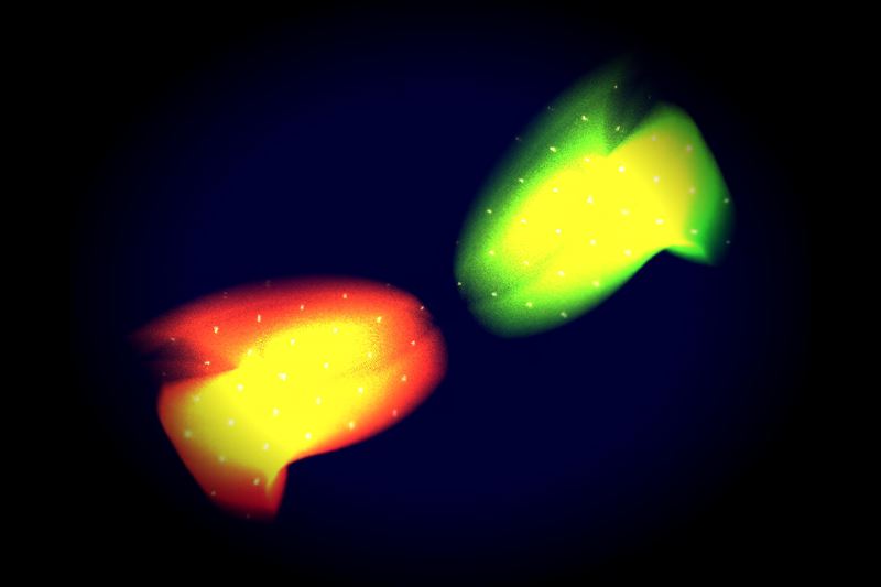
Stormy night
by Seung-Bu Park, Sun-Seon Lee, Jung-Eun Chu
Center for Climate Physics
Hurricanes (typhoons) are feared for their destructive power, but when simulated on computers they exhibit unprecedented natural beauty. This image shows the time-evolution of a pair of computer-generated typhoons over the Indian Ocean and their interaction with a meandering westerly wind jet. The numerical model used for this animation captures the dynamics of the global coupled atmosphere/ocean/land and ice system with a resolution of about 25 km. ICCP scientists conducted the numerical experiment on a supercomputer in the United States. The title "Stormy Night" was inspired by Vincent van Gogh's masterpiece, The Starry Night.
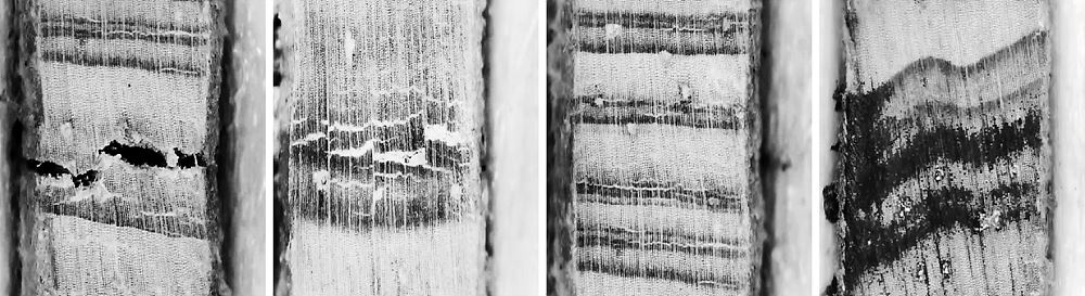
A message from the past
by Axel Timmermann, Karl Stein, Elke Zeller
Center for Climate Physics
"Li circuli delli rami degli alberi segati mostrano il numero delli suoi anni, e quali furono più umidi o più secchi la maggiore o minore loro grossezza."
(The circles of the branches of the sawn trees show the number of their years, and which ones were damp or dry the greater or lesser their thickness.)
Leonardo da Vinci, Trattato della Pittura
Tree growth only occurs in the outermost section of the tree, in a layer under the bark that is only a few cells thick. Outside tropical regions, the rate of tree growth changes throughout the year, creating distinct annual rings. Leonardo da Vinci first related the thickness of the annual rings to the amount of rainfall received during the year. ICCP scientists use tree ring data to gain information about past climates and past changes in ecosystems. The image shows 4 sections of a tree core sampled by Axel Timmermann from a high elevation site in Hawaii. In addition to using the visual images, scientists also analyze stable carbon isotopes within the tree rings, which give them an insight into subseasonal climate variations and changes in photosynthesis.
2016 IBS Art in Science
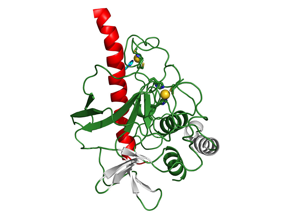
The Beauty of Evolution:the Structure of the Drosha Protein
by Sung Chul Kwon
Center for RNA Research)
A type of regulatory RNA called microRNA involved in almost all biological processes such as development, aging, cancer, differentiation and immune response, is generated by two consecutive processing steps involving Drosha and Dicer proteins. This figure shows the bottom part of a Drosha protein whose structure has been solved 12 years after its first identification. Drosha has the same backbone structure as Dicer (red and green), plus some unique regions (yellow and white). Thanks to this information, we can infer how Drosha diverged from the ancestral Dicer and obtained its new function.
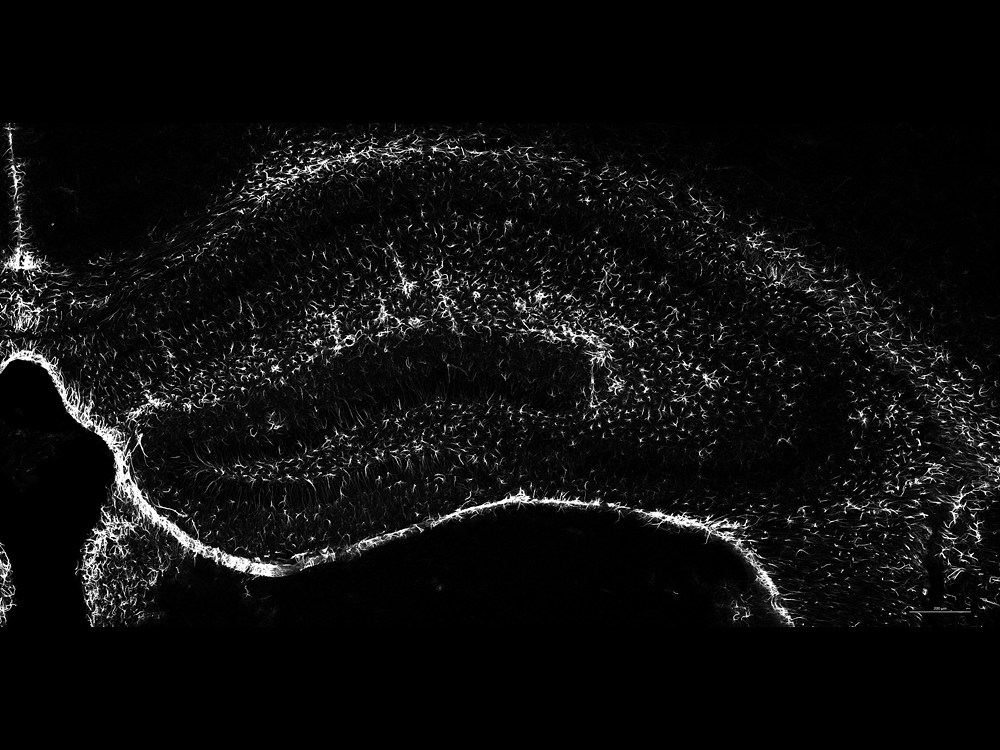
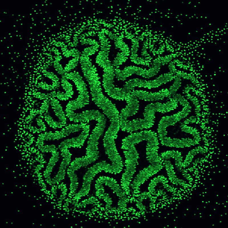
Milky Way in the Brain
by Haram Park
Center for Synaptic Brain Dysfunctions
The hippocampus is the "sea horse" shaped structure located bilaterally within mammalian brain. It is part of the limbic system, and is thought to contribute to information consolidation, spatial memory, and both short and long term memory. In the image, we stained star-shaped astrocytes, known as astroglia in the hippocampal region of the brain with GFAP, glial fibrillary acidic protein. The stained cells are represented in white and they look like white stars set within a contrastingly dark background, akin to the milky way observed in the night sky.
The Pond
by Junyeop Roh
Center for Synaptic Brain Dysfunctions
Claude Monet has been immortalized by his renowned painting of the pond dotted by colorful water lilies. When calcium-binding proteins (Parvalbumin, in red), synaptic proteins (PTP delta, in green) and DNA (in blue) are stained in the layer 2/3 of mouse medial prefrontal cortex region, the resultant image is remarkably reminiscent of the masterpiece. The medial prefrontal cortex has been extensively studied as the region mediating social behavior, decision making and, in humans, personality expression.
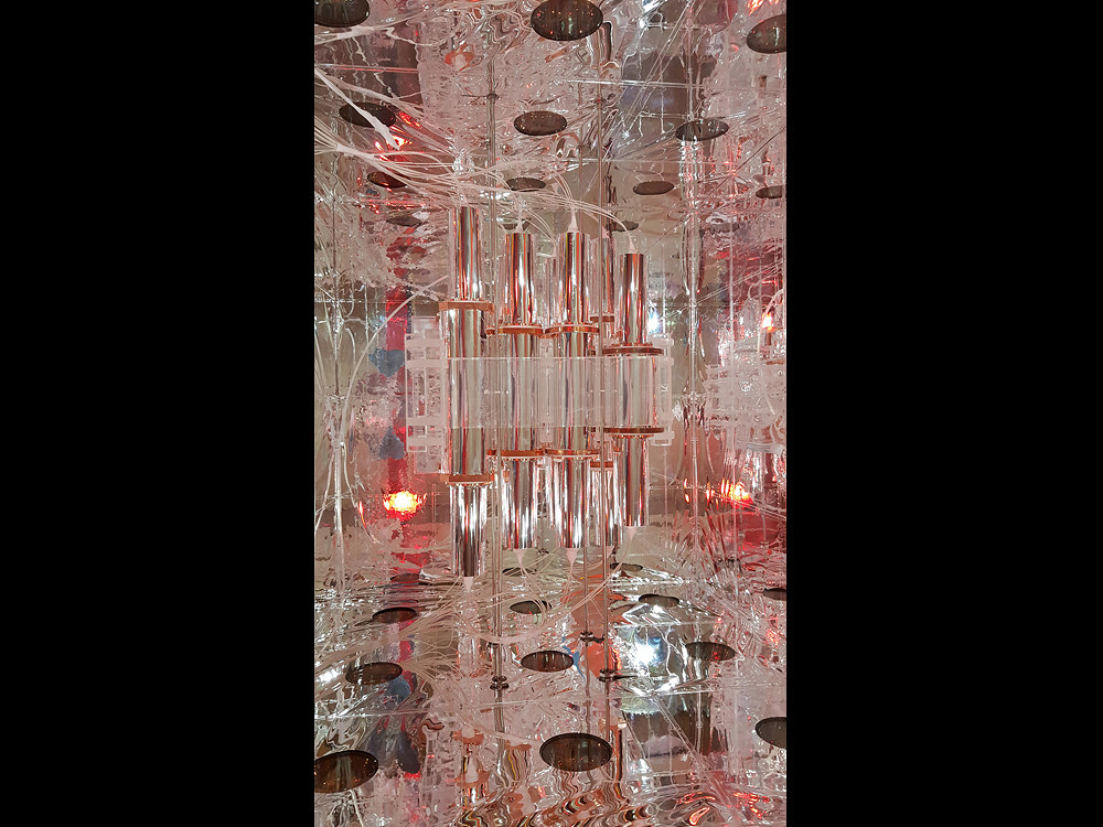
Chamber for the Mystery Matter
by Chang Hyon Ha
Center for Underground Physics
The photo shows the innermost view of the dark matter experiment, called COSINE, situated 700 meters below the surface at Yangyang underground laboratory. The cylindrical cells in the middle center encase ultra-pure NaI(Tl) crystals which are used to collect a faint light induced by the interaction of dark matter and the crystal nuclei. In order to distinguish the signal from external noise entering outside of the crystals, we position the crystals in a large chamber where all surfaces are laminated by reflective films that work like a mirror. Then, photons from the noise or any other light in the chamber can easily bounce back and forth and be contained, creating the exotic image.
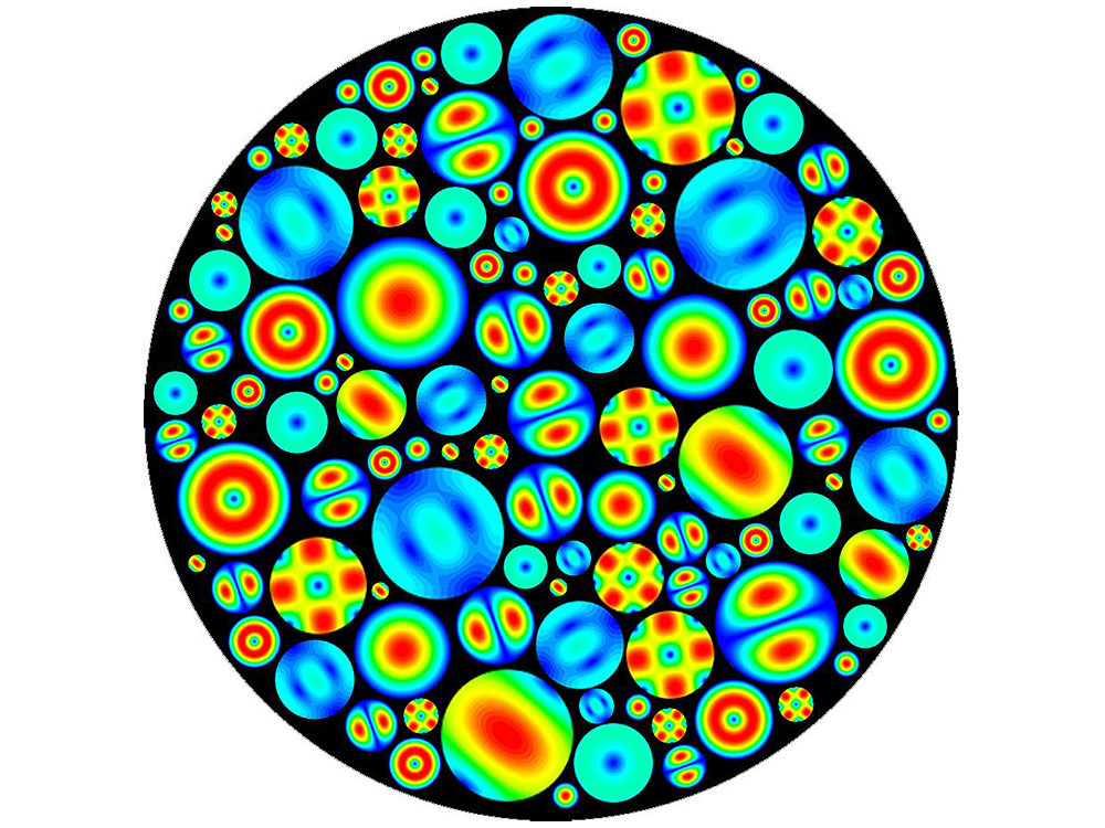
World of Diversity
by Young-Im Kim,Jihoon Choi
Center for Axion and Precision Physics
This work of art shows that various resonant modes are generated in a variety of sizes resonator visually. A resonator is a device that records a specific frequency and vibration using a resonance phenomenon that is amplified when a specific frequency is found. Various studies using such resonance phenomenon is being done. Resonance is a phenomenon that can be found everywhere in our world. In one of the resonator to various resonance modes exist similar to our world people have a variety of looks and emotions.
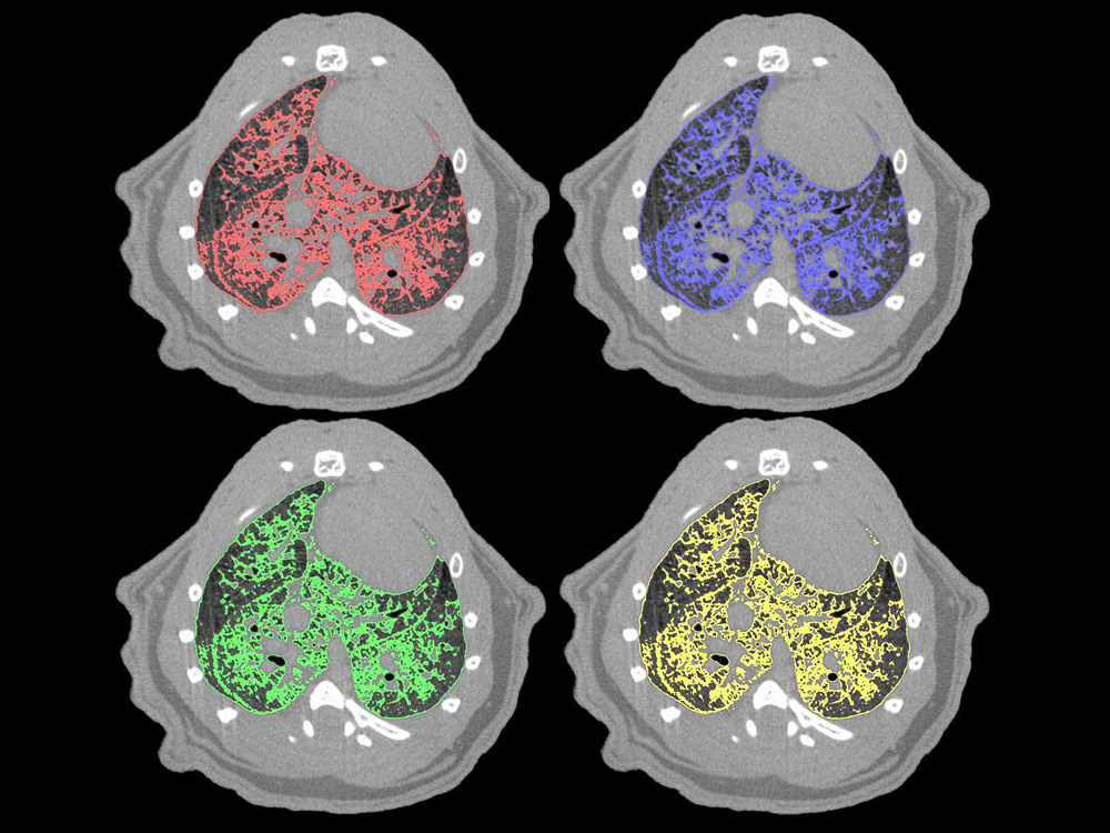
Drowning Lung
by Seung-Jun Lee, Gou Young Koh
Center for Vascular Research
The image is a visualization of vascular leakage in the lungs of a mouse model of sepsis. An infection or injury can lead to sepsis where severe inflammatory response and vascular leakage occur throughout the body. These septic challenges lead to multiple organ failure, including accumulation of fluid in the lungs. We have demonstrated the beneficiary role of the endothelial tyrosine kinase receptor Tie2 in ameliorating vascular leakage and sepsis.
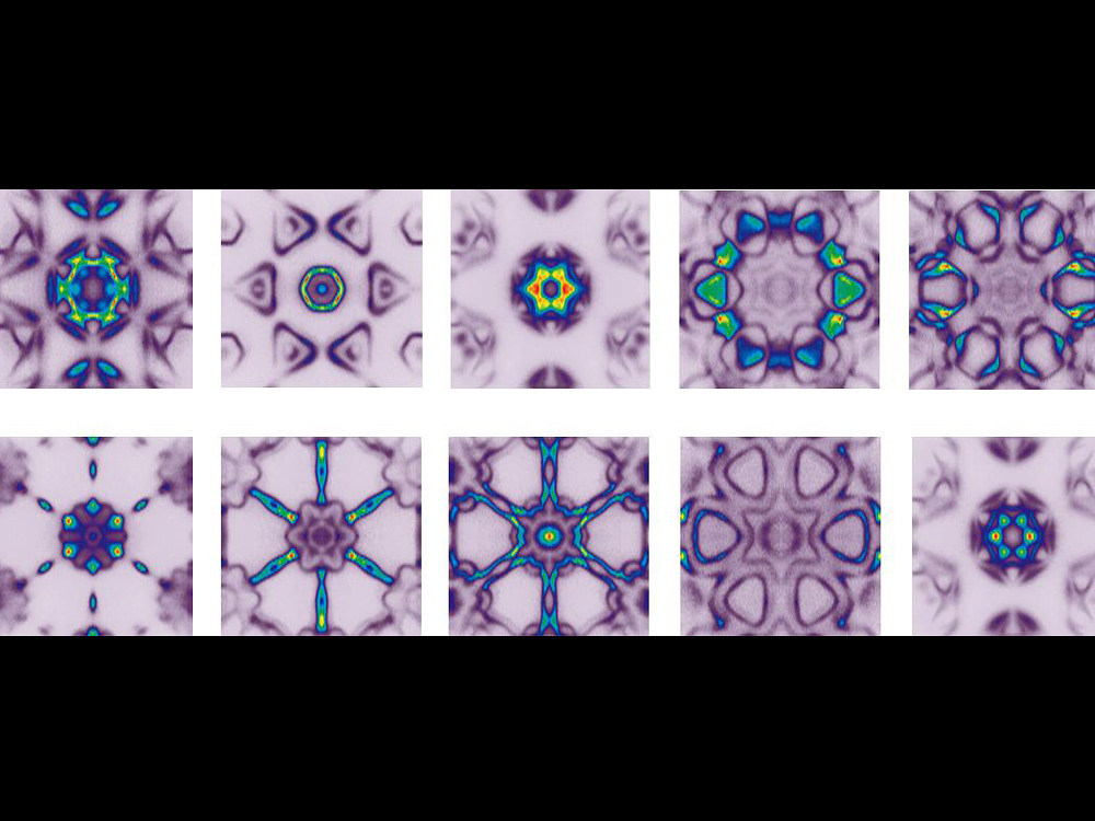
Material World through Kaleidoscope
by Beom Seo Kim
Center for Correlated Electron Systems
Molybdenum di-telluride (MoTe2) has drawn growing attention due to its application possibility. It has six-fold symmetry in the electronic structure due to its crystal structure. The electronic structure of MoTe2 was measured by angle resolved photoemission and images were constructed by folding constant energy contour of the experimental data. Each image represents constant energy contour at a specific binding energy. While the image drastically changes over different binding energies, the six-fold symmetry remains intact. The six-fold symmetry is reminiscent of the colors of a peacock or a view through a kaleidoscope.
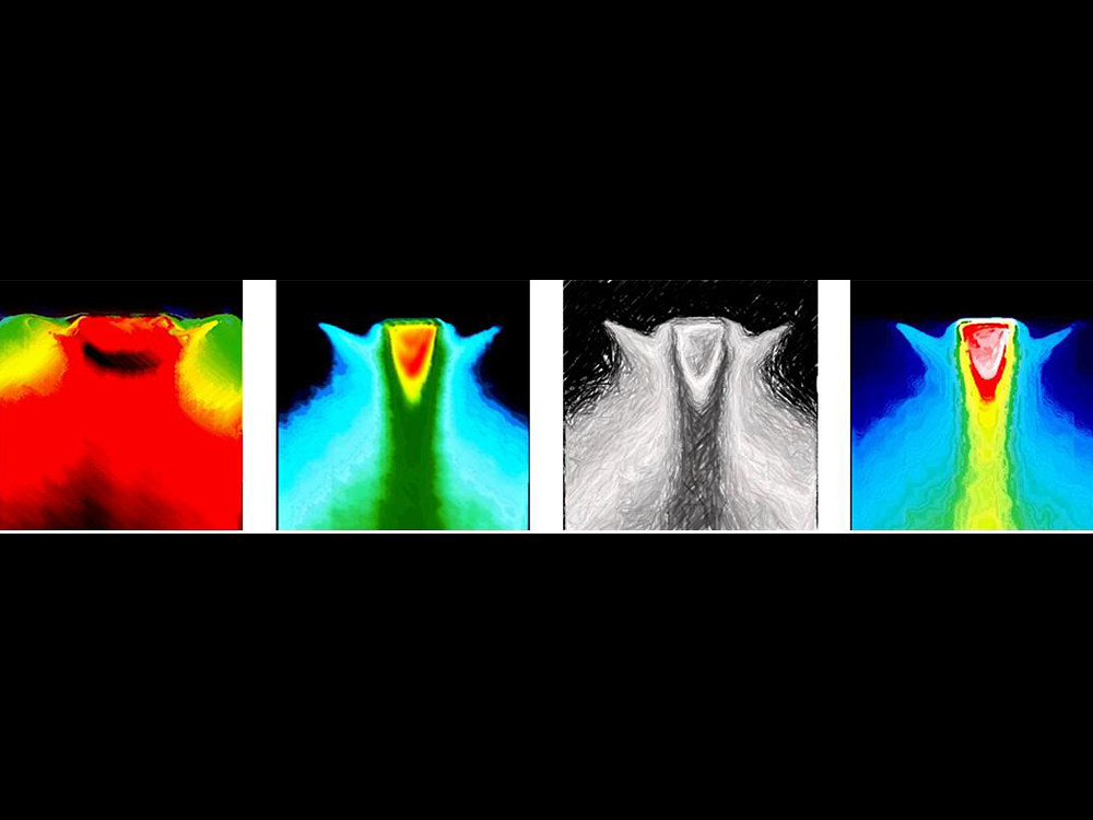
The Scream of Electrons
by Shoresh Soltani
Center for Correlated Electron Systems
Portraits of electron pockets living in a peculiar quantum world on the surface of strontium titanate. These ghostly beings which resemble The Scream by Edvard Munch, reveal themselves when we shed synchrotron light on a flat, shiny and clean surface of an single crystal in very low temperature and ultrahigh vacuum. They are responsible for electrical conductance on the surface of an insulating strontium titanate crystal which is the basis of oxide electronics. Different colors in each image, speak for different electron densities.
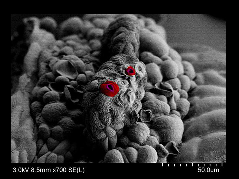
The Nose of Plant
by Taek Han Yoon
Center for Plant Aging Research
Plant's respiratory organs are called 'stomata'. They are responsible for photosynthesis and therefore are essential to the life of most of the species on Earth. Via Field Emission Scanning Electron Microscopy(FE SEM)* imaging technique, we were able to reveal that stomata are shaped like a heart....maybe a hint to Mother Earth’s love?
2015 IBS Art in Science
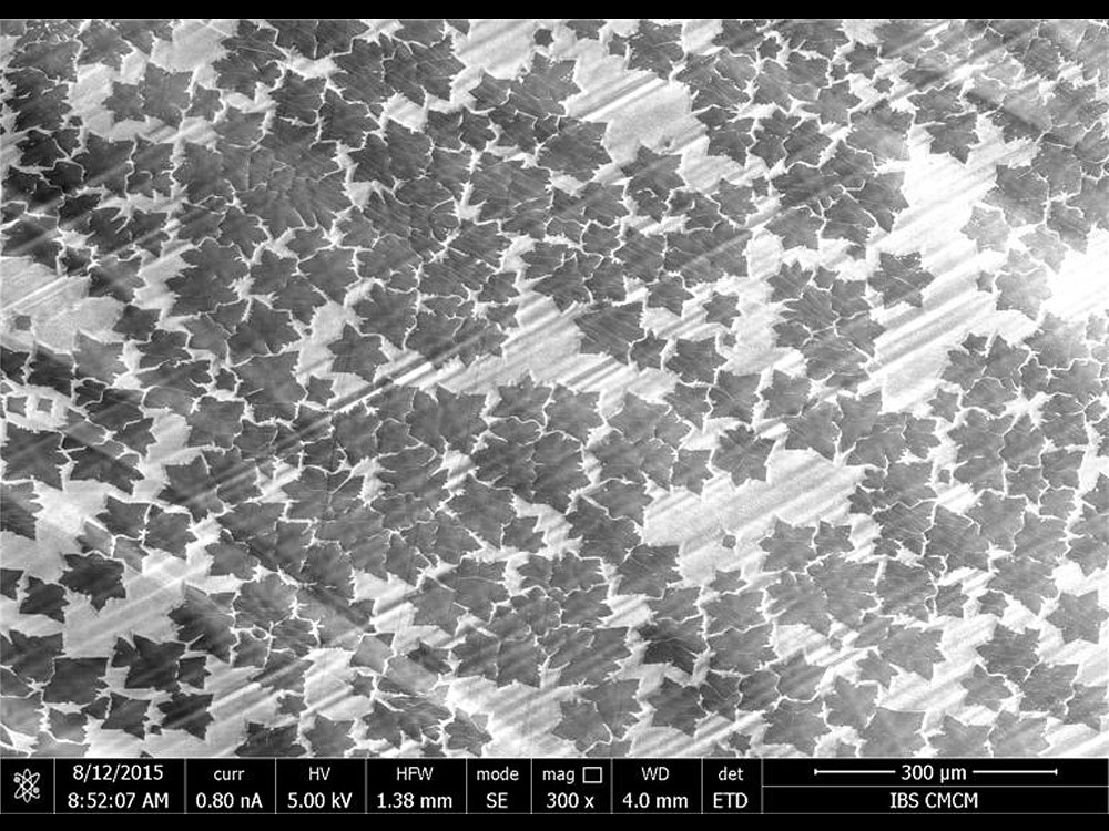
Autumn Graphene Leaves on Copper Snow
by Youngwoo Kwon, Rodney S. Ruoff
Center for Multidimensional Carbon Materials
Copper foil is one the most common substrates for graphene growth. Because of the unique crystalline structure of graphene ('2D honeycomb' structure), growth of graphene happens in certain patterns such as hexagons or, as in this case, hexagrams (also referred to as "sexagrams"). This Image by Scanning Electron Microscope (SEM)* shows the pattern of graphene growth at the micron scale - quite on time to greet the fall season with beautiful autumn/winter leaves on a copper landscape on a campus such as UNIST!
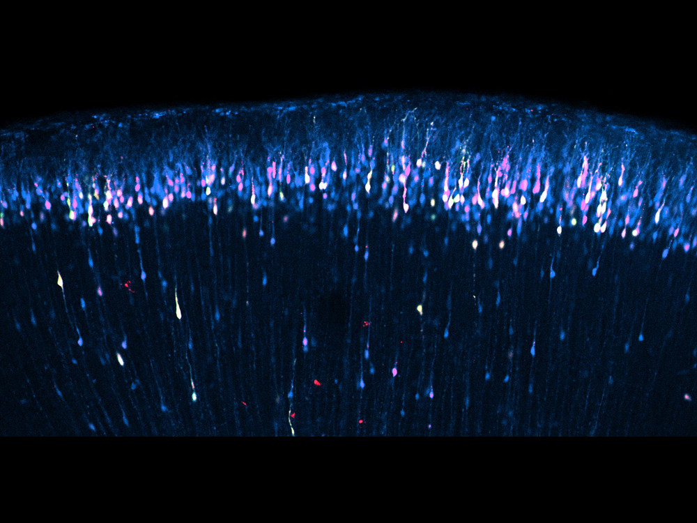
Star Shower in the Brain
by Sangkyu Lee, Hyeonjin Jung
Center for Cognition and Sociality
Brain's outer layer near the skull consists of cerebral cortex neurons. Fluorescent proteins of three different colors (green, red, far-red) were expressed in the neurons under developmental stage by injecting expression plasmids into ventricle regions of uterus (embryonic day 15) followed by electroporation with stick electrodes. This fluorescence image was taken after cross-section of brain sample.
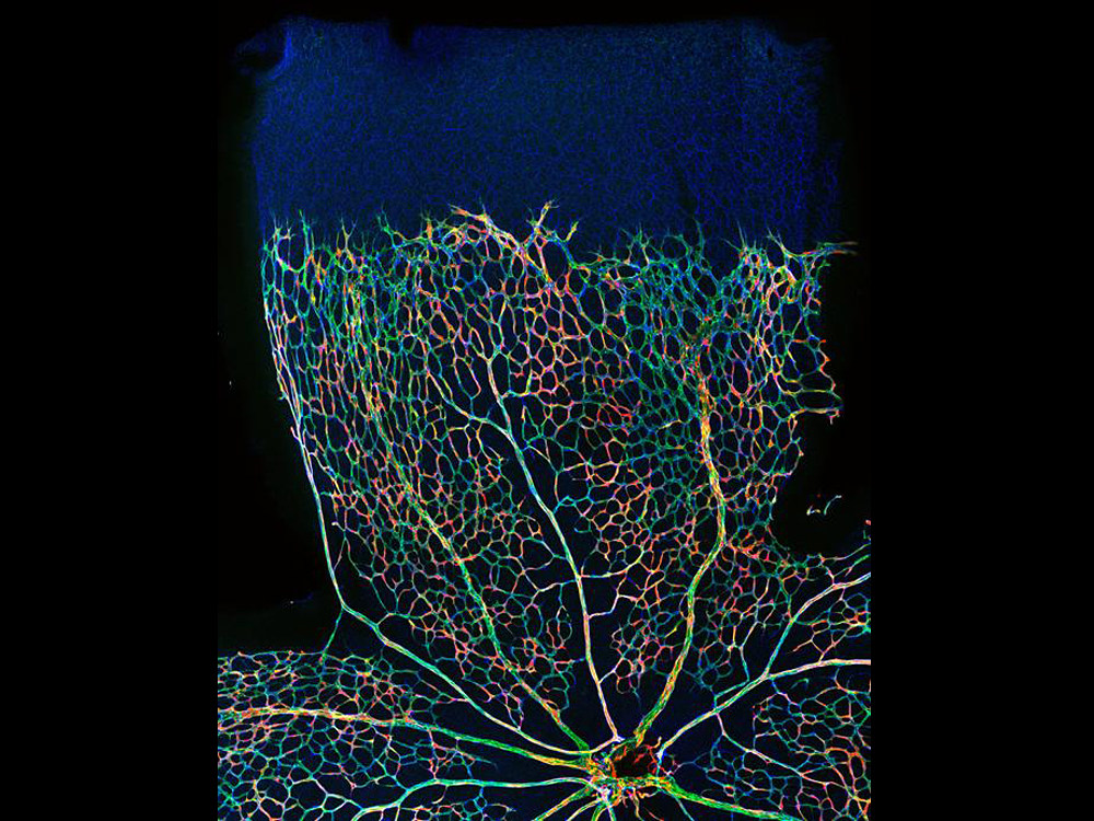
Rainbow in the Eye
by Do Young Park
Center for Vascular Research
Retinal vessels grow from center to periphery after birth. Retinal vessels are composed of vascular endothelial cells and pericytes. Pericytes cover endothelial cells and are important for angiogenesis, survival of endothelial cells, and barrier function. In this image, retinal blood vessels (in green) of mouse retina at postnatal day 5 show sprouting peripherally. Pericytes (red) are shown in mosaic pattern from Cre-induced loxP recombination to make tdtomato signals in pericytes.
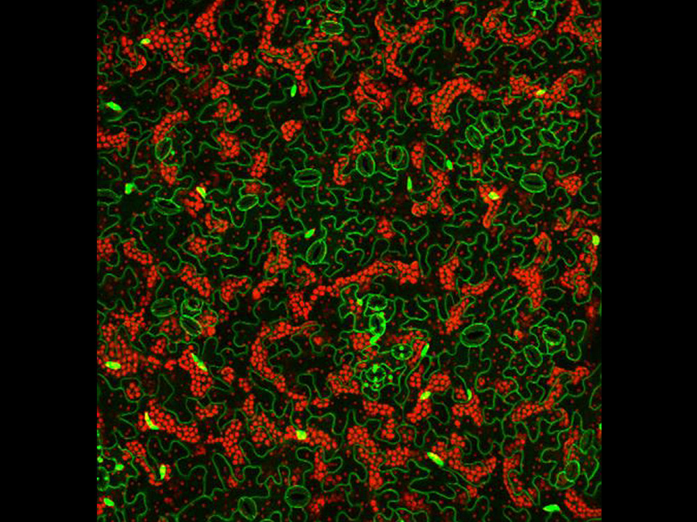
Nature Puzzle
by Sunghyun Hong, Sowook Seo, Soohyun Jung
Center for Plant Aging Research
Fluorescences in Cotyledon epidermis of 2 weeks grown Arabidopsis expressing H2B-GFP (Nucleus localized) and SYP122-GFP (Plasma membrane) were detected by LSM780 fluorescent confocal microscopy. Bright green dots are nuclei of epidermal cells and green lines are membranes. Small paired cells containing smaller green spots are guard cells which control gas exchange by opening and closing the stomata. Red fluorescent spots are chloroplasts which absorb sunlight to produce food and contain various fluorescent compounds such as anthocyanin causing autofluorescence. Shapes of epidermal cells are irregular like pieces of jigsaw puzzle.