| Title of announcement | Reading Pleasure and Pain from the Brain | ||||
|---|---|---|---|---|---|
| Business forms | Expiration date for bidding | ||||
| Department in charge | 전체관리자 | Registration Date | 2024-06-11 | Hits | 430 |
| att. |
![docx 파일명 : [v5] IBS보도자료_영문_이수안_수정2.docx](/images/mimetype/docx.gif) [v5] IBS보도자료_영문_이수안_수정2.docx
[v5] IBS보도자료_영문_이수안_수정2.docx
|
||||
|
|
|||||
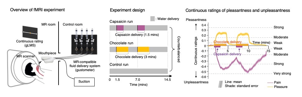
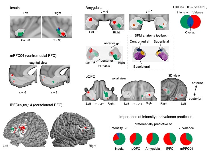
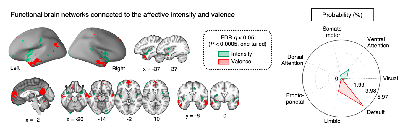
Reading Pleasure and Pain from the Brain- Identifying brain regions that predict pleasure and pain using fMRI-based activity patterns - A team of researchers led by LEE Soo Ahn and WOO Choong-Wan at the Center for Neuroscience Imaging Research (CNIR) within the Institute for Basic Science (IBS), in collaboration with CHOI Myunghwan at Seoul National University and Tor D. WAGER at Dartmouth College, has revealed how the brain processes emotional information of sustained pain and pleasure. Using functional Magnetic Resonance Imaging (fMRI), the team recorded brain activity while participants experienced sustained pain and pleasure induced by capsaicin and chocolate fluids. Through sophisticated machine learning techniques, they unraveled the brain activity patterns that encode pleasant or unpleasant emotions and the magnitude of sustained pain and pleasure. Although pain and pleasure are opposite experiences, they are intricately connected. Previous studies have suggested a set of brain regions that respond to both pain and pleasure. However, most previous studies have been conducted on animals rather than humans, and studies that directly compared the brain representations of pain and pleasure within the same individuals are still lacking. In this study, the research team conducted an experiment that induced sustained pain and pleasure to participants in the MR scanner, by delivering capsaicin and chocolate fluids. While experiencing sustained pain and pleasure, participants reported moment-by-moment changes in subjective pleasantness and unpleasantness. The participants’ subjective reports of pleasantness and unpleasantness gradually increased and persisted during the capsaicin and chocolate fluids deliveries and decreased after the deliveries ended. By inducing dynamic changes in sustained pain and pleasure, the team aimed to identify the brain regions activated by both experiences. The research team collected the brain imaging data and moment-by-moment changes in pleasantness or unpleasantness ratings from 58 participants. The team utilized machine learning techniques to analyze the brain data, and they identified a set of brain regions that responded to both sustained pain and pleasure. Based on the brain activity patterns of these common brain regions, the team developed two predictive models to capture (1) the magnitude of affective experiences regardless of how pleasant or unpleasant they are (i.e., ‘affective intensity’) and (2) the magnitude of pleasantness or unpleasantness (i.e., ‘affective valence’). The researchers found that these models successfully predicted the affective intensity and valence information of sustained pleasure and pain, from both the 58 individuals in the training dataset and 62 new individuals in the independent test dataset. The activity patterns predictive of the affective intensity and valence were spatially distinguishable, and these patterns were connected to distinct functional brain networks. This suggests that the affective intensity and valence information represent multiple aspects of brain mechanisms underlying pain-pleasure interaction. “While there have been separate lines of studies on pain and pleasure, research comparing the experiences of both pain and pleasure within the same individuals has been rarely conducted,” stated Dr. WOO Choong-Wan, associate director of IBS, who led the study. “The brain activity patterns for affective valence and intensity can contribute to the understanding of how pain and pleasure interact, as well as the brain mechanisms underlying depression commonly observed in chronic pain patients.” LEE Soo Ahn, a doctoral candidate and the first author of this study, emphasized, “These results demonstrate that pain and pleasure share the same underlying emotional information on pleasantness and unpleasantness,” adding, “We should focus on the fact that affective valence and intensity information can be represented across multiple brain regions.”
Notes for editors
- References
- Media Contact
- About the Institute for Basic Science (IBS)
|
|||||