


주메뉴
- About IBS 연구원소개
-
Research Centers
연구단소개
- Research Outcomes
- Mathematics
- Physics
- Center for Theoretical Physics of the Universe(Particle Theory and Cosmology Group)
- Center for Theoretical Physics of the Universe(Cosmology, Gravity and Astroparticle Physics Group)
- Center for Exotic Nuclear Studies
- Center for Artificial Low Dimensional Electronic Systems
- Center for Underground Physics
- Center for Axion and Precision Physics Research
- Center for Theoretical Physics of Complex Systems
- Center for Quantum Nanoscience
- Center for Van der Waals Quantum Solids
- Chemistry
- Life Sciences
- Earth Science
- Interdisciplinary
- Institutes
- Korea Virus Research Institute
- News Center 뉴스 센터
- Career 인재초빙
- Living in Korea IBS School-UST
- IBS School 윤리경영

주메뉴
- About IBS
-
Research Centers
- Research Outcomes
- Mathematics
- Physics
- Center for Theoretical Physics of the Universe(Particle Theory and Cosmology Group)
- Center for Theoretical Physics of the Universe(Cosmology, Gravity and Astroparticle Physics Group)
- Center for Exotic Nuclear Studies
- Center for Artificial Low Dimensional Electronic Systems
- Center for Underground Physics
- Center for Axion and Precision Physics Research
- Center for Theoretical Physics of Complex Systems
- Center for Quantum Nanoscience
- Center for Van der Waals Quantum Solids
- Chemistry
- Life Sciences
- Earth Science
- Interdisciplinary
- Institutes
- Korea Virus Research Institute
- News Center
- Career
- Living in Korea
- IBS School


News Center

| Title | Opening a New Chapter of Optical Imaging | ||||
|---|---|---|---|---|---|
| Name | Department of Communications | Registration Date | 2016-08-31 | Hits | 4638 |
| att. |
 thumb.jpg
|
||||
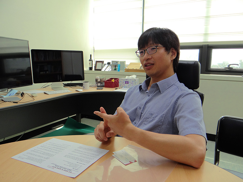
Opening a New Chapter of Optical Imaging- Associate Director CHOI, Wonshik of the Center for Molecular Spectroscopy and Dynamics (CMSD) - Interview PreviewQ: Could you introduce the CMSD? Q: As a physics major, you study bio imaging research. What was the turning point that made you choose the bio imaging field? Q: Your research on Tomographic Phase Microscopy (TPM) was featured in The Economist. Could you tell us more about the coverage?
Optical imaging investigates phenomena that are too small to be seen by the naked eye, but discernible using light microscopes. Since its invention in the early 17th century, the advancement of microscope has been quite slow. The breakthrough in resolving power came only 400 years after the birth of the microscope: By circumventing a presumed scientific limitation, the super-resolved fluorescence microscopy was developed allowing scientists to observe things smaller than roughly half the wavelength of light, i.e. 0.2 micrometers. The 2014 Nobel Prize in Chemistry was awarded to three scientists who broke that limit; bringing optical microscopy down to the nanoscale by exploiting the glow of molecules in response to light. Two main features of light microscopy are its resolution and imaging depth: Resolution is the ability to see fine things; a second attribute of a microscope is its imaging depth, which is the range of depth that a complex specimen is viewed in acceptable focus. The imaging depth has recently become of great interest for the study into molecules inside cells. Making great strides in clarity and accuracy of imaging, optical microscopy may have a wide variety of applications for medicine and industry, as well as research. The CMSD aims to develop various optical imaging tools for observing molecules in real time. Professor Choi, appointed as an associate director last July, is an expert in the area of optical bio-imaging. Choi pursues research to develop ultrahigh-resolution and deep-tissue imaging methods by revealing the mechanism of light transport in a complex scattering medium such as human skin tissues. Spotlighted by The EconomistChoi conducted experiments with the single-atom laser during his Ph. D. course in atomic physics. While working in the MIT Spectroscopy Laboratory as a visiting researcher, Choi came across the bio-imaging field. He was quite drawn to the infinite possibilities of the technique's applications: Though the then technical level required for bio-imaging field was not as advanced as that in atomic physics, its potential impact, Choi thought, seemed quite immediate on people.
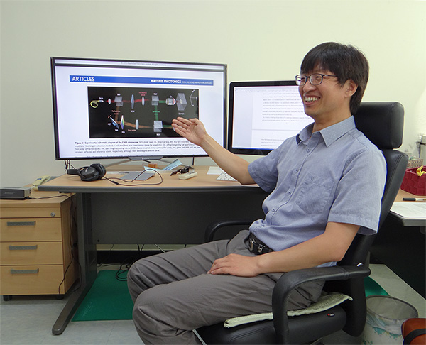
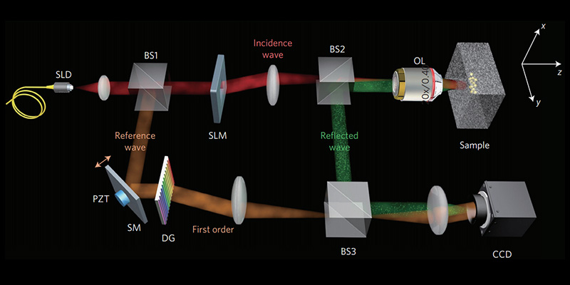
During his postdoctoral course at MIT, Choi invented TPM: A novel optical microscopy method enabling quantitative 3-D imaging of the refractive index of living cells and tissues. Professor Michael Feld, the then head of the Spectroscopy Laboratory conducted experiments to identify cancer cells by measuring refractive index (RI) distribution across a single cell with ultrahigh resolution optical microscopy. Researchers had to dissect the cell to calculate RI of individual organelles, meaning it is impossible to conduct experiments with a live cell. Choi developed a technique for quantitative 3D mapping of RI in living cells and tissues using optical microscopy from variable illumination angles. He explains, "It is somewhat akin to taking a CT scan on a single cell." Choi's invention of TPM was published in Nature Methods in 2007. As an exceptional invention, TPM was unusually featured by the journal The Economist, which described the triumph as saying that TPM may make biology as transparent as the cells it studies. Since his professorship at Korea University began in 2009, Choi presented a series of outstanding research results in the bio-imaging field. Choi and his colleagues demonstrated that a disordered medium can be converted into a lens to overcome the diffraction limit of a conventional imaging system. Choi did not necessarily see the disordered medium as a barrier to overcome; rather he took advantage of multiple scattering induced by the turbid media to extract the original image information from. Another breakthrough resulted in a method to develop the world's thinnest endoscope using fine optical fibers that can resolve a pattern of light that contains a series of shapes. Choi and his colleagues also invented a way to deliver light energy deep within the skin by identifying eigenmodes within the complex media. In addition, his group developed the Collective Accumulation of Single Scattering Microscopy (CASS), which allows scientists to monitor fine cellular changes inside a thick skin. Both of these inventions were published in the journal Nature Photonics. Choi noted, "The discovery that a disordered medium can be converted into a lens is quite meaningful for me since the achievement laid the foundation for my subsequent research results." The paper published in Physical Review Letters was picked as one of the top 10 innovation of 2011 by the journal New Scientist. Optical Imaging, from medical diagnosis to neuroscience and to brain research
Choi regards the invention of CASS as one of the most significant achievements. CASS microscopy extracted collectively the so-called single-scattered waves from the background of strong multiple-scattered waves. The single-scattered waves are those scattered only a single time by the target object, thereby retaining object information. CASS microscopy delivers high-resolution optical imaging as the collective accumulation of single scattering waves works as if clearing the complex media hidden deep within the skin. Choi said, "CASS microscopy represents one of the world's top-notch technologies, which allows for a high-resolution optical imaging of an unprecedented target depth. It can visualize fine cellular details 1.15mm down the skin with a resolution of 1μm." 1 micrometer 10-6 meter. Typical biological cells are about 10-20 µm in size. The research achievements by Choi and his colleagues are expected to find a number of medical applications, especially for diagnosis and operations. For example, 80% of cancer cells usually develop 1-3mm down the outer layer of the skin or organ and the initial development accompanies the enlargement of cell nucleus. Therefore, the currently available technique, such as CT, MRI, and ultrasound scans are limited in their capability to make early detection of cancer due to their low resolving power. On the contrary, the super-depth and high-resolution imaging allows for early diagnosis of cancer and the mapping of cancer cells surrounding tumors, which is useful for removing the malignant cells only: Super-depth endoscopic microscope can identify tumor cells before they grow into an abnormal growth of tissue. "Stem cell research can also benefit from super-depth and high-resolution imaging," Choi added. "The super-depth imaging allows scientists the ability to reveal the growth mechanism of stem cells in the spinal cord." Choi plans to expand the scope of its applications to neuroscience and brain research. California Institute of Technology, Washington University, and the Janelia Research Campus of Howard Hughes Medical Institute are already expediting their efforts to apply super-depth imaging technique in neuroscience. Choi is at the forefront of visualization of subcellular structures with high resolving power. He is now investigating on how to utilize multiple-scattered waves, as well as single-scattered waves. "Single-scattered waves are limited to a certain penetration level, preventing the fundamental achievable imaging depth," explained Choi. "If we are successful in discovering a method to use multiple-scattered waves, it will revolutionize the imaging field." He said that most of his research has focused verifying theoretical research and the IBS support will help pursue research to both discover new theories and explore their medical applications
|
|||||
| Next | |
|---|---|
| before |
- Content Manager
- Public Relations Team : Suh, William Insang 042-878-8137
- Last Update 2023-11-28 14:20

55, Expo-ro, Yuseong-gu, Daejeon, Korea, 34126
Tel. +82-42-878-8114 | Fax. +82-42-878-8079 | E-mail. webmaster@ibs.re.kr
Copyright(c) IBS. All Rights Reserved.
