


주메뉴
- About IBS 연구원소개
-
Research Centers
연구단소개
- Research Outcomes
- Mathematics
- Physics
- Center for Theoretical Physics of the Universe(Particle Theory and Cosmology Group)
- Center for Theoretical Physics of the Universe(Cosmology, Gravity and Astroparticle Physics Group)
- Center for Exotic Nuclear Studies
- Center for Artificial Low Dimensional Electronic Systems
- Center for Underground Physics
- Center for Axion and Precision Physics Research
- Center for Theoretical Physics of Complex Systems
- Center for Quantum Nanoscience
- Center for Van der Waals Quantum Solids
- Chemistry
- Life Sciences
- Earth Science
- Interdisciplinary
- Institutes
- Korea Virus Research Institute
- News Center 뉴스 센터
- Career 인재초빙
- Living in Korea IBS School-UST
- IBS School 윤리경영

주메뉴
- About IBS
-
Research Centers
- Research Outcomes
- Mathematics
- Physics
- Center for Theoretical Physics of the Universe(Particle Theory and Cosmology Group)
- Center for Theoretical Physics of the Universe(Cosmology, Gravity and Astroparticle Physics Group)
- Center for Exotic Nuclear Studies
- Center for Artificial Low Dimensional Electronic Systems
- Center for Underground Physics
- Center for Axion and Precision Physics Research
- Center for Theoretical Physics of Complex Systems
- Center for Quantum Nanoscience
- Center for Van der Waals Quantum Solids
- Chemistry
- Life Sciences
- Earth Science
- Interdisciplinary
- Institutes
- Korea Virus Research Institute
- News Center
- Career
- Living in Korea
- IBS School


News Center

| Title | Unveiling how lymph nodes regulate immune response | ||
|---|---|---|---|
| Embargo date | 2020-02-06 14:38 | Hits | 681 |
| Research Center |
Center for Vascular Research |
||
| Press release | |||
| att. | |||
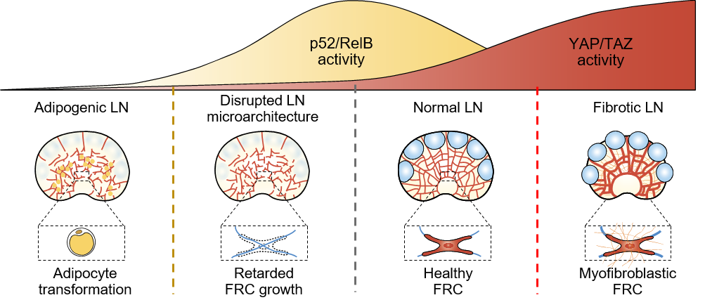
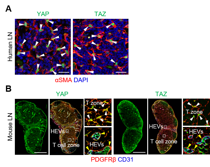
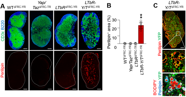
Unveiling how lymph nodes regulate immune response- The Hippo pathway keeps lymph nodes’ development healthy. If impaired, lymph nodes become full of fat cells or fibrosis develops - Pathogens such as severe acute respiratory syndrome (SARS), Middle East respiratory syndrome (MERS), and recently the novel coronavirus in Wuhan, China (2019-nCoV) have been a global threat. Lymph nodes (LNs) fight against infectious diseases by providing a shelter for immune cells to grow and launch an attack against pathogens. However, LNs’ particular inner workings are poorly understood. Scientists led by KOH Gou Young at the Center for Vascular Research, within the Institute for Basic Science (IBS), with collaborators of the Korea Advanced Institute of Science and Technology (KAIST), South Korea, have found that a chain of chemical reactions, known as the Hippo-YAP/TAZ signaling pathway, that plays a dominant role in the formation and maintenance of LNs. Their findings have been reported in the journal Nature Communications. One of the key components of LNs are fibroblastic reticular cells (FRCs), which form LN’s basic infrastructure and trigger immune responses by releasing cytokines, which are proteins important for immunity. Functional FRCs form during LN’s development: a poorly defined population of mesenchymal cells differentiate into FRC precursors, which further develop into mature FRCs with immune functions. Whereas the molecular details involved in the latter process, such as lymphotoxin-β receptor (LTβR) signaling, have been thoroughly described, the details of the commitment steps of FRC development are still unclear. The research team confirmed the importance of the Hippo pathway – a key regulator of cellular proliferation and organ size control – in FRCs’ maturation. The researchers used more than 20 different genetically modified mouse models to characterize the Hippo pathway at specific time points, depleting the proteins YAP/TAZ at various stages of FRC development.
“As I witnessed the enriched expression of YAP/TAZ in fibroblastic reticular cells of lymph nodes, I knew there must be a role of the Hippo pathway in FRCs,” says CHOI Sung Yong, first co-author of this study. By performing a careful examination of the mice’s LNs, the team found that FRCs transform into fat cells when YAP/TAZ are reduced in FRC precursors.
BAE Hosung, first co-author of this study, explains, “It was like a mathematic equation, when we drew out the findings on the blackboard, we were sure that depleting YAP/TAZ in fibroblastic reticular cell precursors would show an effect on the lymph nodes.” The researchers found that YAP/TAZ binding to p52 is required for maintaining FRC identity. JEONG Sun-Hye, first co-author of this study, notes, “I had this basic instinct that YAP/TAZ should bind with key components that regulate fibroblastic reticular cell identity, such as p52.”
Future research will focus on determining whether diseases or conditions that affect systemic immune responses can be linked to alterations in the Hippo signaling pathway in FRCs, and whether modulating Hippo signaling within FRCs could serve as a viable therapeutic option. Beyond their importance in the immune response against flu, FRCs have recently gained considerable recognition for their role in cancer progression and patient outcome. The degree of stromal fibrosis within metastatic LNs is an important prognostic factor that significantly affects disease-free survival of cancer patients. “It definitely warrants more extensive investigation of fibroblastic reticular cells in patients with tumor lymph node metastases prior to clinical investigation,” adds Koh.
Notes for editors - References - Media Contact - About the Institute for Basic Science (IBS) |
|||
|
|
|||

| Next | |
|---|---|
| before |
- Content Manager
- Communications Team : Kwon Ye Seul 042-878-8237
- Last Update 2023-11-28 14:20

55, Expo-ro, Yuseong-gu, Daejeon, Korea, 34126
Tel. +82-42-878-8114 | Fax. +82-42-878-8079 | E-mail. webmaster@ibs.re.kr
Copyright(c) IBS. All Rights Reserved.
